Advertisement

Main menu

  • Home
  • Content
    • Current Issue
    • Accepted Manuscripts
    • Article Preview
    • Past Issue Archive
    • AJNR Case Collection
    • Case of the Week Archive
    • Classic Case Archive
    • Case of the Month Archive
  • Special Collections
    • Spinal CSF Leak Articles (Jan 2020-June 2024)
    • 2024 AJNR Journal Awards
    • Most Impactful AJNR Articles
  • Multimedia
    • AJNR Podcast
    • AJNR Scantastics
    • Video Articles
  • For Authors
    • Submit a Manuscript
    • Author Policies
    • Fast publishing of Accepted Manuscripts
    • Graphical Abstract Preparation
    • Manuscript Submission Guidelines
    • Imaging Protocol Submission
    • Submit a Case for the Case Collection
  • About Us
    • About AJNR
    • Editorial Board
  • More
    • Become a Reviewer/Academy of Reviewers
    • Subscribers
    • Permissions
    • Alerts
    • Feedback
    • Advertisers
    • ASNR Home
  • Other Publications
    • ajnr

User menu

  • Alerts
  • Log in

Search

  • Advanced search
American Journal of Neuroradiology

American Journal of Neuroradiology

ASHNR American Society of Functional Neuroradiology ASHNR American Society of Pediatric Neuroradiology ASSR
  • Alerts
  • Log in

Advanced Search

  • Home
  • Content
    • Current Issue
    • Accepted Manuscripts
    • Article Preview
    • Past Issue Archive
    • AJNR Case Collection
    • Case of the Week Archive
    • Classic Case Archive
    • Case of the Month Archive
  • Special Collections
    • Spinal CSF Leak Articles (Jan 2020-June 2024)
    • 2024 AJNR Journal Awards
    • Most Impactful AJNR Articles
  • Multimedia
    • AJNR Podcast
    • AJNR Scantastics
    • Video Articles
  • For Authors
    • Submit a Manuscript
    • Author Policies
    • Fast publishing of Accepted Manuscripts
    • Graphical Abstract Preparation
    • Manuscript Submission Guidelines
    • Imaging Protocol Submission
    • Submit a Case for the Case Collection
  • About Us
    • About AJNR
    • Editorial Board
  • More
    • Become a Reviewer/Academy of Reviewers
    • Subscribers
    • Permissions
    • Alerts
    • Feedback
    • Advertisers
    • ASNR Home
  • Follow AJNR on Twitter
  • Visit AJNR on Facebook
  • Follow AJNR on Instagram
  • Join AJNR on LinkedIn
  • RSS Feeds

Welcome to the new AJNR, Updated Hall of Fame, and more. Read the full announcements.


AJNR is seeking candidates for the position of Associate Section Editor, AJNR Case Collection. Read the full announcement.


Diffuse Pediatric-Type High-Grade Glioma H3-Wild-Type and IDH-Wild-Type, Optico-Chiasmatic Region

  • Background:
    • Pediatric high-grade gliomas are relatively uncommon tumors and are considered a separate entity in the new WHO Classification of CNS Tumors. Although they are qualified as “diffuse,” a circumscribed pattern of growth may be encountered. Regardless of morphology, they are considered biologically aggressive tumors with a dismal prognosis. The presence of extensive meningeal spread of the tumor at presentation is a poor prognostic indicator.
  • Clinical Presentation:
    • Clinical features may vary depending on the location of the tumor.
  • Key Diagnostic Features:
    • Clinical suspicion of high-grade tumor is based on the short duration of symptoms and lack of constitutional symptoms to suggest infective etiology.
    • Imaging features are not very specific.
    • Inhomogeneously enhancing masses, often with rim enhancement have been described. Lesions may be well-demarcated and may have mild perilesional edema. 
    • Diffusion restriction is not a consistent feature.
  • Differential Diagnosis:
    • Tuberculosis: Constitutional symptoms often present, T2 hypointense lesions with peripheral enhancement with associated basal exudates and leptomeningeal enhancement
    • Lymphoma: T2 iso-/hypointense lesions along the ventricular margins with diffusion restriction and homo-/heterogeneous enhancement
    • Germ cell tumors: Lesions of T2 heterogeneous signal with foci of blooming and patchy diffusion restriction. Elevated serum levels of tumor markers like human chorionic gonadotrophin and alpha-fetoprotein may be present.
  • Management:
    • Tumor resection followed by radiotherapy and chemotherapy
October 31, 2024

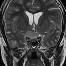
A 12-year-old boy presents with gradual right-sided visual loss for 3 months, headache for 1 month, and left-sided vision loss for 3 weeks.

View Case
  • Read more about 10312024
  • Comments
Advertisement
Subscribe to RSS - Diffuse Pediatric-Type High-Grade Glioma H3-Wild-Type and IDH-Wild-Type, Optico-Chiasmatic Region

Indexed Content

  • Current Issue
  • Accepted Manuscripts
  • Article Preview
  • Past Issues
  • Editorials
  • Editors Choice
  • Fellow Journal Club
  • Letters to the Editor

Cases

  • Case Collection
  • Archive - Case of the Week
  • Archive - Case of the Month
  • Archive - Classic Case

Special Collections

  • Special Collections

Resources

  • News and Updates
  • Turn around Times
  • Submit a Manuscript
  • Author Policies
  • Manuscript Submission Guidelines
  • Evidence-Based Medicine Level Guide
  • Publishing Checklists
  • Graphical Abstract Preparation
  • Imaging Protocol Submission
  • Submit a Case
  • Become a Reviewer/Academy of Reviewers
  • Get Peer Review Credit from Publons

Multimedia

  • AJNR Podcast
  • AJNR SCANtastic
  • Video Articles

About Us

  • About AJNR
  • Editorial Board
  • Not an AJNR Subscriber? Join Now
  • Alerts
  • Feedback
  • Advertise with us
  • Librarian Resources
  • Permissions
  • Terms and Conditions

American Society of Neuroradiology

  • Not an ASNR Member? Join Now

© 2025 by the American Society of Neuroradiology All rights, including for text and data mining, AI training, and similar technologies, are reserved.
Print ISSN: 0195-6108 Online ISSN: 1936-959X

Powered by HighWire