Advertisement

Main menu

  • Home
  • Content
    • Current Issue
    • Accepted Manuscripts
    • Article Preview
    • Past Issue Archive
    • AJNR Case Collection
    • Case of the Week Archive
    • Classic Case Archive
    • Case of the Month Archive
  • Special Collections
    • Spinal CSF Leak Articles (Jan 2020-June 2024)
    • 2024 AJNR Journal Awards
    • Most Impactful AJNR Articles
  • Multimedia
    • AJNR Podcast
    • AJNR Scantastics
    • Video Articles
  • For Authors
    • Submit a Manuscript
    • Author Policies
    • Fast publishing of Accepted Manuscripts
    • Graphical Abstract Preparation
    • Manuscript Submission Guidelines
    • Imaging Protocol Submission
    • Submit a Case for the Case Collection
  • About Us
    • About AJNR
    • Editorial Board
  • More
    • Become a Reviewer/Academy of Reviewers
    • Subscribers
    • Permissions
    • Alerts
    • Feedback
    • Advertisers
    • ASNR Home
  • Other Publications
    • ajnr

User menu

  • Alerts
  • Log in

Search

  • Advanced search
American Journal of Neuroradiology

American Journal of Neuroradiology

ASHNR American Society of Functional Neuroradiology ASHNR American Society of Pediatric Neuroradiology ASSR
  • Alerts
  • Log in

Advanced Search

  • Home
  • Content
    • Current Issue
    • Accepted Manuscripts
    • Article Preview
    • Past Issue Archive
    • AJNR Case Collection
    • Case of the Week Archive
    • Classic Case Archive
    • Case of the Month Archive
  • Special Collections
    • Spinal CSF Leak Articles (Jan 2020-June 2024)
    • 2024 AJNR Journal Awards
    • Most Impactful AJNR Articles
  • Multimedia
    • AJNR Podcast
    • AJNR Scantastics
    • Video Articles
  • For Authors
    • Submit a Manuscript
    • Author Policies
    • Fast publishing of Accepted Manuscripts
    • Graphical Abstract Preparation
    • Manuscript Submission Guidelines
    • Imaging Protocol Submission
    • Submit a Case for the Case Collection
  • About Us
    • About AJNR
    • Editorial Board
  • More
    • Become a Reviewer/Academy of Reviewers
    • Subscribers
    • Permissions
    • Alerts
    • Feedback
    • Advertisers
    • ASNR Home
  • Follow AJNR on Twitter
  • Visit AJNR on Facebook
  • Follow AJNR on Instagram
  • Join AJNR on LinkedIn
  • RSS Feeds

Welcome to the new AJNR, Updated Hall of Fame, and more. Read the full announcements.


AJNR is seeking candidates for the position of Associate Section Editor, AJNR Case Collection. Read the full announcement.


Epidural Lipomatosis with Post-operative Cord Infarct

  • Background
    • ​Epidural lipomatosis is a rare condition due to overgrowth of adipose tissue in the extradural space, most commonly found in the thoracic spine. 
    • The underlying mechanism remains unknown, however 4 main underlying causes can be identified; the most prevalent being exogenous steroid use, but also includes endogenous elevation of steroids, obesity, and other idiopathic causes. 
  • Clinical Presentation
    • ​Chronic back pain, gradual lower limb weakness, and neurological deficits related to local mass effect of lipomatosis and resultant spinal cord and/or nerve root compression.
  • Key Diagnostic Features
    • ​Epidural fat follows fat signal on all MR sequences, has homogeneous high signal intensity on T1- and T2-weighed images, with suppressed signal on STIR and fat-saturated MR images.
    • Mean sagittal thickness of epidural fat greater than or equal to 7 mm is suggestive of epidural lipomatosis.
    • Along the thoracic spine, fat is predominantly found dorsally with ventral displacement of the thecal sac.
    • At the lumbar level, the fat is circumferential, with a trefoil or Y-shaped appearance.
  • Differential Diagnosis (for epidural process)​
    • ​Subacute epidural hematoma: Can have similar MR appearance to epidural lipomatosis but use of fat-suppression techniques in conjunction with T1- and T2-weighted sequences can help differentiate. Acute epidural hemorrhage is isointense on T1-weighted MR images initially and later becomes hyperintense on T1- and T2-weighted MR images when subacute. Chronicity of symptoms with lack of trauma and negative coagulation panel is also more suggestive of epidural lipomatosis. 
    • Spinal lipoma/spinal angiolipoma: Encapsulated spinal lipoma occurs commonly in the thoracic spine within epidural space dorsal to thecal sac, and presents as a focal lesion without extension to adjacent vertebral levels whereas spinal angiolipoma can be composed of adipose and vascular elements with potential for invasion of surrounding structures, e.g. bone. 
    • Epidural tumor/metastases: Intermediate to high signal on T2WI and STIR with gadolinium enhancement. E.g. epidural liposcarcoma presents with irregular thickened septa on MR images which can enhance following contrast, with areas of hyperintense signal on T2-weighted MR.
  • Differential Diagnosis (for extensive cord T2 hyperintensity)
    • ​Cord ischemia/infarct: Abnormal T2 hyperintense signal within the cord, presenting as focal cord enlargement, without enhancement.  While MR may be normal within first few hours after onset of symptoms,  these delayed imaging findings are not uncommon and can appear several days post initial infarct. 
    • Demyelinating disease: Includes acute disseminated encephalomyelitis (ADEM) and transverse myelitis (TM). ADEM appears as areas of increased T2/FLAIR with edema and localized mass effect, with enhancement uncommon. TM appears hypo- or isointense on T1, hyperintense on T2, with variable enhancement.
    • Type 1 dural AVF: Hyperintensities with peripheral sparing on T2WI with possible cord extension of 5-7 vertebral levels and conus involvement. Spinal cord edema in dural AVF is usually homogenous with a tapered margin. Dilated intervertebral vein or engorged venous flow voids on T2-weighted images are suggestive. 
    • Spinal cord intramedullary tumor (if timing applicable): Manifests as expansion of the cord with varying signal intensities and enhancement depending on the tumor type. In children, astrocytomas are most common type – they are ill defined, are T1 hypo- or isointense, T2 hyperintense. and have heterogeneous or no enhancement.
    • Radiation myelopathy (if history applicable): Presents as swelling of the cord with T2 hyperintensity with or without areas of enhancement. 
  • Treatment
    • ​Dependent on severity of symptoms, ranging from conservative therapy such as weight loss and steroid tapering, to more invasive interventions such as surgical decompression with multilevel laminectomy and fusion when there is cord compression and radiculopathy.
    • A rare, post-surgical complication of such decompression includes cord ischemia/infarct, such as that seen in this case. Possible mechanisms for post-decompression cord infarct consist of, but are not limited to, reperfusion injury such as hypotension and/or sub-therapeutic MAP, intra-operative manipulation, epidural hematoma, hemorrhage, and persistent and/or new cord compression. 
June 6, 2019

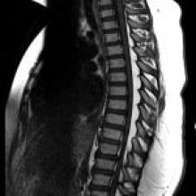
A 9-year-old boy with a BMI of 40, worsening bilateral lower extremity weakness, and gait abnormality for three months. Preoperative physical exam shows bilateral lower extremity hyperreflexia, clonus, and positive Babinski. Four days postoperatively, he developed bilateral leg paralysis and a transient episode of hypotension.

View Case
  • Read more about 06062019
  • Comments
Advertisement
Subscribe to RSS - Epidural Lipomatosis with Post-operative Cord Infarct

Indexed Content

  • Current Issue
  • Accepted Manuscripts
  • Article Preview
  • Past Issues
  • Editorials
  • Editors Choice
  • Fellow Journal Club
  • Letters to the Editor

Cases

  • Case Collection
  • Archive - Case of the Week
  • Archive - Case of the Month
  • Archive - Classic Case

Special Collections

  • Special Collections

Resources

  • News and Updates
  • Turn around Times
  • Submit a Manuscript
  • Author Policies
  • Manuscript Submission Guidelines
  • Evidence-Based Medicine Level Guide
  • Publishing Checklists
  • Graphical Abstract Preparation
  • Imaging Protocol Submission
  • Submit a Case
  • Become a Reviewer/Academy of Reviewers
  • Get Peer Review Credit from Publons

Multimedia

  • AJNR Podcast
  • AJNR SCANtastic
  • Video Articles

About Us

  • About AJNR
  • Editorial Board
  • Not an AJNR Subscriber? Join Now
  • Alerts
  • Feedback
  • Advertise with us
  • Librarian Resources
  • Permissions
  • Terms and Conditions

American Society of Neuroradiology

  • Not an ASNR Member? Join Now

© 2025 by the American Society of Neuroradiology All rights, including for text and data mining, AI training, and similar technologies, are reserved.
Print ISSN: 0195-6108 Online ISSN: 1936-959X

Powered by HighWire