Advertisement

Main menu

  • Home
  • Content
    • Current Issue
    • Accepted Manuscripts
    • Article Preview
    • Past Issue Archive
    • AJNR Case Collection
    • Case of the Week Archive
    • Classic Case Archive
    • Case of the Month Archive
  • Special Collections
    • Spinal CSF Leak Articles (Jan 2020-June 2024)
    • 2024 AJNR Journal Awards
    • Most Impactful AJNR Articles
  • Multimedia
    • AJNR Podcast
    • AJNR Scantastics
    • Video Articles
  • For Authors
    • Submit a Manuscript
    • Author Policies
    • Fast publishing of Accepted Manuscripts
    • Graphical Abstract Preparation
    • Manuscript Submission Guidelines
    • Imaging Protocol Submission
    • Submit a Case for the Case Collection
  • About Us
    • About AJNR
    • Editorial Board
  • More
    • Become a Reviewer/Academy of Reviewers
    • Subscribers
    • Permissions
    • Alerts
    • Feedback
    • Advertisers
    • ASNR Home
  • Other Publications
    • ajnr

User menu

  • Alerts
  • Log in

Search

  • Advanced search
American Journal of Neuroradiology

American Journal of Neuroradiology

ASHNR American Society of Functional Neuroradiology ASHNR American Society of Pediatric Neuroradiology ASSR
  • Alerts
  • Log in

Advanced Search

  • Home
  • Content
    • Current Issue
    • Accepted Manuscripts
    • Article Preview
    • Past Issue Archive
    • AJNR Case Collection
    • Case of the Week Archive
    • Classic Case Archive
    • Case of the Month Archive
  • Special Collections
    • Spinal CSF Leak Articles (Jan 2020-June 2024)
    • 2024 AJNR Journal Awards
    • Most Impactful AJNR Articles
  • Multimedia
    • AJNR Podcast
    • AJNR Scantastics
    • Video Articles
  • For Authors
    • Submit a Manuscript
    • Author Policies
    • Fast publishing of Accepted Manuscripts
    • Graphical Abstract Preparation
    • Manuscript Submission Guidelines
    • Imaging Protocol Submission
    • Submit a Case for the Case Collection
  • About Us
    • About AJNR
    • Editorial Board
  • More
    • Become a Reviewer/Academy of Reviewers
    • Subscribers
    • Permissions
    • Alerts
    • Feedback
    • Advertisers
    • ASNR Home
  • Follow AJNR on Twitter
  • Visit AJNR on Facebook
  • Follow AJNR on Instagram
  • Join AJNR on LinkedIn
  • RSS Feeds

Welcome to the new AJNR, Updated Hall of Fame, and more. Read the full announcements.


AJNR is seeking candidates for the position of Associate Section Editor, AJNR Case Collection. Read the full announcement.


Extraventricular Neurocytoma

  • Background:
    • Neurocytomas are uncommon neoplasms. Most neurocytomas present as intraventricular masses near the foramen of Monro. Extraventricular neurocytoma (EVN) by definition are located within the brain parenchyma. Children and young adults are more frequently affected (median age 34 years). EVNs show no obvious sex predilection.
    • EVNs are mainly found in the frontal (46%) and parietal (23%) regions, but can be located anywhere along the neuroaxis.
  • Clinical Presentation:
    • Symptoms are non-specific. They can present with headache, dizziness, visual impairment, seizures, or focal defictis, depending on the size and localization of the tumor.
  • Key Radiologic Features:
    • These tumors frequently present as a well-circumscribed (75%) mass, with cystic degeneration (58%), mild or moderate perilesional edema (51%), calcification, and hemorrhage. Enhancement is common (92%), but the degree and pattern of enhancement are variable.
    • MRS shows increased Cho/Cr ratio and decreased NAA/Cr ratio, and PWI demonstrates elevated cerebral blood volume (CVB), as other enhancing brain neoplasms.
    • The imaging characteristics of EVNs have significant overlap with oligodendrogliomas. Both present similar morphologic features and perfusion patterns (elevated CVB even in low grade lesions, and Choline/Cr ratio).
    • Therefore the diagnosis requires biopsy.
  • Histopathologic Features:
    • Positive antibody reaction to synaptophysin, glial fibrillary acidic protein (GFAP), neurofilament and NeuN.
  • Differential Diagnoses:
    • Oligodendroglioma, astrocytoma, dysembryoplastic neuroepithelial tumor: As stated before, the imaging characteristics are non-specific for EVNs, as there is significant overlap with other CNS tumors.
  • Treatment:
    • After complete resection most neurocytomas do not recur. Subtotal resection is associated with an increased likelihood of recurrence.
September 1, 2016

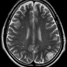
A 46-year-old patient presents with a history of epilepsy, but otherwise no other relevant past medical history.

View Case
  • Read more about 09012016
  • Comments
Advertisement
Subscribe to RSS - Extraventricular Neurocytoma

Indexed Content

  • Current Issue
  • Accepted Manuscripts
  • Article Preview
  • Past Issues
  • Editorials
  • Editors Choice
  • Fellow Journal Club
  • Letters to the Editor

Cases

  • Case Collection
  • Archive - Case of the Week
  • Archive - Case of the Month
  • Archive - Classic Case

Special Collections

  • Special Collections

Resources

  • News and Updates
  • Turn around Times
  • Submit a Manuscript
  • Author Policies
  • Manuscript Submission Guidelines
  • Evidence-Based Medicine Level Guide
  • Publishing Checklists
  • Graphical Abstract Preparation
  • Imaging Protocol Submission
  • Submit a Case
  • Become a Reviewer/Academy of Reviewers
  • Get Peer Review Credit from Publons

Multimedia

  • AJNR Podcast
  • AJNR SCANtastic
  • Video Articles

About Us

  • About AJNR
  • Editorial Board
  • Not an AJNR Subscriber? Join Now
  • Alerts
  • Feedback
  • Advertise with us
  • Librarian Resources
  • Permissions
  • Terms and Conditions

American Society of Neuroradiology

  • Not an ASNR Member? Join Now

© 2025 by the American Society of Neuroradiology All rights, including for text and data mining, AI training, and similar technologies, are reserved.
Print ISSN: 0195-6108 Online ISSN: 1936-959X

Powered by HighWire