
Article Figures & Data
Figures
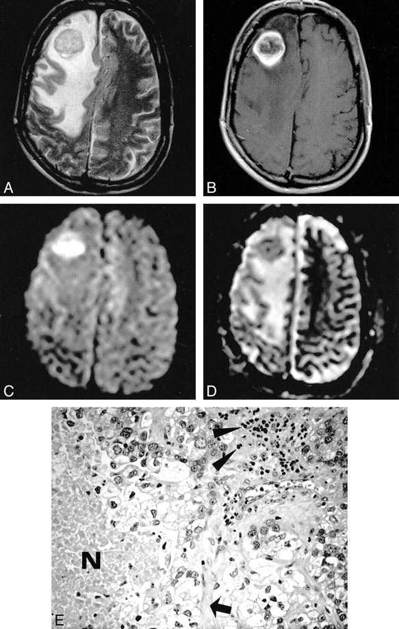
- fig 1.
Metastatic adenocarcinoma. The T2-weighted image (A) shows a hypointense mass lesion surrounded by massive edema. On the contrast-enhanced T1-weighted image (B), there is ring enhancement presumably due to central necrosis. With diffusion weighting (C), the central part of the tumor becomes markedly hyperintense, while the ADC map (D) reveals low values, indicating restricted diffusion. Histopathologic section (E): Note that besides tumor cells, additional geographic necrosis (N), lymphocytes (arrowheads), and connective tissue (arrow) are present (hematoxylin and eosin stain; original magnification, × 200)
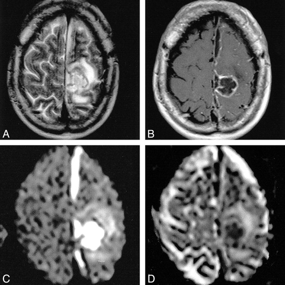
- fig 2.
Pyogenic brain abscess. On the T2-weighted image (A), the lesion has a thick hypointense wall with a hyperintense center, consistent with necrosis. Dense ring enhancement is noted on the contrast- enhanced T1-weighted image (B). The DW image (C) and the ADC map (D) reveal restricted diffusion within the center
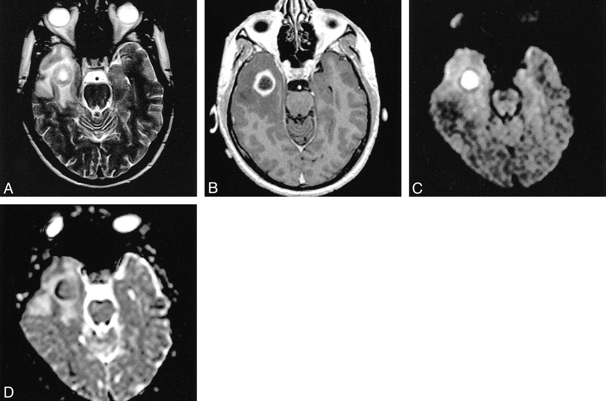
- fig 3.
Pyogenic brain abscess with parafalcial subdural empyema. On T2-weighted image (A) the lesion has a thick hypointense wall. The contrast-enhanced T1-weighted image (B) shows ring enhancement. Evidence for restricted diffusion within the abscess cavity and the parafalcial empyema is provided by the DW image (C) and the ADC map (D)
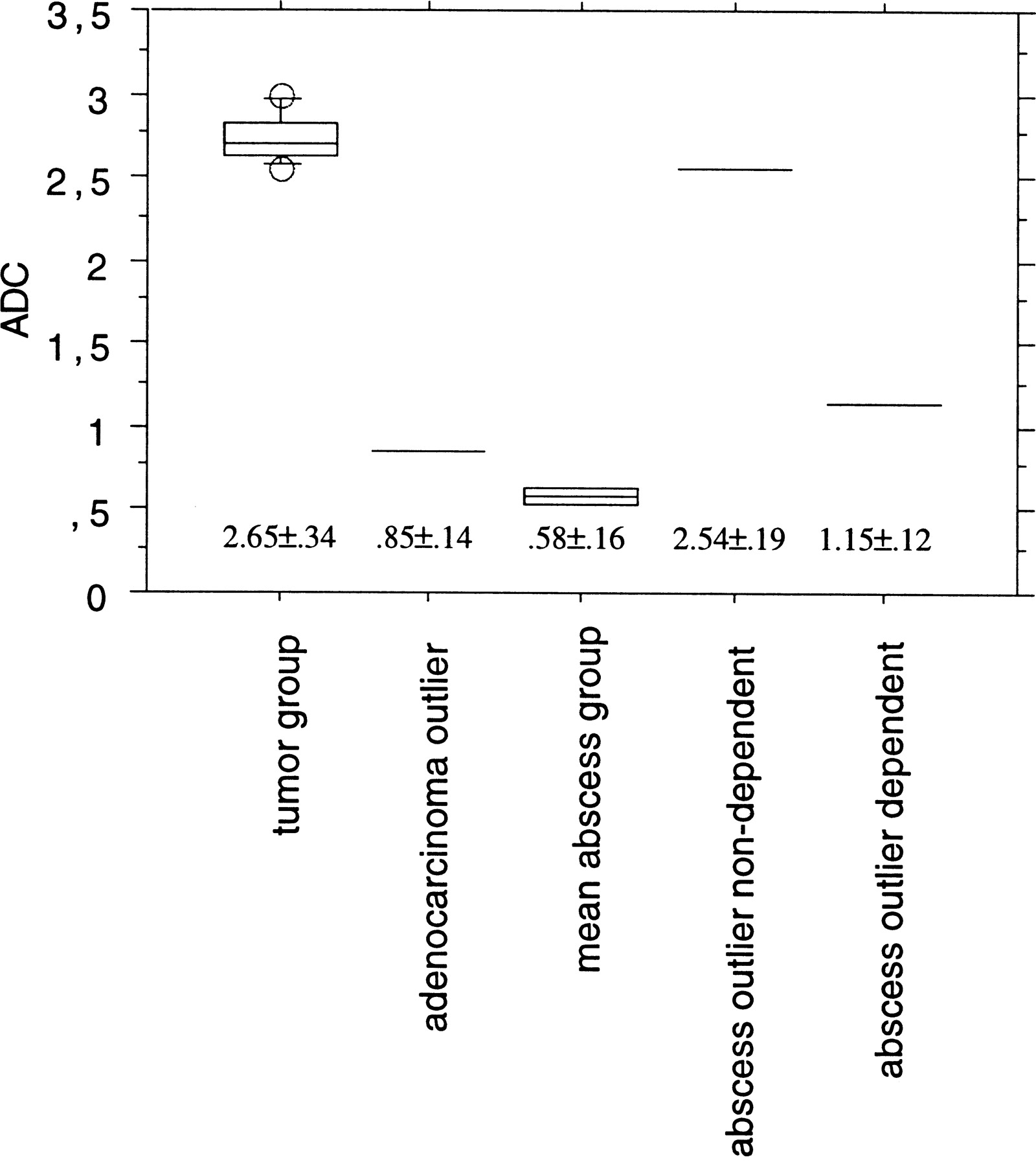
- fig 4.
Mean ADC values ± SD (×10−3 mm2/s) of the mean tumor group (n = 13), adenocarcinoma outlier (n = 1), mean abscess group (n = 2), and nondependent and dependent portions of the abscess outlier (n = 1)
- fig 5.
Postoperative brain abscess. The T2-weighted image (A) shows a hyperintense cavity surrounded by massive edema. The contrast-enhanced T1-weighted image (B) shows ring enhancement. With diffusion weighting (C), the gravity-dependent portion was markedly hyperintense (arrow) and showed markedly hypointense signal on the ADC map, (arrow), indicating restricted diffusion (D). The DW imaging intensity of the nondependent portion was hypointense with corresponding marked hyperintensity on the ADC map


In this issue


Jump to section
Related Articles
Cited By...
- "Dazed and diffused": making sense of diffusion abnormalities in neurologic pathologies
- Differentiation of Brain Abscesses from Necrotic Glioblastomas and Cystic Metastatic Brain Tumors with Diffusion Tensor Imaging
- Unusual findings in cerebral abscess: report of two cases.
- Glioblastoma multiforme with atypical diffusion-weighted MR findings
- Differentiation of Toxoplasmosis and Lymphoma in AIDS Patients by Using Apparent Diffusion Coefficients