
Article Figures & Data
Figures
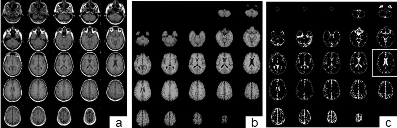
- Fig 1.
Semiautomated method of obtaining BPF. A, Raw T1-weighted 2D spin-echo noncontrast axial sequence. B, After masking (removal) of extracranial tissue. The segmented image (C) results from separation of the parenchyma (black) and CSF (white) into two compartments. The image surrounded by the white square (C) is used to identify normal-appearing white matter for the thresholding technique (see Methods). Adapted from Bermel et al (25).
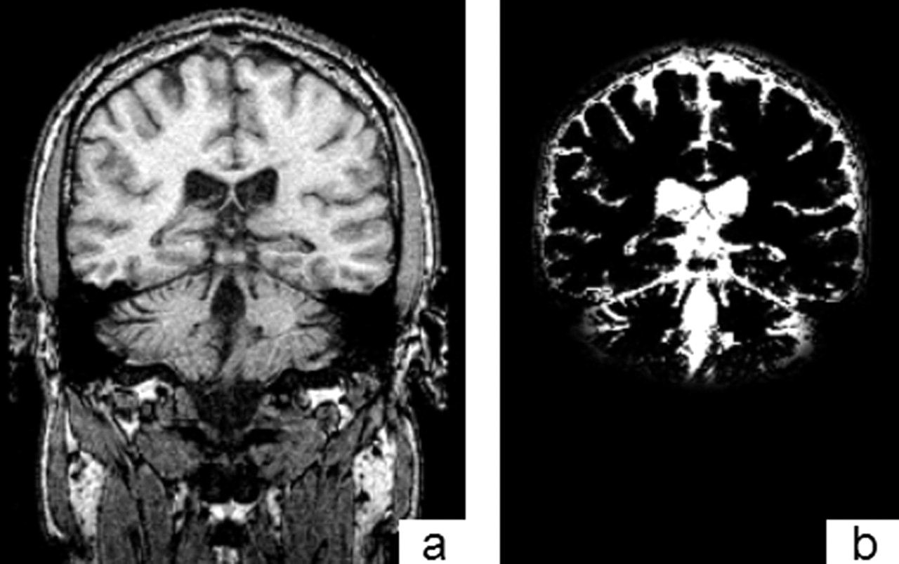
- Fig 3.
Automated method of obtaining BPF by using SPM-99 (see Methods). A T1-weighted 3D section showed the source image (A) and the resulting image after masking and segmentation into parenchyma or CSF (B).
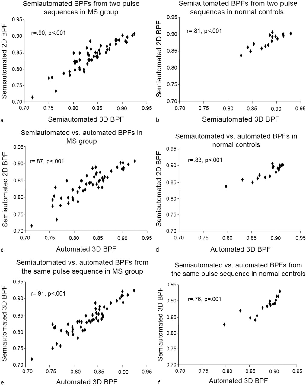
- Fig 4.
Scatterplots of semiautomated-2D versus semiautomated-3D BPF in the MS (A) and control (B) groups. Semiautomated 2D versus automated 3D BPF in the MS (C) and control (D) groups. Semiautomated 3D versus automated 3D BPF in the MS (E) and control (F) groups. BPFs derived by the two methods were highly correlated within the MS group and control group. The lower intercorrelation in the control versus MS group in all three methods is most likely related to restricted range.
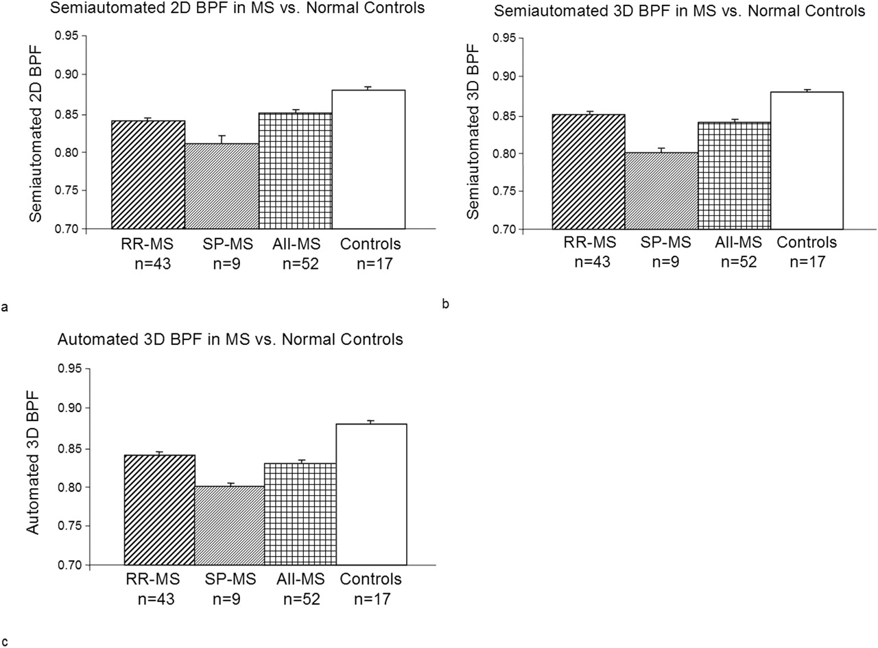
- Fig 5.
2D semiautomated (A), 3D semiautomated (B), and 3D automated (C) BPFs (mean and standard error) in MS and control groups. The three BPFs were similar in demonstrating whole-brain atrophy in the MS versus the control group. Analysis of covariance adjusted for age showed that the semiautomated (2D, P < .001; 3D, P = .04) and automated (3D, P = .002) BPFs were lower in MS than in control group. The three BPFs showed a similar and higher degree of atrophy in secondary progressive versus relapsing-remitting patients, approaching statistical significance (see Results).
- Fig 6.
Scatterplots of BPF versus third ventricular width in patients with MS (n = 52), showing 2D semiautomated (A), 3D semiautomated (B), and 3D automated (C) BPFs. The semiautomated and automated 3D BPFs showed similarly robust inverse correlations with third ventricular width, which indicates a relationship between whole-brain atrophy and central brain atrophy.
- Fig 7.
Scatterplots of BPF versus bicaudate ratio in patients with MS (n = 52), showing 2D semiautomated (A), 3D semiautomated (B), and 3D automated (C) BPFs. The semiautomated and automated BPFs showed identically robust inverse correlations with bicaudate ratio, which indicates an association between whole-brain atrophy and subcortical brain atrophy.
- Fig 8.
Scatterplots of BPF versus total brain T1-hypointense lesion volume in patients with MS (n = 52), showing 2D semiautomated (A), 3D semiautomated (B), and 3D automated (C) BPFs. The semiautomated and automated BPFs showed similarly moderate inverse correlations with T1-hypointense lesion volume.
- Fig 9.
Scatterplots of BPF versus whole-brain FLAIR hyperintense lesion volume in patients with MS (n = 52), showing 2D semiautomated (A), 3D semiautomated (B), and 3D automated (C) BPFs. The automated 3D BPF showed a somewhat higher correlation with that of whole-brain FLAIR hyperintense lesion volume than did the semiautomated BPFs.


Tables
No. of Cases Semiautomated Automated 2D 3D 2D 3D Mean SD Mean SD Mean SD Mean SD Relapsing-remitting MS 43 0.85 0.04 0.85 0.04 NR NR 0.84 0.05 Secondary progressive MS 9 0.81 0.04 0.80 0.03 NS NR 0.80 0.04 All MS 52 0.84 0.04 0.84 0.05 NR NR 0.83 0.05 Controls 17 0.88 0.02 0.88 0.03 NR NR 0.88 0.03 Note.—2D and 3D sequences according to Methods section; MS, multiple sclerosis; NR, segmentation not reliable; SD, standard deviation; semiautomated and automated algorithms according to Methods section.
- TABLE 2:
Comparing two methods of measuring brain parenchymal fraction and their association with clinical and MR imaging variables in 52 patients with multiple sclerosis
Semiautomated Automated 2D 3D 2D 3D Third ventricular width r = −.82, P < .001 r = −.79, P < .001 NR r = −.81, P < .001 Bicaudate ratio r = −.74, P < .001 r = −.74, P < .001 NR r = −.74, P < .001 T1 hypointense lesion volume r = −.38, P = .006 r = −.44, P = .001 NR r = −.48, P < .001 FLAIR hyperintense lesion volume r = −.24, P = .09 r = −.35, P = .01 NR r = −.44, P = .001 EDSS r = −.44, P < .001 r = −.47, P < .001 NR r = −.33, P = .008 Disease duration r = −.50, P < .001 r = −.39, P = .002 NR r = −.53, P < .001
In this issue


Jump to section
Related Articles
Cited By...
- Automated Determination of Brain Parenchymal Fraction in Multiple Sclerosis
- Reliability of Longitudinal Brain Volume Loss Measurements between 2 Sites in Patients with Multiple Sclerosis: Comparison of 7 Quantification Techniques
- Thalamic atrophy and cognition in multiple sclerosis
- Gray and white matter brain atrophy and neuropsychological impairment in multiple sclerosis
- The use of magnetic resonance imaging in the diagnosis and long-term management of multiple sclerosis