Advertisement

Main menu

  • Home
  • Content
    • Current Issue
    • Accepted Manuscripts
    • Article Preview
    • Past Issue Archive
    • AJNR Case Collection
    • Case of the Week Archive
    • Classic Case Archive
    • Case of the Month Archive
  • Special Collections
    • Spinal CSF Leak Articles (Jan 2020-June 2024)
    • 2024 AJNR Journal Awards
    • Most Impactful AJNR Articles
  • Multimedia
    • AJNR Podcast
    • AJNR Scantastics
    • Video Articles
  • For Authors
    • Submit a Manuscript
    • Author Policies
    • Fast publishing of Accepted Manuscripts
    • Graphical Abstract Preparation
    • Manuscript Submission Guidelines
    • Imaging Protocol Submission
    • Submit a Case for the Case Collection
  • About Us
    • About AJNR
    • Editorial Board
  • More
    • Become a Reviewer/Academy of Reviewers
    • Subscribers
    • Permissions
    • Alerts
    • Feedback
    • Advertisers
    • ASNR Home
  • Other Publications
    • ajnr

User menu

  • Alerts
  • Log in

Search

  • Advanced search
American Journal of Neuroradiology

American Journal of Neuroradiology

ASHNR American Society of Functional Neuroradiology ASHNR American Society of Pediatric Neuroradiology ASSR
  • Alerts
  • Log in

Advanced Search

  • Home
  • Content
    • Current Issue
    • Accepted Manuscripts
    • Article Preview
    • Past Issue Archive
    • AJNR Case Collection
    • Case of the Week Archive
    • Classic Case Archive
    • Case of the Month Archive
  • Special Collections
    • Spinal CSF Leak Articles (Jan 2020-June 2024)
    • 2024 AJNR Journal Awards
    • Most Impactful AJNR Articles
  • Multimedia
    • AJNR Podcast
    • AJNR Scantastics
    • Video Articles
  • For Authors
    • Submit a Manuscript
    • Author Policies
    • Fast publishing of Accepted Manuscripts
    • Graphical Abstract Preparation
    • Manuscript Submission Guidelines
    • Imaging Protocol Submission
    • Submit a Case for the Case Collection
  • About Us
    • About AJNR
    • Editorial Board
  • More
    • Become a Reviewer/Academy of Reviewers
    • Subscribers
    • Permissions
    • Alerts
    • Feedback
    • Advertisers
    • ASNR Home
  • Follow AJNR on Twitter
  • Visit AJNR on Facebook
  • Follow AJNR on Instagram
  • Join AJNR on LinkedIn
  • RSS Feeds

Welcome to the new AJNR, Updated Hall of Fame, and more. Read the full announcements.


AJNR is seeking candidates for the position of Associate Section Editor, AJNR Case Collection. Read the full announcement.


Getting new auth cookie, if you see this message a lot, tell someone!
Research ArticlePediatrics

Imaging Characteristics of Cerebrovascular Arteriopathy and Stroke in Hutchinson-Gilford Progeria Syndrome

V.M. Silvera, L.B. Gordon, D.B. Orbach, S.E. Campbell, J.T. Machan and N.J. Ullrich
American Journal of Neuroradiology May 2013, 34 (5) 1091-1097; DOI: https://doi.org/10.3174/ajnr.A3341
V.M. Silvera
aFrom the Departments of Radiology (V.M.S., D.B.O.)
  • Find this author on Google Scholar
  • Find this author on PubMed
  • Search for this author on this site
L.B. Gordon
bAnesthesia (L.B.G.)
dDepartment of Pediatrics (L.B.G.), Hasbro Children's Hospital, Providence, Rhode Island
eWarren Alpert Medical School (L.B.G., J.T.M.)
  • Find this author on Google Scholar
  • Find this author on PubMed
  • Search for this author on this site
D.B. Orbach
aFrom the Departments of Radiology (V.M.S., D.B.O.)
  • Find this author on Google Scholar
  • Find this author on PubMed
  • Search for this author on this site
S.E. Campbell
fCenter for Gerontology and Healthcare Research (S.E.C.), Brown University, Providence, Rhode Island
  • Find this author on Google Scholar
  • Find this author on PubMed
  • Search for this author on this site
J.T. Machan
eWarren Alpert Medical School (L.B.G., J.T.M.)
gDepartments of Biostatistics, (J.T.M.)
hOrthopaedics, Surgery, and Psychology (J.T.M.), Rhode Island Hospital, Providence, Rhode Island.
  • Find this author on Google Scholar
  • Find this author on PubMed
  • Search for this author on this site
N.J. Ullrich
cNeurology (N.J.U.), Boston Children's Hospital, Harvard Medical School, Boston, Massachusetts
  • Find this author on Google Scholar
  • Find this author on PubMed
  • Search for this author on this site
  • Article
  • Figures & Data
  • Supplemental
  • Info & Metrics
  • Responses
  • References
  • PDF
Loading

Article Figures & Data

Figures

  • Tables
  • Fig 1.
    • Download figure
    • Open in new tab
    • Download powerpoint
    Fig 1.

    Kaplan-Meier model of stroke-free probability depicts the youngest ages of patients at which imaging demonstrated ≥1 infarct, acute or chronic. The solid line shows the estimated stroke-free probability; dashed lines, the 95% CIs; and the dotted lines, the estimated age at which 50% of children have radiographically detectable stroke.

  • Fig 2.
    • Download figure
    • Open in new tab
    • Download powerpoint
    Fig 2.

    Graphic representation of infarct distribution.
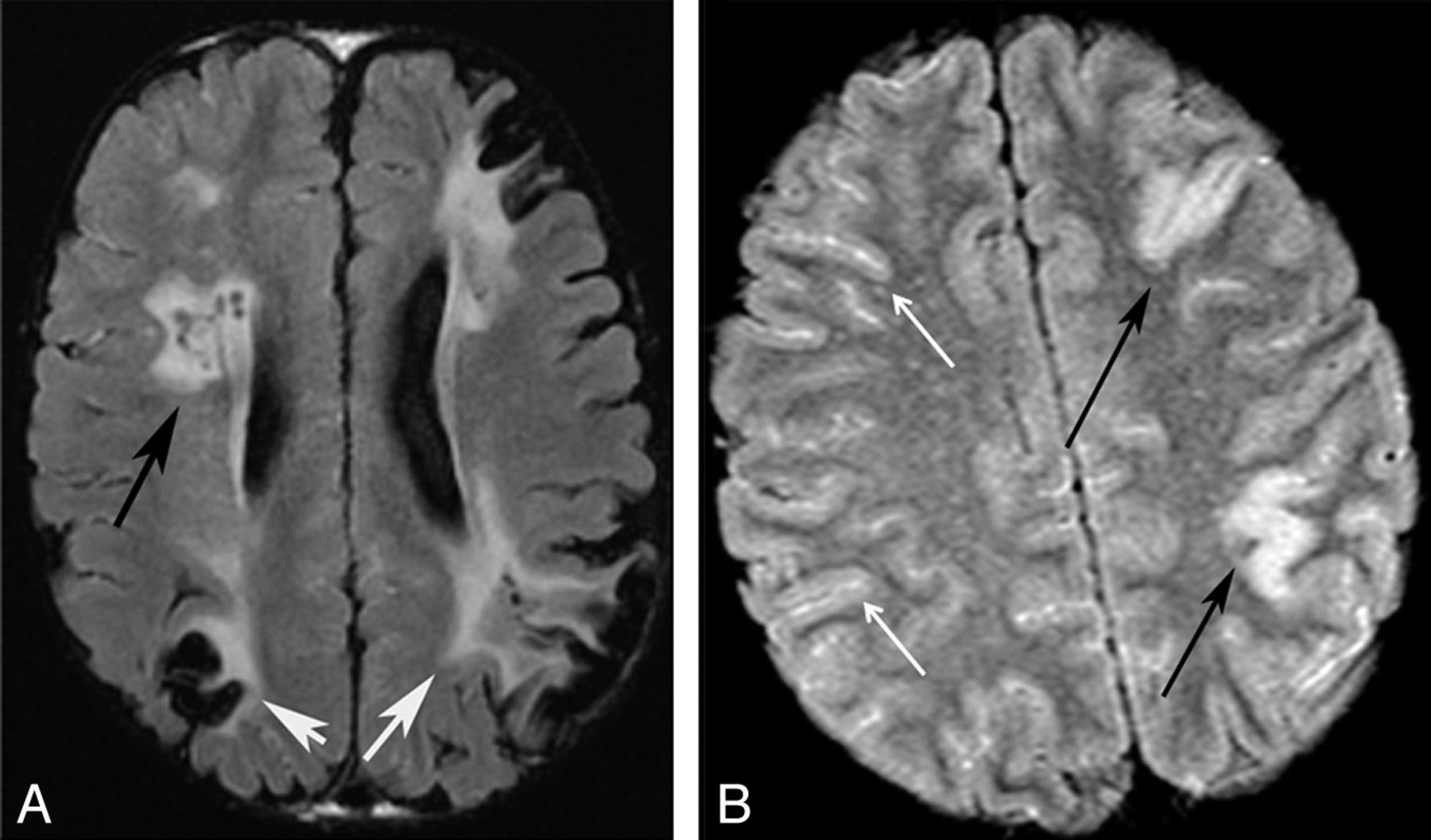
  • Fig 3.
    • Download figure
    • Open in new tab
    • Download powerpoint
    Fig 3.

    Patterns of infarction. Axial FLAIR images of 2 different patients. A, Chronic watershed (white arrows) and white matter infarcts (black arrow). B, Acute gyral infarcts (black arrows). Bright signal in the sulci indicates slow cortical collateral flow (white arrows).

  • Fig 4.
    • Download figure
    • Open in new tab
    • Download powerpoint
    Fig 4.

    Arterial calcification. CTA reformatted images of the same patient demonstrate right VA calcification (A) and ICA and external carotid artery (B) calcifications.

  • Fig 5.
    • Download figure
    • Open in new tab
    • Download powerpoint
    Fig 5.

    VA stenoses and collaterals. A, Lateral projection of a conventional angiogram demonstrates a short-segment high-grade stenosis of the distal V2 segment of the VA. B, Lateral projection of a 3D model of the cervical MRA in a different patient demonstrates a high-grade stenosis of the V2 segment of the VA (single white arrow), an enlarged deep cervical artery (double white arrows), and enlargement of the occipital artery (curved white arrow).

  • Fig 6.
    • Download figure
    • Open in new tab
    • Download powerpoint
    Fig 6.

    Collateral vessels. A, Coronal T2WI of an enlarged ASA (white arrow). B, Collapsed view of an MRA in a different patient demonstrates M1 (single black arrow) and A1 stenoses (black double arrows), internal maxillary artery collaterals (black wavy arrows), subfrontal collaterals (single white arrow), and an enlarged ASA and posterior spinal artery (white double arrows).

  • Fig 7.
    • Download figure
    • Open in new tab
    • Download powerpoint
    Fig 7.

    ICA stenosis. 3D model of the MRA of the circle of Willis demonstrates bilateral short-segment high-grade stenoses of the cavernous ICAs (white arrows).

  • Fig 8.
    • Download figure
    • Open in new tab
    • Download powerpoint
    Fig 8.

    Collateral vessels. A, Axial T2WI shows collateral vessels in the basal cistern (arrow). Source images from an MRA in a different patient demonstrate subfrontal (B) and perisplenial (C) collateral vessels (arrows.)

Tables

  • Figures
  • Indications for neuroimaging

    IndicationsaNo. of Scans
    Acute neurologic symptoms suspicious for TIA/stroke50
    Screening/diagnostic31
    Follow-up on prior imaging21
    Seizure13
    Unknown8
    Trauma7
    Headaches5
    Otherb5
    Extra-axial hemorrhage/subdural hematoma3
    Papilledema2
    Growth hormone initiation1
    • ↵a Some patients had >1 indication for imaging and/or multiple studies done at 1 study date.

    • ↵b Eye pain, stent placement, cyst evaluation.

PreviousNext
Back to top

In this issue

American Journal of Neuroradiology: 34 (5)
American Journal of Neuroradiology
Vol. 34, Issue 5
1 May 2013
  • Table of Contents
  • Index by author
Advertisement
Print
Download PDF
Email Article

Thank you for your interest in spreading the word on American Journal of Neuroradiology.

NOTE: We only request your email address so that the person you are recommending the page to knows that you wanted them to see it, and that it is not junk mail. We do not capture any email address.

Enter multiple addresses on separate lines or separate them with commas.
Imaging Characteristics of Cerebrovascular Arteriopathy and Stroke in Hutchinson-Gilford Progeria Syndrome
(Your Name) has sent you a message from American Journal of Neuroradiology
(Your Name) thought you would like to see the American Journal of Neuroradiology web site.
CAPTCHA
This question is for testing whether or not you are a human visitor and to prevent automated spam submissions.
Cite this article
V.M. Silvera, L.B. Gordon, D.B. Orbach, S.E. Campbell, J.T. Machan, N.J. Ullrich
Imaging Characteristics of Cerebrovascular Arteriopathy and Stroke in Hutchinson-Gilford Progeria Syndrome
American Journal of Neuroradiology May 2013, 34 (5) 1091-1097; DOI: 10.3174/ajnr.A3341

Citation Manager Formats

  • BibTeX
  • Bookends
  • EasyBib
  • EndNote (tagged)
  • EndNote 8 (xml)
  • Medlars
  • Mendeley
  • Papers
  • RefWorks Tagged
  • Ref Manager
  • RIS
  • Zotero
0 Responses
Respond to this article
Share
Bookmark this article
Imaging Characteristics of Cerebrovascular Arteriopathy and Stroke in Hutchinson-Gilford Progeria Syndrome
V.M. Silvera, L.B. Gordon, D.B. Orbach, S.E. Campbell, J.T. Machan, N.J. Ullrich
American Journal of Neuroradiology May 2013, 34 (5) 1091-1097; DOI: 10.3174/ajnr.A3341
del.icio.us logo Twitter logo Facebook logo Mendeley logo
  • Tweet Widget
  • Facebook Like
  • Google Plus One
Purchase

Jump to section

  • Article
    • Abstract
    • ABBREVIATIONS:
    • Materials and Methods
    • Results
    • Discussion
    • Conclusions
    • Acknowledgments
    • Footnotes
    • References
  • Figures & Data
  • Supplemental
  • Info & Metrics
  • Responses
  • References
  • PDF

Related Articles

  • PubMed
  • Google Scholar

Cited By...

  • Phosphorylated Lamin A/C in the nuclear interior binds active enhancers associated with abnormal transcription in progeria
  • Moving from gene discovery to clinical trials in Hutchinson-Gilford progeria syndrome
  • Neurologic features of Hutchinson-Gilford progeria syndrome after lonafarnib treatment
  • Crossref (51)
  • Google Scholar

This article has been cited by the following articles in journals that are participating in Crossref Cited-by Linking.

  • Progeria: A Paradigm for Translational Medicine
    Leslie B. Gordon, Frank G. Rothman, Carlos López-Otín, Tom Misteli
    Cell 2014 156 3
  • Hutchinson-Gilford Progeria Syndrome: A premature aging disease caused by LMNA gene mutations
    Susana Gonzalo, Ray Kreienkamp, Peter Askjaer
    Ageing Research Reviews 2017 33
  • Neurocutaneous Syndromes
    Nicole J. Ullrich, Leslie B. Gordon
    2015 132
  • Aging in the Cardiovascular System: Lessons from Hutchinson-Gilford Progeria Syndrome
    Magda R. Hamczyk, Lara del Campo, Vicente Andrés
    Annual Review of Physiology 2018 80 1
  • Phosphorylated Lamin A/C in the Nuclear Interior Binds Active Enhancers Associated with Abnormal Transcription in Progeria
    Kohta Ikegami, Stefano Secchia, Omar Almakki, Jason D. Lieb, Ivan P. Moskowitz
    Developmental Cell 2020 52 6
  • Generation of a Hutchinson–Gilford progeria syndrome monkey model by base editing
    Fang Wang, Weiqi Zhang, Qiaoyan Yang, Yu Kang, Yanling Fan, Jingkuan Wei, Zunpeng Liu, Shaoxing Dai, Hao Li, Zifan Li, Lizhu Xu, Chu Chu, Jing Qu, Chenyang Si, Weizhi Ji, Guang-Hui Liu, Chengzu Long, Yuyu Niu
    Protein & Cell 2020 11 11
  • Neurologic features of Hutchinson-Gilford progeria syndrome after lonafarnib treatment
    Nicole J. Ullrich, Mark W. Kieran, David T. Miller, Leslie B. Gordon, Yoon-Jae Cho, V. Michelle Silvera, Anita Giobbie-Hurder, Donna Neuberg, Monica E. Kleinman
    Neurology 2013 81 5
  • Accelerated Vascular Aging as a Paradigm for Hypertensive Vascular Disease: Prevention and Therapy
    Matthias Barton, Marc Husmann, Matthias R. Meyer
    Canadian Journal of Cardiology 2016 32 5
  • Hutchinson–Gilford Progeria Syndrome: A Premature Aging Disease
    Muhammad Saad Ahmed, Sana Ikram, Nousheen Bibi, Asif Mir
    Molecular Neurobiology 2017
  • An Upregulation in the Expression of Vanilloid Transient Potential Channels 2 Enhances Hypotonicity-Induced Cytosolic Ca2+ Rise in Human Induced Pluripotent Stem Cell Model of Hutchinson Gillford Progeria
    Chun-Yin Lo, Yung-Wui Tjong, Jenny Chung-Yee Ho, Chung-Wah Siu, Sin-Ying Cheung, Nelson L. Tang, Shan Yu, Hung-Fat Tse, Xiaoqiang Yao, Alexander G. Obukhov
    PLoS ONE 2014 9 1

More in this TOC Section

  • Comparison of Image Quality and Radiation Dose in Pediatric Temporal Bone CT Using Photon-Counting Detector CT and Energy-Integrating Detector CT
  • SyMRI & MR Fingerprinting in Brainstem Myelination
  • Pons&Vermis Localization on Fetal MRI Using U-Net
Show more Pediatrics

Similar Articles

Advertisement

Indexed Content

  • Current Issue
  • Accepted Manuscripts
  • Article Preview
  • Past Issues
  • Editorials
  • Editors Choice
  • Fellow Journal Club
  • Letters to the Editor

Cases

  • Case Collection
  • Archive - Case of the Week
  • Archive - Case of the Month
  • Archive - Classic Case

Special Collections

  • Special Collections

Resources

  • News and Updates
  • Turn around Times
  • Submit a Manuscript
  • Author Policies
  • Manuscript Submission Guidelines
  • Evidence-Based Medicine Level Guide
  • Publishing Checklists
  • Graphical Abstract Preparation
  • Imaging Protocol Submission
  • Submit a Case
  • Become a Reviewer/Academy of Reviewers
  • Get Peer Review Credit from Publons

Multimedia

  • AJNR Podcast
  • AJNR SCANtastic
  • Video Articles

About Us

  • About AJNR
  • Editorial Board
  • Not an AJNR Subscriber? Join Now
  • Alerts
  • Feedback
  • Advertise with us
  • Librarian Resources
  • Permissions
  • Terms and Conditions

American Society of Neuroradiology

  • Not an ASNR Member? Join Now

© 2025 by the American Society of Neuroradiology All rights, including for text and data mining, AI training, and similar technologies, are reserved.
Print ISSN: 0195-6108 Online ISSN: 1936-959X

Powered by HighWire