Advertisement

Main menu

  • Home
  • Content
    • Current Issue
    • Accepted Manuscripts
    • Article Preview
    • Past Issue Archive
    • AJNR Case Collection
    • Case of the Week Archive
    • Classic Case Archive
    • Case of the Month Archive
  • Special Collections
    • Spinal CSF Leak Articles (Jan 2020-June 2024)
    • 2024 AJNR Journal Awards
    • Most Impactful AJNR Articles
  • Multimedia
    • AJNR Podcast
    • AJNR Scantastics
    • Video Articles
  • For Authors
    • Submit a Manuscript
    • Author Policies
    • Fast publishing of Accepted Manuscripts
    • Graphical Abstract Preparation
    • Manuscript Submission Guidelines
    • Imaging Protocol Submission
    • Submit a Case for the Case Collection
  • About Us
    • About AJNR
    • Editorial Board
  • More
    • Become a Reviewer/Academy of Reviewers
    • Subscribers
    • Permissions
    • Alerts
    • Feedback
    • Advertisers
    • ASNR Home
  • Other Publications
    • ajnr

User menu

  • Alerts
  • Log in

Search

  • Advanced search
American Journal of Neuroradiology

American Journal of Neuroradiology

ASHNR American Society of Functional Neuroradiology ASHNR American Society of Pediatric Neuroradiology ASSR
  • Alerts
  • Log in

Advanced Search

  • Home
  • Content
    • Current Issue
    • Accepted Manuscripts
    • Article Preview
    • Past Issue Archive
    • AJNR Case Collection
    • Case of the Week Archive
    • Classic Case Archive
    • Case of the Month Archive
  • Special Collections
    • Spinal CSF Leak Articles (Jan 2020-June 2024)
    • 2024 AJNR Journal Awards
    • Most Impactful AJNR Articles
  • Multimedia
    • AJNR Podcast
    • AJNR Scantastics
    • Video Articles
  • For Authors
    • Submit a Manuscript
    • Author Policies
    • Fast publishing of Accepted Manuscripts
    • Graphical Abstract Preparation
    • Manuscript Submission Guidelines
    • Imaging Protocol Submission
    • Submit a Case for the Case Collection
  • About Us
    • About AJNR
    • Editorial Board
  • More
    • Become a Reviewer/Academy of Reviewers
    • Subscribers
    • Permissions
    • Alerts
    • Feedback
    • Advertisers
    • ASNR Home
  • Follow AJNR on Twitter
  • Visit AJNR on Facebook
  • Follow AJNR on Instagram
  • Join AJNR on LinkedIn
  • RSS Feeds

Welcome to the new AJNR, Updated Hall of Fame, and more. Read the full announcements.


AJNR is seeking candidates for the position of Associate Section Editor, AJNR Case Collection. Read the full announcement.


LetterLetter

Klippel-Feil Syndrome and Anterior Cervical Meningomyelocele: A Rare Case Report

G. Balachandran
American Journal of Neuroradiology October 2009, 30 (9) E130; DOI: https://doi.org/10.3174/ajnr.A1730
G. Balachandran
  • Find this author on Google Scholar
  • Find this author on PubMed
  • Search for this author on this site

We report a case of Klippel-Feil syndrome associated with an anterior cervical meningomyelocele.

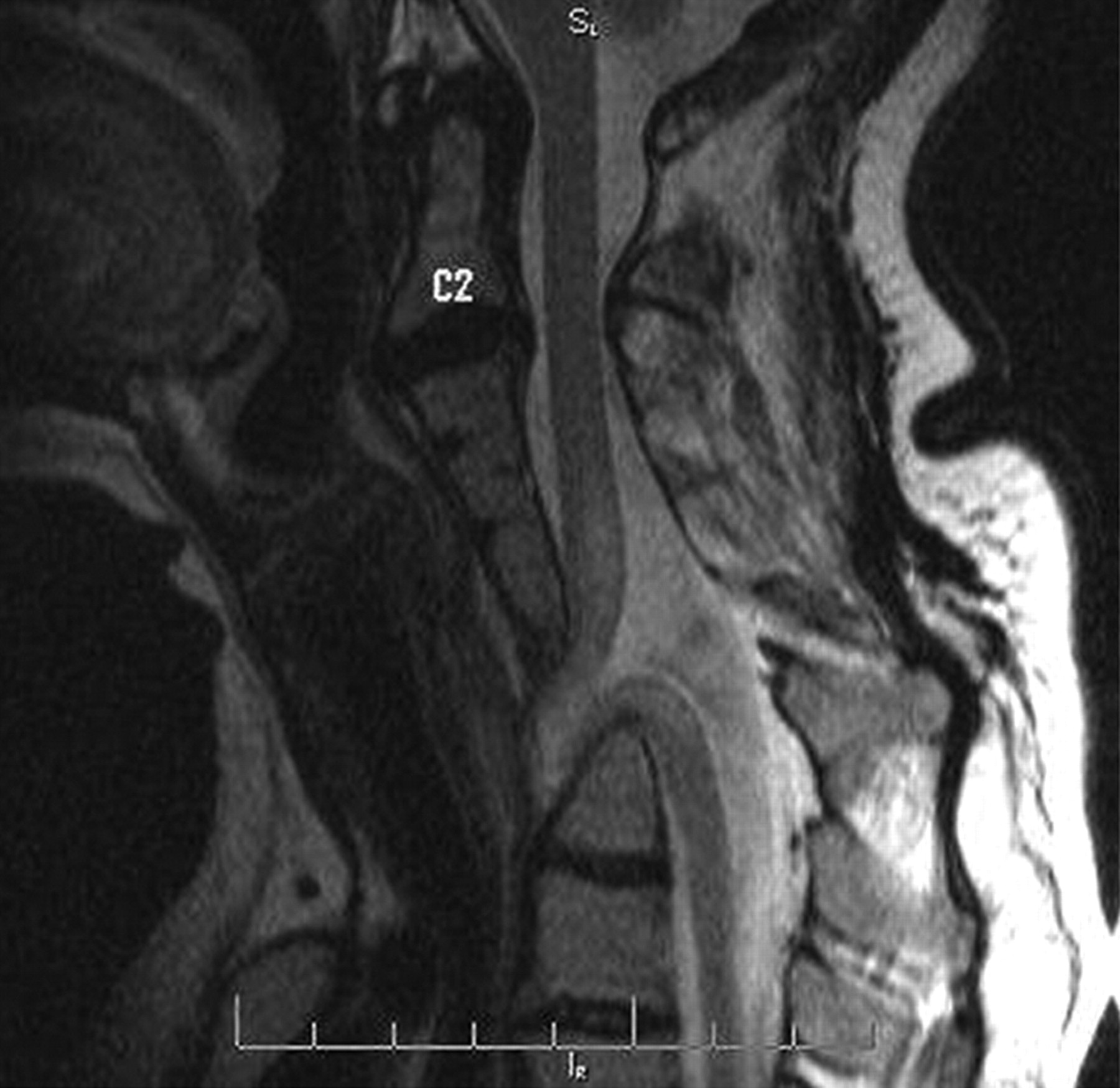
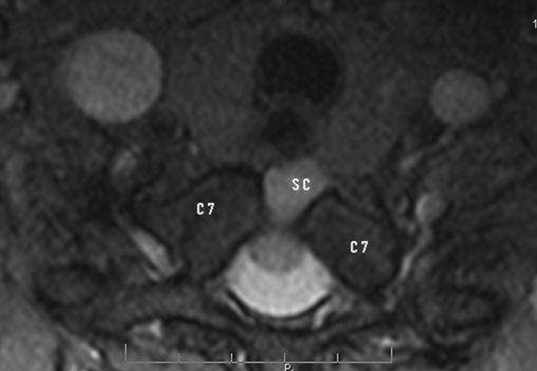
A 36-year old female patient was referred for evaluation of bilateral symmetric small muscle wasting in both hands. MR images (Fig 1) showed fusion of cervical vertebrae and neural arches. There was an anterior meningomyelocele at the level of C6 and C7. The cervical spinal cord was kinked, made an anterior loop, and was dragged anteriorly within the widened posterior thecal sac, stretching the nerve roots within the thecal sac. Part of spinal cord (Fig 2) was lying within the spinal canal and part of it was lying outside the anterior vertebral bodies at the level of C6.

Fig 1.
  • Download figure
  • Open in new tab
  • Download powerpoint
Fig 1.

T2-weighted sagittal view of the cervical spine shows fused cervical vertebrae and an anterior meningomyelocele.

Fig 2.
  • Download figure
  • Open in new tab
  • Download powerpoint
Fig 2.

T2-weighted transverse view of the cervical spine shows herniation of spinal cord (SC) through an anterior defect in the C7 body.

The Klippel-Feil deformity is a complex of osseous and visceral anomalies, which include low hairline, platybasia, fused cervical vertebrae with a short neck, and deafness. Associated central nervous system abnormalities include occipital cephalocele, Chiari I malformation, syrinx, microcephaly, and hydrocephalus. A meningomyelocele is a herniation of the meninges along with the cord through a foramina or defect in the vertebral column and is frequently (> 80%) located posteriorly in the lumbosacral area. An anterior meningomyelocele is rare and is generally described in the thoracic or sacral region.

  • Copyright © American Society of Neuroradiology
Advertisement

Indexed Content

  • Current Issue
  • Accepted Manuscripts
  • Article Preview
  • Past Issues
  • Editorials
  • Editors Choice
  • Fellow Journal Club
  • Letters to the Editor

Cases

  • Case Collection
  • Archive - Case of the Week
  • Archive - Case of the Month
  • Archive - Classic Case

Special Collections

  • Special Collections

Resources

  • News and Updates
  • Turn around Times
  • Submit a Manuscript
  • Author Policies
  • Manuscript Submission Guidelines
  • Evidence-Based Medicine Level Guide
  • Publishing Checklists
  • Graphical Abstract Preparation
  • Imaging Protocol Submission
  • Submit a Case
  • Become a Reviewer/Academy of Reviewers
  • Get Peer Review Credit from Publons

Multimedia

  • AJNR Podcast
  • AJNR SCANtastic
  • Video Articles

About Us

  • About AJNR
  • Editorial Board
  • Not an AJNR Subscriber? Join Now
  • Alerts
  • Feedback
  • Advertise with us
  • Librarian Resources
  • Permissions
  • Terms and Conditions

American Society of Neuroradiology

  • Not an ASNR Member? Join Now

© 2025 by the American Society of Neuroradiology All rights, including for text and data mining, AI training, and similar technologies, are reserved.
Print ISSN: 0195-6108 Online ISSN: 1936-959X

Powered by HighWire