Advertisement

Main menu

  • Home
  • Content
    • Current Issue
    • Accepted Manuscripts
    • Article Preview
    • Past Issue Archive
    • AJNR Case Collection
    • Case of the Week Archive
    • Classic Case Archive
    • Case of the Month Archive
  • Special Collections
    • Spinal CSF Leak Articles (Jan 2020-June 2024)
    • 2024 AJNR Journal Awards
    • Most Impactful AJNR Articles
  • Multimedia
    • AJNR Podcast
    • AJNR Scantastics
    • Video Articles
  • For Authors
    • Submit a Manuscript
    • Author Policies
    • Fast publishing of Accepted Manuscripts
    • Graphical Abstract Preparation
    • Manuscript Submission Guidelines
    • Imaging Protocol Submission
    • Submit a Case for the Case Collection
  • About Us
    • About AJNR
    • Editorial Board
  • More
    • Become a Reviewer/Academy of Reviewers
    • Subscribers
    • Permissions
    • Alerts
    • Feedback
    • Advertisers
    • ASNR Home
  • Other Publications
    • ajnr

User menu

  • Alerts
  • Log in

Search

  • Advanced search
American Journal of Neuroradiology

American Journal of Neuroradiology

ASHNR American Society of Functional Neuroradiology ASHNR American Society of Pediatric Neuroradiology ASSR
  • Alerts
  • Log in

Advanced Search

  • Home
  • Content
    • Current Issue
    • Accepted Manuscripts
    • Article Preview
    • Past Issue Archive
    • AJNR Case Collection
    • Case of the Week Archive
    • Classic Case Archive
    • Case of the Month Archive
  • Special Collections
    • Spinal CSF Leak Articles (Jan 2020-June 2024)
    • 2024 AJNR Journal Awards
    • Most Impactful AJNR Articles
  • Multimedia
    • AJNR Podcast
    • AJNR Scantastics
    • Video Articles
  • For Authors
    • Submit a Manuscript
    • Author Policies
    • Fast publishing of Accepted Manuscripts
    • Graphical Abstract Preparation
    • Manuscript Submission Guidelines
    • Imaging Protocol Submission
    • Submit a Case for the Case Collection
  • About Us
    • About AJNR
    • Editorial Board
  • More
    • Become a Reviewer/Academy of Reviewers
    • Subscribers
    • Permissions
    • Alerts
    • Feedback
    • Advertisers
    • ASNR Home
  • Follow AJNR on Twitter
  • Visit AJNR on Facebook
  • Follow AJNR on Instagram
  • Join AJNR on LinkedIn
  • RSS Feeds

Welcome to the new AJNR, Updated Hall of Fame, and more. Read the full announcements.


AJNR is seeking candidates for the position of Associate Section Editor, AJNR Case Collection. Read the full announcement.


Getting new auth cookie, if you see this message a lot, tell someone!
Research ArticleFunctional Vignette

Memory Part 2: The Role of the Medial Temporal Lobe

F.D. Raslau, I.T. Mark, A.P. Klein, J.L. Ulmer, V. Mathews and L.P. Mark
American Journal of Neuroradiology May 2015, 36 (5) 846-849; DOI: https://doi.org/10.3174/ajnr.A4169
F.D. Raslau
aFrom the Department of Radiology (F.D.R.), University of Kentucky, Lexington, Kentucky
  • Find this author on Google Scholar
  • Find this author on PubMed
  • Search for this author on this site
I.T. Mark
bMorsani College of Medicine (I.T.M.), University of South Florida, Tampa, Florida
  • Find this author on Google Scholar
  • Find this author on PubMed
  • Search for this author on this site
A.P. Klein
cDepartment of Radiology (A.P.K., J.L.U., V.M., L.P.M.), Medical College of Wisconsin, Milwaukee, Wisconsin.
  • Find this author on Google Scholar
  • Find this author on PubMed
  • Search for this author on this site
J.L. Ulmer
cDepartment of Radiology (A.P.K., J.L.U., V.M., L.P.M.), Medical College of Wisconsin, Milwaukee, Wisconsin.
  • Find this author on Google Scholar
  • Find this author on PubMed
  • Search for this author on this site
V. Mathews
cDepartment of Radiology (A.P.K., J.L.U., V.M., L.P.M.), Medical College of Wisconsin, Milwaukee, Wisconsin.
  • Find this author on Google Scholar
  • Find this author on PubMed
  • Search for this author on this site
L.P. Mark
cDepartment of Radiology (A.P.K., J.L.U., V.M., L.P.M.), Medical College of Wisconsin, Milwaukee, Wisconsin.
  • Find this author on Google Scholar
  • Find this author on PubMed
  • Search for this author on this site
  • ORCID record for L.P. Mark
  • Article
  • Figures & Data
  • Info & Metrics
  • Responses
  • References
  • PDF
Loading

The medial temporal lobe plays a central role in memory processing and is more than just the hippocampus.1 The hippocampal formation, which forms the upper segment of the medial temporal lobe, is a heterogeneous structure consisting of the Ammon horn or Cornus Ammonis (Cornus Ammonis area 1 to Cornus Ammonis area 4) and the dentate gyrus appearing as 2 interlocking U's on a coronal image. The collapsed portion of the temporal horn of the lateral ventricle forms the superior border of the hippocampal formation. The slightly more dilated lateral aspect of the temporal horn forms the lateral margin, while the ambient cistern is at the medial border.2

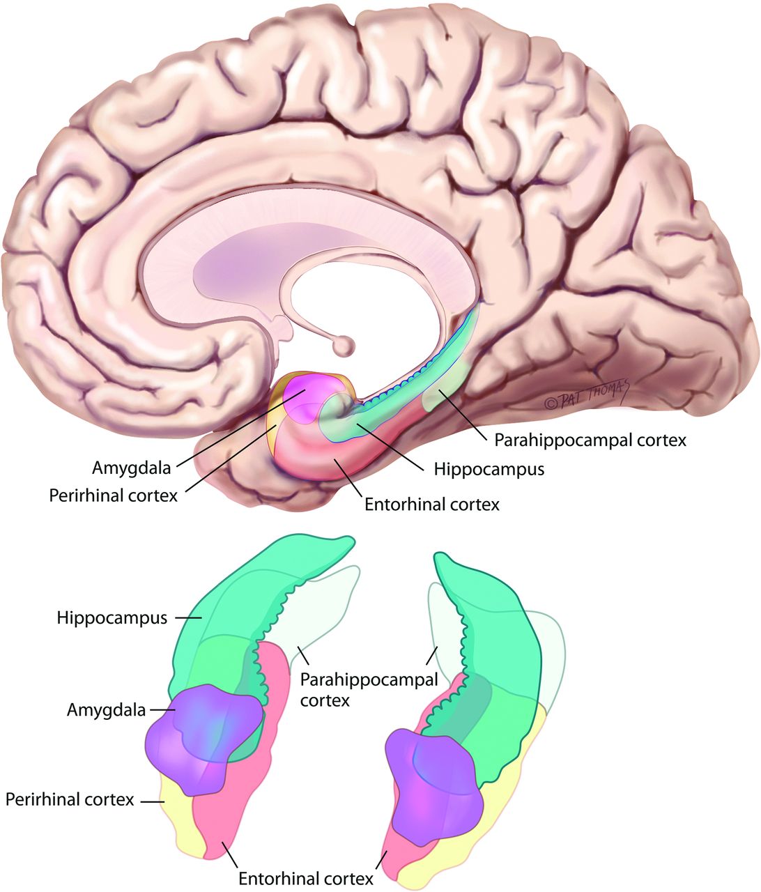
The parahippocampal gyrus, positioned just inferior to the hippocampus, forms the other major component of the medial temporal lobe (Fig 1). The parahippocampal gyrus is the most medial of the group of 3 gyri that form the inferior surface of the temporal lobe. The collateral sulcus forms the lateral border of the parahippocampal gyrus, while the ambient cistern is at the medial margin. The parahippocampal gyrus can be subdivided into anterior and posterior components. The anterior portion, sometimes referred as the rhinal cortex,3⇓–5 consists of medial and lateral parts, the entorhinal (Brodmann area 28) and perirhinal (Brodmann area 35) cortices, respectively. The border between the entorhinal and perirhinal cortices is located at the junction between the inferior surface of the parahippocampal gyrus and the inferior aspect of the collateral sulcus (Fig 2).6,7 The superior aspect of the collateral sulcus demarcates the position of the lateral margin of the perirhinal cortex. In other words, the perirhinal cortex forms the medial bank of the collateral sulcus.8 The posterior component of the parahippocampal gyrus consists of the parahippocampal cortex, which is not synonymous with the parahippocampal gyrus. The entorhinal, perirhinal, and parahippocampal cortices together, therefore, make up the parahippocampal gyrus. The subiculum is transitional cortex that bridges the Ammon horn of the hippocampal formation (allocortex) with the parahippocampal gyrus (isocortex).2 The subiculum can be further subdivided into the subiculum proper, presubiculum, and parasubiculum.

Fig 1.
  • Download figure
  • Open in new tab
  • Download powerpoint
Fig 1.

The medial temporal lobe consists of the hippocampal formation (blue-green) superiorly and the parahippocampal gyrus inferiorly. The entorhinal (brown) and perirhinal (yellow) cortices form the medial and lateral components, respectively, of the anterior portion of the parahippocampal gyrus, while the parahippocampal cortex (off-white) forms the posterior portion. Adapted with permission from Purves D, Brannon E, Cabeza R, et al. Principles of Cognitive Neuroscience. Sunderland, MA: Sinauer Associates; 2008.

Fig 2.
  • Download figure
  • Open in new tab
  • Download powerpoint
Fig 2.

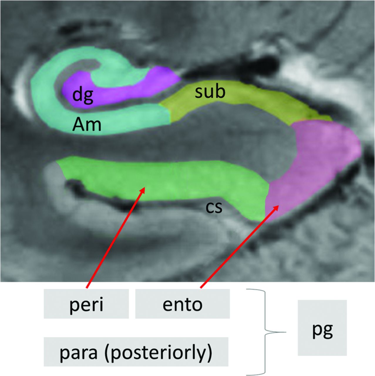
Coronal T2-weighted MR image of the right medial temporal lobe. The Ammon horn (Am) is blue, interlocking with the dentate gyrus (dg) in lavender. The perirhinal cortex (peri) in green is lateral to the entorhinal cortex (ento) in pink. The subiculum (sub) in yellow links the Ammon horn with the entorhinal cortex. Note that the sulcus just lateral to the perirhinal cortex is the collateral sulcus (cs) and the apex or superior most portion of the collateral sulcus marks the position of the lateral margin of the perirhinal cortex, while the inferior aspect of the sulcus marks the border between the perirhinal and entorhinal cortices. The parahippocampal gyus (pg) consists of the perirhinal, entorhinal, and parahippocampal (para) cortices.

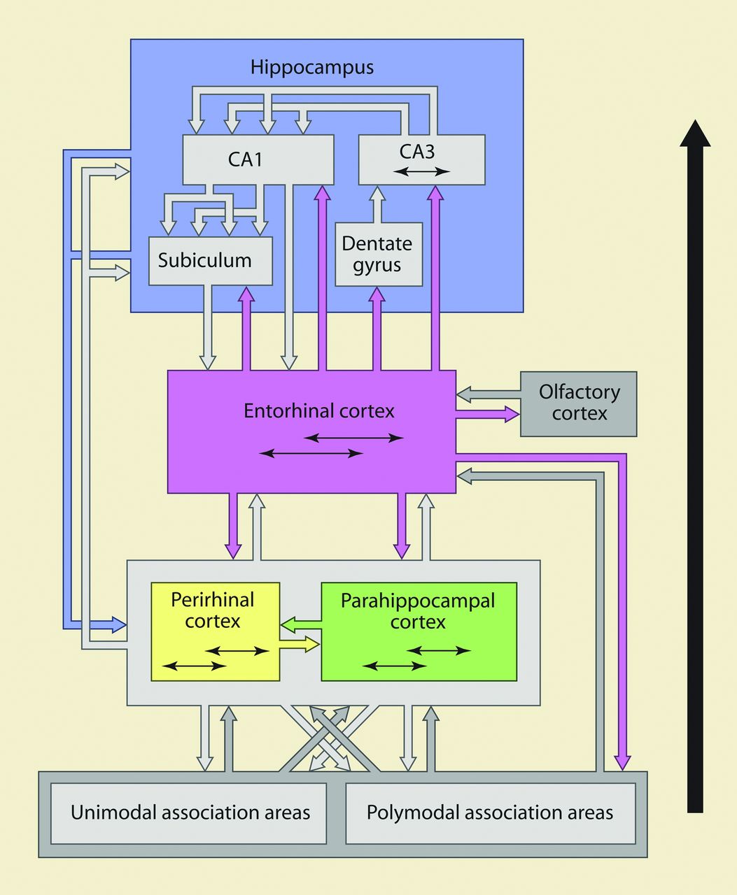
The organization of the medial temporal lobe suggests a hierarchic format in which information is initially collected through the perirhinal and parahippocampal cortices, passes to the entorhinal cortex, and ultimately reaches the hippocampal formation, which forms major output projections via the fornix (Fig 3).9 The parahippocampal gyrus, however, does not merely funnel information to the hippocampus.10 A large network of connections both within and among the subregions of the parahippocampal gyrus performs extensive information processing on its own and among subregions before any of the information reaches the hippocampus. Exteroceptive (external to the organism) information is processed by the parahippocampal gyrus via the ventral and dorsal streams (Fig 4). The ventral stream from the occipital lobe consists of visual information in terms of object recognition, while the dorsal stream from the parietal lobe carries spatial context information to the parahippocampal gyrus. Interoceptive (internal to the organism) signals carrying information such as emotions and motivation from the medial prefrontal cortex, nucleus accumbens, and amygdala project to the rostral hippocampal formation and rhinal cortex regions. This information from multiple sources is combined and ultimately output via the fornix from the hippocampal formation.

Fig 3.
  • Download figure
  • Open in new tab
  • Download powerpoint
Fig 3.

The net flow of information is from the perirhinal and parahippocampal cortices to the entorhinal cortex and then to the hippocampal formation, but considerable information processing occurs within and among the subregions of the parahippocampal gyrus before hippocampal formation involvement. Adapted with permission from Lavenex P, Amaral DG. Hippocampal-neocortical interaction: a hierarchy of associativity. Hippocampus 2000;10:420–30, John Wiley and Sons.

Fig 4.
  • Download figure
  • Open in new tab
  • Download powerpoint
Fig 4.

Medial temporal lobe processing of exteroceptive and interceptive signals for memory formation. The ventral stream from the occipital lobe projects information about object recognition, while the dorsal stream from the parietal lobe conveys spatial information. The rostral interceptive signals convey emotions and motivations.

Closer inspection of the medial temporal subregions has revealed additional layers of complexity and organization. The entorhinal cortex, for example, can be seen to have distinctive medial and lateral regions that differ histologically and physiologically.11 The medial entorhinal cortex (Brodmann area 28b) is actively involved in the processing of spatial information from the dorsal stream, whereas the lateral entorhinal cortex (Brodmann area 28a) does so with the object-recognition information from the ventral stream. Furthermore, the rhinal cortex functionally differentiates familiar and novel information input, where more familiar items are given fewer resources for encoding compared with new items. The rhinal cortex, therefore, functions as a gatekeeper of the declarative memory system by optimizing memory-encoding resources to novel information.12

In recent years, investigators have also postulated 2 cortical systems (anterior and posterior temporal) for memory-guided behavior involving the perirhinal and parahippocampal cortices.13 The anterior temporal system consists of the perirhinal cortex, temporopolar cortex, lateral orbital frontal cortex, and amygdala, while the posterior temporal system includes the parahippocampal cortex, retrosplenial cortex (Brodmann areas 29 and 30), anterior thalamic nuclei, mammillary bodies, pre- and parasubiculum, and components of the so-called default network, of which the retrosplenial cortex is a part, including the posterior cingulate gyrus, precuneus, angular gyrus, and ventral medial prefrontal cortex. The anterior system is more involved in object and face recognition, conceptual identity, and salience, while the posterior system focuses on scene recognition, location, trajectory, temporal context and order, and situations.

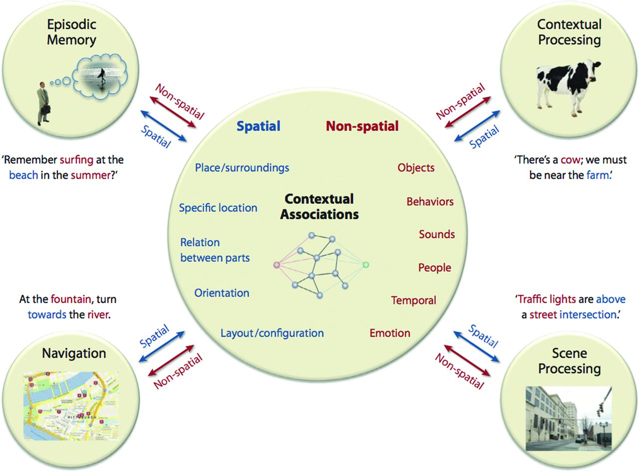
Semantic dementia involves more of the anterior temporal system, and patients often show the deficits in fine-grain object recognition. Alzheimer disease often involves more of the posterior temporal system and is frequently associated with deficits in scene discrimination. In this same fashion, detailed evaluation reveals that the parahippocampal cortex functions are much more complex than just the processing of spatial layout information. The parahippocampal cortex is part of a larger network that connects regions of the frontal, parietal, and temporal lobes. This includes auditory association areas of the superior temporal gyrus, the polymodal association areas (such as the retrosplenial cortex, lateral inferior parietal lobule, dorsal bank of the superior temporal sulcus), temporal pole, perirhinal cortex, parahippocampal cortex itself, entorhinal cortex, medial prefrontal cortex, dorsal lateral prefrontal cortex, orbital prefrontal cortex, insula, and so forth. It is best to think of the major role of the parahippocampal cortex as facilitating contextual associations, which are the principal elements underlying many higher level cognitive processes (Fig 5).14

Fig 5.
  • Download figure
  • Open in new tab
  • Download powerpoint
Fig 5.

The parahippocampal cortex functions involve more than just spatial processing. The connections with the different areas of the frontal, parietal, and temporal lobes, including the default network, position the parahippocampal cortex as a critical component in processing contextual associations, which are fundamental aspects of higher cognitive functions. Adapted with permission from Aminoff EM, Kveraga K, Bar M. The role of the parahippocampal cortex in cognition. Trends Cogn Sci 2013;17:379–90, Elsevier.

In summary, the medial temporal lobe occupies a central position in the intersection of multiple neuronal networks. Its anatomic complexity within its subregions and with other cerebral structures reflects the multifaceted nature of memory. Its functions, however, are more than those associated with declarative memory and are now known to be wide-ranging and include higher level cognitive functions, especially with the connections with the retrosplenial cortex and default network as demonstrated by resting-state fMRI studies.

The next Functional Vignette will be the last installment on memory and will showcase clinical cases in which memory is affected when key anatomic structures are involved.

Acknowledgments

The authors express our deepest gratitude to Jean Augustinack, PhD, Harvard Medical School, Massachusetts General Hospital, Martinos Faculty, for her invaluable advice on the anatomy of the perirhinal and entorhinal cortices.

REFERENCES

  1. 1.↵
    1. Amaral DG
    . Introduction: what is where in the medial temporal lobe? Hippocampus 1999;9:1–6
    CrossRefPubMed
  2. 2.↵
    1. Duvernoy HM,
    2. Cattin TP,
    3. Naidic C, et al
    . The Human Hippocampus: Functional Anatomy, Vascularization and Serial Sections with MRI. 3rd ed. Berlin: Springer-Verlag; 2005:232
  3. 3.↵
    1. Meunier M,
    2. Hadfield W,
    3. Bachevalier J, et al
    . Effects of rhinal cortex lesions combined with hippocampectomy on visual recognition memory in rhesus monkeys. J Neurophysiol 1996;75:1190–1205
    Abstract/FREE Full Text
  4. 4.↵
    1. Murray EA,
    2. Mishkin M
    . Visual recognition in monkeys following rhinal cortical ablations combined with either amygdalectomy or hippocampectomy. J Neurosci 1986;6:1991–2003
    Abstract
  5. 5.↵
    1. Liu Z,
    2. Richmond BJ,
    3. Murray EA, et al
    . DNA targeting of rhinal cortex D2 receptor protein reversibly blocks learning of cues that predict reward. Proc Natl Acad Sci U S A 2004;101:2336–41
  6. 6.↵
    1. Augustinack JC,
    2. Huber KE,
    3. Stevens AA, et al
    . Predicting the location of human perirhinal cortex, Brodmann's area 35, from MRI. Neuroimage 2013;64:32–42
    CrossRefPubMed
  7. 7.↵
    1. Augustinack JC,
    2. van der Kouwe AJ,
    3. Fischl B
    . Medial temporal cortices in ex vivo magnetic resonance imaging. J Comp Neurol 2013;521:4177–88
    CrossRefPubMed
  8. 8.↵
    1. Feczko E,
    2. Augustinack JC,
    3. Fischl B, et al
    . An MRI-based method for measuring volume, thickness and surface area of entorhinal, perirhinal, and posterior parahippocampal cortex. Neurobiol Aging 2009;30;420–31
    CrossRefPubMed
  9. 9.↵
    1. Lavenex P,
    2. Amaral DG
    . Hippocampal-neocortical interaction: a hierarchy of associativity. Hippocampus 2000;10:420–30
    CrossRefPubMed
  10. 10.↵
    1. Purves D,
    2. Brannon E,
    3. Cabeza R, et al
    . Principles of Cognitive Neuroscience. Sunderland, MA: Sinauer Associates; 2008
  11. 11.↵
    1. Burgalossi A,
    2. Brecht M
    . Cellular, columnar and modular organization of spatial representations in medial entorhinal cortex. Curr Opin Neurobiol 2014;24:47–54
    CrossRefPubMed
  12. 12.↵
    1. Fernández G,
    2. Tendolkar I
    . The rhinal cortex: ‘gatekeeper’ of the declarative memory system. Trends Cogn Sci 2006;10:358–62
    CrossRefPubMed
  13. 13.↵
    1. Ranganath C,
    2. Ritchey M
    . Two cortical systems for memory-guided behavior. Nat Rev Neurosci 2012;13:713–26
    CrossRefPubMed
  14. 14.↵
    1. Aminoff EM,
    2. Kveraga K,
    3. Bar M
    . The role of the parahippocampal cortex in cognition. Trends Cogn Sci 2013;17:379–90
    CrossRefPubMed
  • © 2015 by American Journal of Neuroradiology
PreviousNext
Back to top

In this issue

American Journal of Neuroradiology: 36 (5)
American Journal of Neuroradiology
Vol. 36, Issue 5
1 May 2015
  • Table of Contents
  • Index by author
  • Complete Issue (PDF)
Advertisement
Print
Download PDF
Email Article

Thank you for your interest in spreading the word on American Journal of Neuroradiology.

NOTE: We only request your email address so that the person you are recommending the page to knows that you wanted them to see it, and that it is not junk mail. We do not capture any email address.

Enter multiple addresses on separate lines or separate them with commas.
Memory Part 2: The Role of the Medial Temporal Lobe
(Your Name) has sent you a message from American Journal of Neuroradiology
(Your Name) thought you would like to see the American Journal of Neuroradiology web site.
CAPTCHA
This question is for testing whether or not you are a human visitor and to prevent automated spam submissions.
Cite this article
F.D. Raslau, I.T. Mark, A.P. Klein, J.L. Ulmer, V. Mathews, L.P. Mark
Memory Part 2: The Role of the Medial Temporal Lobe
American Journal of Neuroradiology May 2015, 36 (5) 846-849; DOI: 10.3174/ajnr.A4169

Citation Manager Formats

  • BibTeX
  • Bookends
  • EasyBib
  • EndNote (tagged)
  • EndNote 8 (xml)
  • Medlars
  • Mendeley
  • Papers
  • RefWorks Tagged
  • Ref Manager
  • RIS
  • Zotero
0 Responses
Respond to this article
Share
Bookmark this article
Memory Part 2: The Role of the Medial Temporal Lobe
F.D. Raslau, I.T. Mark, A.P. Klein, J.L. Ulmer, V. Mathews, L.P. Mark
American Journal of Neuroradiology May 2015, 36 (5) 846-849; DOI: 10.3174/ajnr.A4169
del.icio.us logo Twitter logo Facebook logo Mendeley logo
  • Tweet Widget
  • Facebook Like
  • Google Plus One
Purchase

Jump to section

  • Article
    • Acknowledgments
    • REFERENCES
  • Figures & Data
  • Info & Metrics
  • Responses
  • References
  • PDF

Related Articles

  • No related articles found.
  • PubMed
  • Google Scholar

Cited By...

  • Individualised quantitative susceptibility mapping reveals abnormal hippocampal iron markers in acute mild traumatic brain injury
  • Magnetic susceptibility of the hippocampal subfields and basal ganglia in acute mild traumatic brain injury
  • Distribution of paramagnetic and diamagnetic cortical substrates following mild Traumatic Brain Injury: A depth- and curvature-based quantitative susceptibility mapping study
  • Effects of mixed metal exposures on MRI diffusion features in the medial temporal lobe
  • AUTS2 expression within mammalian lineage: a predictor of neural networks involved in Autism Spectrum Disorders
  • AUTS2 gene dosage affects synaptic AMPA receptors via a local dendritic spine AUTS2-TTC3-AKT-mTORC1 signaling dysfunction
  • Regular Tai Chi Practice Is Associated with Improved Memory as well as Structural and Functional Integrity of the Hippocampal Formation in the Elderly
  • Crossref (39)
  • Google Scholar

This article has been cited by the following articles in journals that are participating in Crossref Cited-by Linking.

  • Apolipoprotein E and Alzheimer's Disease: Findings, Hypotheses, and Potential Mechanisms
    Nicole Koutsodendris, Maxine R. Nelson, Antara Rao, Yadong Huang
    Annual Review of Pathology: Mechanisms of Disease 2022 17 1
  • Interictal epileptiform activity outside the seizure onset zone impacts cognition
    Hoameng Ung, Christian Cazares, Ameya Nanivadekar, Lohith Kini, Joost Wagenaar, Danielle Becker, Abba Krieger, Timothy Lucas, Brian Litt, Kathryn A. Davis
    Brain 2017 140 8
  • Hormone replacement therapy is associated with improved cognition and larger brain volumes in at-risk APOE4 women: results from the European Prevention of Alzheimer’s Disease (EPAD) cohort
    Rasha N. M. Saleh, Michael Hornberger, Craig W. Ritchie, Anne Marie Minihane
    Alzheimer's Research & Therapy 2023 15 1
  • Medial Temporal Lobe Subregional Atrophy in Aging and Alzheimer's Disease: A Longitudinal Study
    Léa Chauveau, Elizabeth Kuhn, Cassandre Palix, Francesca Felisatti, Valentin Ourry, Vincent de La Sayette, Gaël Chételat, Robin de Flores
    Frontiers in Aging Neuroscience 2021 13
  • Regular Tai Chi Practice Is Associated With Improved Memory as Well as Structural and Functional Alterations of the Hippocampus in the Elderly
    Chunlin Yue, Qian Yu, Yanjie Zhang, Fabian Herold, Jian Mei, Zhaowei Kong, Stephane Perrey, Jiao Liu, Notger G. Müller, Zonghao Zhang, Yuliu Tao, Arthur Kramer, Benjamin Becker, Liye Zou
    Frontiers in Aging Neuroscience 2020 12
  • Anatomy and White Matter Connections of the Parahippocampal Gyrus
    Yueh-Hsin Lin, Vukshitha Dhanaraj, Alana E. Mackenzie, Isabella M. Young, Onur Tanglay, Robert G. Briggs, Arpan R. Chakraborty, Jorge Hormovas, R. Dineth Fonseka, Sihyong J. Kim, Jacky T. Yeung, Charles Teo, Michael E. Sughrue
    World Neurosurgery 2021 148
  • Amygdala activity and amygdala-hippocampus connectivity: Metabolic diseases, dementia, and neuropsychiatric issues
    Juhyun Song
    Biomedicine & Pharmacotherapy 2023 162
  • The effects of physical exercise on parahippocampal function
    PD Loprinzi
    Physiology International 2019 106 2
  • Understanding the Relationship Between Age-Related Hearing Loss and Alzheimer’s Disease: A Narrative Review
    Hadeel Y. Tarawneh, Dona M.P. Jayakody, Hamid R. Sohrabi, Ralph N. Martins, Wilhelmina H.A.M. Mulders
    Journal of Alzheimer's Disease Reports 2022 6 1
  • Association Between Amygdala Volume and Trajectories of Neuropsychiatric Symptoms in Alzheimer's Disease and Dementia With Lewy Bodies
    Alberto Jaramillo-Jimenez, Lasse M. Giil, Diego A. Tovar-Rios, Miguel Germán Borda, Daniel Ferreira, Kolbjørn Brønnick, Ketil Oppedal, Dag Aarsland
    Frontiers in Neurology 2021 12

More in this TOC Section

  • Memory Part 1: Overview
Show more Functional Vignette

Similar Articles

Advertisement

Indexed Content

  • Current Issue
  • Accepted Manuscripts
  • Article Preview
  • Past Issues
  • Editorials
  • Editors Choice
  • Fellow Journal Club
  • Letters to the Editor

Cases

  • Case Collection
  • Archive - Case of the Week
  • Archive - Case of the Month
  • Archive - Classic Case

Special Collections

  • Special Collections

Resources

  • News and Updates
  • Turn around Times
  • Submit a Manuscript
  • Author Policies
  • Manuscript Submission Guidelines
  • Evidence-Based Medicine Level Guide
  • Publishing Checklists
  • Graphical Abstract Preparation
  • Imaging Protocol Submission
  • Submit a Case
  • Become a Reviewer/Academy of Reviewers
  • Get Peer Review Credit from Publons

Multimedia

  • AJNR Podcast
  • AJNR SCANtastic
  • Video Articles

About Us

  • About AJNR
  • Editorial Board
  • Not an AJNR Subscriber? Join Now
  • Alerts
  • Feedback
  • Advertise with us
  • Librarian Resources
  • Permissions
  • Terms and Conditions

American Society of Neuroradiology

  • Not an ASNR Member? Join Now

© 2025 by the American Society of Neuroradiology All rights, including for text and data mining, AI training, and similar technologies, are reserved.
Print ISSN: 0195-6108 Online ISSN: 1936-959X

Powered by HighWire