
Article Figures & Data
Figures
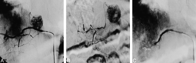
- fig 1.
Case 7: 27-year-old woman with a giant cell tumor in the L3 vertebral body who underwent embolization 2 days before surgery.
A, Preembolization angiogram of the right second lumbar artery shows hypervascular supply to the tumor.
B, Selective injection of the dorsispinal artery at L2 (arrow) shows the extraspinal longitudinal anastomoses from L1 to L4 with tumor supply by L2 and L3.
C, Superselective injection of the feeding artery near the dorsispinal branch was followed by infusion of PVA particles.
D, Postembolization angiogram shows nearly total occlusion of the feeding artery and patency of the normal branches.
- fig 2.
Case 10: 55-year-old man with a paraganglioma in the T12 vertebral body who underwent embolization 7 days before surgery.
A and B, Angiogram of the right segmental artery at T12 (A) and selective angiogram with microcatheter (B) show the posterior radiculomedullary artery (small arrows) originating from the same trunk as the feeding artery (large arrow) to the tumor.
C, Pedicular injection after superselective catheterization and infusion of PVA particles and Gelfoam pieces in the feeder show complete occlusion of the feeder (large arrow) and patency of the posterior radiculomedullary artery (small arrows).
- fig 3.
Case 6: 31-year-old man with an intramedullary hemangioblastoma at T8–T9 who underwent embolization the day before surgery.
A, Preembolization angiogram shows that the posterior radiculomedullary artery (arrow), mainly supplying the intradural tumor, originates from the right ninth intercostal artery.
B, Angiogram after embolization with coils (large arrow) shows occlusion of the intercostal artery distal to the feeder, and the presence of another normal branch originating just distal to the feeder. Infusion of PVA particles with preferential flow and infusion of Gelfoam was then performed through the microcatheter (small arrow) close to the feeder.
C, Postembolization angiogram shows complete occlusion of both the feeder and the normal branch.
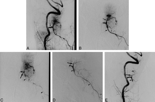
- fig 4.
Case 1: 41-year-old woman with a meningioma at C1–C2 who underwent embolization the day before surgery.
A, Right vertebral angiogram before embolization shows a C2 branch supplying the intradural tumor (arrow).
B, Selective microcatheter injection of the common trunk of the feeder (large arrow) and the normal branch (small arrow) is performed before embolization.
C, Late arterial phase of selective injection shows that most of the contrast medium in the normal branch is washed out (small arrows) because the wedged microcatheter caused flow reversal in the branch. Note that dense contrast medium remains in the trunk (large arrow) just distal to the tip of the microcatheter.
D, Injection after infusion of PVA particles in the trunk shows almost complete occlusion of the feeder (large arrow) and patency of the normal branch (small arrows).
E, Right vertebral angiogram after embolization with Gelfoam pieces shows complete occlusion of both the feeder (large arrow) and the normal branch (small arrow).


Tables

Clinical data and results in 18 patients treated with preoperative transarterial embolization

In this issue


Jump to section
Related Articles
Cited By...
- Assessing Vascularity of Osseous Spinal Metastases with Dual-Energy CT-DSA: A Pilot Study Compared with Catheter Angiography
- Concomitant origin of the anterior or posterior spinal artery with the feeder of a spinal dural arteriovenous fistula (SDAVF)
- The efficacy and risks of preoperative embolization of spinal tumors
- Blood loss in spinal tumour surgery and surgery for metastatic spinal disease: A meta-analysis
- Vascular anatomy of the spinal cord
- Retrospective Analysis of Preoperative Embolization of Spinal Tumors
- Seven-year survival after intralesional resection and adjuvant radiotherapy for a giant-cell tumour of the sixth cervical vertebra
- Spinal Metastases from Renal Cell Carcinoma: Effect of Preoperative Particle Embolization on Intraoperative Blood Loss