
Article Figures & Data
Figures
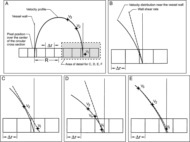
- fig 1.
Velocity distribution and shear rate at the vessel wall.
A, View of the velocity profile along the horizontal row of shaded pixels. Note that the boundary between the vessel lumen and the vessel wall will not coincide with a pixel edge; thus, in general, edge pixels will contain signals from flowing fluid and the stationary vessel wall.
B–E, Detail near the vessel wall showing the actual velocity gradient. Calculation of wall shear rate using LE (C), LE* (D), and QE (E). Velocity measurements in the edge pixels and the two adjacent pixels are denoted by v1, v2, and v3, respectively.
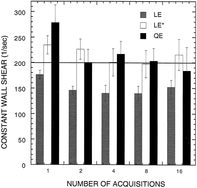
- fig 2.
Measured shear rates for a constant-flow experiment in the phantom. The predicted shear rate at the wall was 198 s−1. Experiments were repeated using different numbers of signal averages (NEX). Error bars represent ±1 SE
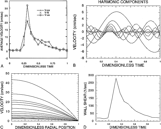
- fig 3.
Measured shear rates for the pulsatile flow experiment in the phantom.
A, Pulsatile average velocity waveform measured by retrospectively gated 2D PC MR imaging (V-mr), reconstructed from a Fourier decomposition of harmonic components (V-ift) and independently measured using an ultrasonic flow probe (V-us).
B, Fourier harmonic components of the pulsatile average velocity wave form measured by MR imaging used to calculate the pulsatile velocity profile.
C, Velocity profile, v(r,t), from the center of the vessel to the wall at 16 frames over the cardiac cycle. Each profile is the sum of harmonic components at one frame.
D, Wall shear rate over the cardiac cycle, and the slope of the velocity profile at the vessel wall.
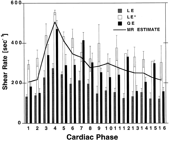
- fig 4.
Measured shear rates for pulsatile flow experiment in the phantom. An ultrasonic flow probe provided an independent estimate of average velocity
- fig 5.
Measured shear rates obtained in a volunteer.
- fig 6.
Scatter plots for the in vivo experiments. The values of wall shear rate measured using PC MR data are plotted against reference values. Measured values were obtained by applying LE (A), LE* (B), and QE (C) algorithms to the PC MR data. Reference wall shear rates were obtained using the procedure shown in figure 4 and described in the text


