Advertisement

Main menu

  • Home
  • Content
    • Current Issue
    • Accepted Manuscripts
    • Article Preview
    • Past Issue Archive
    • AJNR Case Collection
    • Case of the Week Archive
    • Classic Case Archive
    • Case of the Month Archive
  • Special Collections
    • Spinal CSF Leak Articles (Jan 2020-June 2024)
    • 2024 AJNR Journal Awards
    • Most Impactful AJNR Articles
  • Multimedia
    • AJNR Podcast
    • AJNR Scantastics
    • Video Articles
  • For Authors
    • Submit a Manuscript
    • Author Policies
    • Fast publishing of Accepted Manuscripts
    • Graphical Abstract Preparation
    • Manuscript Submission Guidelines
    • Imaging Protocol Submission
    • Submit a Case for the Case Collection
  • About Us
    • About AJNR
    • Editorial Board
  • More
    • Become a Reviewer/Academy of Reviewers
    • Subscribers
    • Permissions
    • Alerts
    • Feedback
    • Advertisers
    • ASNR Home
  • Other Publications
    • ajnr

User menu

  • Alerts
  • Log in

Search

  • Advanced search
American Journal of Neuroradiology

American Journal of Neuroradiology

ASHNR American Society of Functional Neuroradiology ASHNR American Society of Pediatric Neuroradiology ASSR
  • Alerts
  • Log in

Advanced Search

  • Home
  • Content
    • Current Issue
    • Accepted Manuscripts
    • Article Preview
    • Past Issue Archive
    • AJNR Case Collection
    • Case of the Week Archive
    • Classic Case Archive
    • Case of the Month Archive
  • Special Collections
    • Spinal CSF Leak Articles (Jan 2020-June 2024)
    • 2024 AJNR Journal Awards
    • Most Impactful AJNR Articles
  • Multimedia
    • AJNR Podcast
    • AJNR Scantastics
    • Video Articles
  • For Authors
    • Submit a Manuscript
    • Author Policies
    • Fast publishing of Accepted Manuscripts
    • Graphical Abstract Preparation
    • Manuscript Submission Guidelines
    • Imaging Protocol Submission
    • Submit a Case for the Case Collection
  • About Us
    • About AJNR
    • Editorial Board
  • More
    • Become a Reviewer/Academy of Reviewers
    • Subscribers
    • Permissions
    • Alerts
    • Feedback
    • Advertisers
    • ASNR Home
  • Follow AJNR on Twitter
  • Visit AJNR on Facebook
  • Follow AJNR on Instagram
  • Join AJNR on LinkedIn
  • RSS Feeds

Welcome to the new AJNR, Updated Hall of Fame, and more. Read the full announcements.


AJNR is seeking candidates for the position of Associate Section Editor, AJNR Case Collection. Read the full announcement.


Research ArticleHEAD AND NECK

High-Resolution MR Cisternography of the Cerebellopontine Angle: 2D versus 3D Fast Spin-Echo Sequences

Eriko Iwayama, Shinji Naganawa, Tokiko Ito, Hiroshi Fukatsu, Mitsuru Ikeda, Takeo Ishigaki and Nobuyasu Ichinose
American Journal of Neuroradiology May 1999, 20 (5) 889-895;
Eriko Iwayama
  • Find this author on Google Scholar
  • Find this author on PubMed
  • Search for this author on this site
Shinji Naganawa
  • Find this author on Google Scholar
  • Find this author on PubMed
  • Search for this author on this site
Tokiko Ito
  • Find this author on Google Scholar
  • Find this author on PubMed
  • Search for this author on this site
Hiroshi Fukatsu
  • Find this author on Google Scholar
  • Find this author on PubMed
  • Search for this author on this site
Mitsuru Ikeda
  • Find this author on Google Scholar
  • Find this author on PubMed
  • Search for this author on this site
Takeo Ishigaki
  • Find this author on Google Scholar
  • Find this author on PubMed
  • Search for this author on this site
Nobuyasu Ichinose
  • Find this author on Google Scholar
  • Find this author on PubMed
  • Search for this author on this site
  • Article
  • Figures & Data
  • Info & Metrics
  • Responses
  • References
  • PDF
Loading

Article Figures & Data

Figures

  • Tables
  • fig 1.
    • Download figure
    • Open in new tab
    • Download powerpoint
    fig 1.

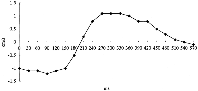
    Flow velocity and flow pattern of CSF in the prepontine cistern obtained in a volunteer with cine phase velocity mapping. The average peak velocity of CSF in four volunteers was 1.2 cm/s.

  • fig 2.
    • Download figure
    • Open in new tab
    • Download powerpoint
    fig 2.

    A and B, Single-section 3-mm-thick 2D image (A) and 30-mm-slab 3-mm-section 3D image (B). The 2D image shows loss of signal intensity in the prepontine cistern (arrows) compared with the 3D image (arrows)
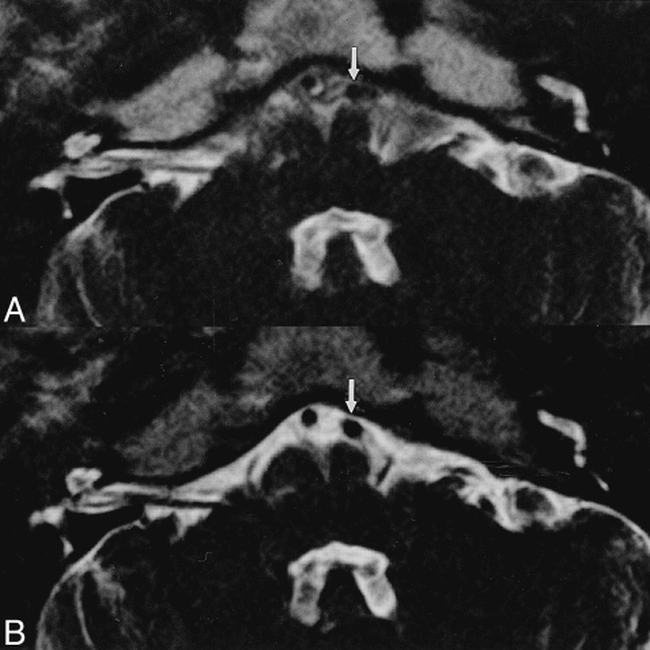
  • fig 3.
    • Download figure
    • Open in new tab
    • Download powerpoint
    fig 3.

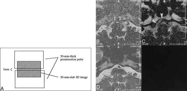
    A, Schematic diagram of 30-mm-slab 3D images (shaded area) with intraslab presaturation pulses except for the 1-mm-slab center volume.

    B, 30-mm-slab 1-mm-section 3D image with intraslab presaturation pulses show signal loss (arrows).

    C, Single-section 3-mm-thick 2D image shows some signal loss (arrowheads).

    D, 30-mm-slab 1-mm-section 3D image without presaturation pulse shows no signal loss in the prepontine cistern.

  • fig 4.
    • Download figure
    • Open in new tab
    • Download powerpoint
    fig 4.

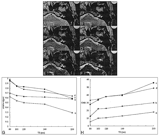
    A–H, Comparison of various TEs in single-section 3-mm-thick 2D images: TE = 80 (A), TE = 100 (B), TE = 120 (C), TE = 160 (D), TE = 254 (E); and 30-mm-slab 1-mm-section 3D image (F). In all four volunteers, CSF signal relative to that of the water phantom decreased gradually as TE increased in single-section 3-mm-thick 2D images (G). In all four volunteers, CNR between the water phantom and the cerebellar peduncle increased gradually as TE increased (H)
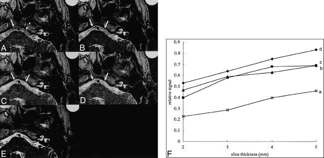
  • fig 5.
    • Download figure
    • Open in new tab
    • Download powerpoint
    fig 5.

    A–F, Comparison of various section thicknesses for single-section 2D images: 2 mm (A), 3 mm (B), 4 mm (C), 5 mm (D); and 30-mm-slab 1-mm-section 3D image (E). In all four volunteers, CSF signal relative to that of the water phantom increased gradually as section thickness increased (arrows in A–E) (F)

  • fig 6.
    • Download figure
    • Open in new tab
    • Download powerpoint
    fig 6.

    Images obtained in a healthy volunteer for comparison of 2D and 3D FSE sequences.

    A, Multisection 3-mm-thick 2D image.

    B, 30-mm-slab 1-mm-section 3D image. The visibility of the vertebral artery is poor in the 2D image (arrow) but excellent in the 3D image (arrow).

  • fig 7.
    • Download figure
    • Open in new tab
    • Download powerpoint
    fig 7.

    57-year-old man with right-sided trigeminal neuralgia.

    A and B, Multisection 3-mm-thick 2D image (A) and 30-mm-slab 1-mm-section 3D image (B). A mass (white arrowheads) presumed to be an epidermoid cyst compressing the trigeminal nerve (black arrowheads) is depicted in the right cerebellopontine angle. Although the tumor is isointense with CSF on the 3D image (large arrow), the tumor can be recognized more clearly on the 2D image, owing to signal loss in CSF (small arrows).

Tables

  • Figures
  • Table 1:
    • View popup
    • Download powerpoint
    Table 1:

    The peak velocity (cm/s) of CSF in four volunteers

  • Table 2:
    • View popup
    • Download powerpoint
    Table 2:

    Relative signal intensity* on single-section 3-mm-thick 2D images, 30-mm-slab 3-mm-section 3D images, 30-mm-slab 3D images with intraslab presaturation pulses except in the 1-mm-slab center volume, and 30-mm-slab 1-mm-section 3D images without a presaturation pulse in four volunteers

  • Table 3:
    • View popup
    • Download powerpoint
    Table 3:

    The visibility grading of vertebrobasilar artery, vestibulocochlear nerve, and trigeminal nerve as seen on 2D versus 3D images (n = 17)
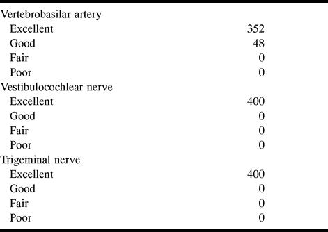
  • Table 4:
    • View popup
    • Download powerpoint
    Table 4:

    The visibility grading of vertebrobasilar artery, vestibulocochlear nerve, and trigeminal nerve on 3D images (n = 400)

PreviousNext
Back to top

In this issue

American Journal of Neuroradiology
Vol. 20, Issue 5
1 May 1999
  • Table of Contents
  • Index by author
Advertisement
Print
Download PDF
Email Article

Thank you for your interest in spreading the word on American Journal of Neuroradiology.

NOTE: We only request your email address so that the person you are recommending the page to knows that you wanted them to see it, and that it is not junk mail. We do not capture any email address.

Enter multiple addresses on separate lines or separate them with commas.
High-Resolution MR Cisternography of the Cerebellopontine Angle: 2D versus 3D Fast Spin-Echo Sequences
(Your Name) has sent you a message from American Journal of Neuroradiology
(Your Name) thought you would like to see the American Journal of Neuroradiology web site.
CAPTCHA
This question is for testing whether or not you are a human visitor and to prevent automated spam submissions.
Cite this article
Eriko Iwayama, Shinji Naganawa, Tokiko Ito, Hiroshi Fukatsu, Mitsuru Ikeda, Takeo Ishigaki, Nobuyasu Ichinose
High-Resolution MR Cisternography of the Cerebellopontine Angle: 2D versus 3D Fast Spin-Echo Sequences
American Journal of Neuroradiology May 1999, 20 (5) 889-895;

Citation Manager Formats

  • BibTeX
  • Bookends
  • EasyBib
  • EndNote (tagged)
  • EndNote 8 (xml)
  • Medlars
  • Mendeley
  • Papers
  • RefWorks Tagged
  • Ref Manager
  • RIS
  • Zotero
0 Responses
Respond to this article
Share
Bookmark this article
High-Resolution MR Cisternography of the Cerebellopontine Angle: 2D versus 3D Fast Spin-Echo Sequences
Eriko Iwayama, Shinji Naganawa, Tokiko Ito, Hiroshi Fukatsu, Mitsuru Ikeda, Takeo Ishigaki, Nobuyasu Ichinose
American Journal of Neuroradiology May 1999, 20 (5) 889-895;
del.icio.us logo Twitter logo Facebook logo Mendeley logo
  • Tweet Widget
  • Facebook Like
  • Google Plus One

Jump to section

  • Article
    • Abstract
    • Methods
    • Results
    • Discussion
    • Conclusion
    • Footnotes
    • References
  • Figures & Data
  • Info & Metrics
  • Responses
  • References
  • PDF

Related Articles

  • No related articles found.
  • PubMed
  • Google Scholar

Cited By...

  • No citing articles found.
  • Crossref
  • Google Scholar

This article has not yet been cited by articles in journals that are participating in Crossref Cited-by Linking.

More in this TOC Section

  • Correlation of Apparent Diffusion Coefficient at 3T with Prognostic Parameters of Retinoblastoma
  • Parathyroid Lesions: Characterization with Dual-Phase Arterial and Venous Enhanced CT of the Neck
  • Comparison of Contrast Effect on the Cochlear Perilymph after Intratympanic and Intravenous Gadolinium Injection
Show more Head and Neck

Similar Articles

Advertisement

Indexed Content

  • Current Issue
  • Accepted Manuscripts
  • Article Preview
  • Past Issues
  • Editorials
  • Editors Choice
  • Fellow Journal Club
  • Letters to the Editor

Cases

  • Case Collection
  • Archive - Case of the Week
  • Archive - Case of the Month
  • Archive - Classic Case

Special Collections

  • Special Collections

Resources

  • News and Updates
  • Turn around Times
  • Submit a Manuscript
  • Author Policies
  • Manuscript Submission Guidelines
  • Evidence-Based Medicine Level Guide
  • Publishing Checklists
  • Graphical Abstract Preparation
  • Imaging Protocol Submission
  • Submit a Case
  • Become a Reviewer/Academy of Reviewers
  • Get Peer Review Credit from Publons

Multimedia

  • AJNR Podcast
  • AJNR SCANtastic
  • Video Articles

About Us

  • About AJNR
  • Editorial Board
  • Not an AJNR Subscriber? Join Now
  • Alerts
  • Feedback
  • Advertise with us
  • Librarian Resources
  • Permissions
  • Terms and Conditions

American Society of Neuroradiology

  • Not an ASNR Member? Join Now

© 2025 by the American Society of Neuroradiology All rights, including for text and data mining, AI training, and similar technologies, are reserved.
Print ISSN: 0195-6108 Online ISSN: 1936-959X

Powered by HighWire