Advertisement

Main menu

  • Home
  • Content
    • Current Issue
    • Accepted Manuscripts
    • Article Preview
    • Past Issue Archive
    • AJNR Case Collection
    • Case of the Week Archive
    • Classic Case Archive
    • Case of the Month Archive
  • Special Collections
    • Spinal CSF Leak Articles (Jan 2020-June 2024)
    • 2024 AJNR Journal Awards
    • Most Impactful AJNR Articles
  • Multimedia
    • AJNR Podcast
    • AJNR Scantastics
    • Video Articles
  • For Authors
    • Submit a Manuscript
    • Author Policies
    • Fast publishing of Accepted Manuscripts
    • Graphical Abstract Preparation
    • Manuscript Submission Guidelines
    • Imaging Protocol Submission
    • Submit a Case for the Case Collection
  • About Us
    • About AJNR
    • Editorial Board
  • More
    • Become a Reviewer/Academy of Reviewers
    • Subscribers
    • Permissions
    • Alerts
    • Feedback
    • Advertisers
    • ASNR Home
  • Other Publications
    • ajnr

User menu

  • Alerts
  • Log in

Search

  • Advanced search
American Journal of Neuroradiology

American Journal of Neuroradiology

ASHNR American Society of Functional Neuroradiology ASHNR American Society of Pediatric Neuroradiology ASSR
  • Alerts
  • Log in

Advanced Search

  • Home
  • Content
    • Current Issue
    • Accepted Manuscripts
    • Article Preview
    • Past Issue Archive
    • AJNR Case Collection
    • Case of the Week Archive
    • Classic Case Archive
    • Case of the Month Archive
  • Special Collections
    • Spinal CSF Leak Articles (Jan 2020-June 2024)
    • 2024 AJNR Journal Awards
    • Most Impactful AJNR Articles
  • Multimedia
    • AJNR Podcast
    • AJNR Scantastics
    • Video Articles
  • For Authors
    • Submit a Manuscript
    • Author Policies
    • Fast publishing of Accepted Manuscripts
    • Graphical Abstract Preparation
    • Manuscript Submission Guidelines
    • Imaging Protocol Submission
    • Submit a Case for the Case Collection
  • About Us
    • About AJNR
    • Editorial Board
  • More
    • Become a Reviewer/Academy of Reviewers
    • Subscribers
    • Permissions
    • Alerts
    • Feedback
    • Advertisers
    • ASNR Home
  • Follow AJNR on Twitter
  • Visit AJNR on Facebook
  • Follow AJNR on Instagram
  • Join AJNR on LinkedIn
  • RSS Feeds

Welcome to the new AJNR, Updated Hall of Fame, and more. Read the full announcements.


AJNR is seeking candidates for the position of Associate Section Editor, AJNR Case Collection. Read the full announcement.


LetterLetter

Three-Dimensional Power Doppler Imaging of Vertebrobasilar Circulation in Adults

Mastoshi Koga, Kazumi Kimura, Masahiro Yasaka, Ryoichi Otsubo, Yasuhiro Hasegawa, Kazuo Minematsu and Takenori Yamaguchi
American Journal of Neuroradiology May 1999, 20 (5) 943-944;
Mastoshi Koga
  • Find this author on Google Scholar
  • Find this author on PubMed
  • Search for this author on this site
Kazumi Kimura
  • Find this author on Google Scholar
  • Find this author on PubMed
  • Search for this author on this site
Masahiro Yasaka
  • Find this author on Google Scholar
  • Find this author on PubMed
  • Search for this author on this site
Ryoichi Otsubo
  • Find this author on Google Scholar
  • Find this author on PubMed
  • Search for this author on this site
Yasuhiro Hasegawa
  • Find this author on Google Scholar
  • Find this author on PubMed
  • Search for this author on this site
Kazuo Minematsu
  • Find this author on Google Scholar
  • Find this author on PubMed
  • Search for this author on this site
Takenori Yamaguchi
  • Find this author on Google Scholar
  • Find this author on PubMed
  • Search for this author on this site
  • Article
  • Figures & Data
  • Info & Metrics
  • Responses
  • References
  • PDF
Loading

Development of a three-dimensional (3D) sonographic technique has enabled delineation of fetal surface features, abdominal organs, and coronary arterial walls (1–5). Power Doppler imaging is more sensitive in detecting blood flow signals than is color Doppler. Kenton and colleagues reported that power Doppler offered significant advantages over color Doppler in the imaging of intracranial cerebral arteries (6). By a combination of power Doppler and 3D technology, it is possible not only to image small vessels, but also to construct 3D images of vascular structures. In this communication we show vertebrobasilar arteries imaged by 3D power-Doppler imaging.

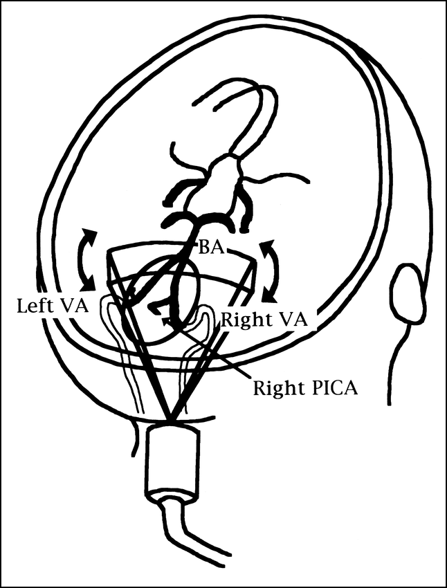
We applied a 2D and 3D power Doppler examination for the imaging of a 22-year-old healthy woman with the HDI 3000 (Advanced Technology Laboratories, Botnell, WA). The 3D imaging rendered a volumetric vessel appearance with power Doppler. Using power Doppler, we acquired flow data in a 2D slice through a volume of the vessel and represented spatial relationships within that 2D plane. We reconstructed the 2D plane data into the 3D power Doppler image. During this process, parenchymal data were subtracted. The transducer was a digital broadband steered phased array with frequencies ranging from 2–3 MHz for B-mode imaging and Doppler functions. Our patient was examined in the lateral decubitus position. The transducer was placed between the squama occipitalis and the palpable spinous process of the first cervical vertebra, and the Doppler beam was aimed at the bridge of the nose. We detected blood flow signals from the vertebral arteries (VAs), the posterior inferior cerebellar arteries (PICAs), and the basilar arteries (BAs) in real time, which were displayed as color signals within a subsector of the black-and-white image by using 2D power Doppler. Then, the transducer was locked back and tilted very slowly to image VAs (V3–V4 segments), PICAs (bilateral views), and the BA (proximal view) for 15 seconds, and collect and store 74 images in the computer memory. Subsequently parenchymal data were subtracted and a rendering of the vascular anatomy was composed with a 3D digital system. Then, 3D power Doppler images of the vertebrobasilar arteries were reconstructed. In addition, 15 images could be displayed selectively on a video monitor in dynamic partial rotation to enable viewing from different angles within the spatial range of 110°. Figure 1 shows the transducer placement and the anatomic structures.

Figure
  • Download figure
  • Open in new tab
  • Download powerpoint

FIG 1. Schema shows relationship between transducer and vascular anatomy. The transducer was placed between the squama occipitalis and the palpable spinous process of the first cervical vertebra. After detecting blood flow signals from the VA, bilateral PICAs, and the BA by 2D power-Doppler imaging, the transducer was locked back and tilted very slowly to assess these arteries. Subsequently, by using a 3D digital system, 3D power-Doppler images of the vertebrobasilar arteries were reconstructed after rendering

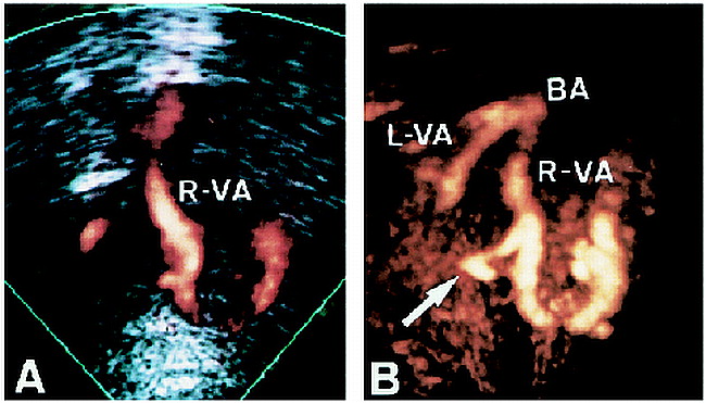
The 2D power Doppler image showed the right VA (V3–V4), part of the right PICA, and the left VA (Fig 2A), but it could not show the entire vertebrobasilar system continuously. The 3D power Doppler method, however, clearly and continuously imaged the VA, and displayed a 3D image of the BA (Fig 2B).

Figure
  • Download figure
  • Open in new tab
  • Download powerpoint

FIG 2. 2D and 3D power-Doppler images.

A, 2D power-Doppler image shows the right VA (segments V3–V4), part of the PICA, and the left VA.

B, 3D power-Doppler study clearly depicts the Vas and the PICA branching from the right VA (arrow).

Clinical application of the 3D power Doppler method, when applied with meticulous technique and improved technology, may enable visualization of vascular diseases such as aneurysms, arteriovenous malformations, and other vascular disorders of the vertebrobasilar system.

Acknowledgments

This work was supported by the Research Grant for Cardiovascular Diseases (8-C4, 9-A2) from the Ministry of Health and Welfare. The authors would like to thank Mr. Juni Warabino for technical assistance and Ms. Yuki Iwakiri for cooperation with the present study.

References

  1. ↵
    Fishman EK, Magid D, Ney DR, et al. Three-dimensional imaging. Radiology 1991;181:321-337
    PubMed
  2. Rankin RN, Fenster A, Downey DB, Munk PL, Levin MF, Vellet AD. Three-dimensional sonographic reconstruction: techniques and diagnostic applications. AJR Am J Roentgenol 1993;161:695-702
    PubMed
  3. Picot PA, Rickey DW, Mitchell R, Rankin RN, Fenster A. Three-dimensional colour Doppler imaging. Ultrasound in Med Biol 1993;19:95-104
    CrossRefPubMed
  4. Steinke W, Meairs S, Ries S, Hennerici M. Sonographic assessment of carotid artery stenosis. comparison of power Doppler imaging and color Doppler flow imaging. Stroke 1996;27:91-94
    Abstract/FREE Full Text
  5. Pretorious DH, Nelson TR. Fetal face visualization using three-dimensional ultrasonography. J Ultrasound Med 1995;14:349-356
    Abstract
  6. ↵
    Kenton AR, Martin PJ, Evans DH. Power Doppler: an advance over colour Doppler for transcranial images? Ultrasound in Med Biol 1996;22:313-317
    CrossRefPubMed
  • Copyright © American Society of Neuroradiology
PreviousNext
Back to top

In this issue

American Journal of Neuroradiology
Vol. 20, Issue 5
1 May 1999
  • Table of Contents
  • Index by author
Advertisement
Print
Download PDF
Email Article

Thank you for your interest in spreading the word on American Journal of Neuroradiology.

NOTE: We only request your email address so that the person you are recommending the page to knows that you wanted them to see it, and that it is not junk mail. We do not capture any email address.

Enter multiple addresses on separate lines or separate them with commas.
Three-Dimensional Power Doppler Imaging of Vertebrobasilar Circulation in Adults
(Your Name) has sent you a message from American Journal of Neuroradiology
(Your Name) thought you would like to see the American Journal of Neuroradiology web site.
CAPTCHA
This question is for testing whether or not you are a human visitor and to prevent automated spam submissions.
Cite this article
Mastoshi Koga, Kazumi Kimura, Masahiro Yasaka, Ryoichi Otsubo, Yasuhiro Hasegawa, Kazuo Minematsu, Takenori Yamaguchi
Three-Dimensional Power Doppler Imaging of Vertebrobasilar Circulation in Adults
American Journal of Neuroradiology May 1999, 20 (5) 943-944;

Citation Manager Formats

  • BibTeX
  • Bookends
  • EasyBib
  • EndNote (tagged)
  • EndNote 8 (xml)
  • Medlars
  • Mendeley
  • Papers
  • RefWorks Tagged
  • Ref Manager
  • RIS
  • Zotero
0 Responses
Respond to this article
Share
Bookmark this article
Three-Dimensional Power Doppler Imaging of Vertebrobasilar Circulation in Adults
Mastoshi Koga, Kazumi Kimura, Masahiro Yasaka, Ryoichi Otsubo, Yasuhiro Hasegawa, Kazuo Minematsu, Takenori Yamaguchi
American Journal of Neuroradiology May 1999, 20 (5) 943-944;
del.icio.us logo Twitter logo Facebook logo Mendeley logo
  • Tweet Widget
  • Facebook Like
  • Google Plus One

Jump to section

  • Article
    • Acknowledgments
    • References
  • Figures & Data
  • Info & Metrics
  • Responses
  • References
  • PDF

Related Articles

  • No related articles found.
  • PubMed
  • Google Scholar

Cited By...

  • No citing articles found.
  • Crossref
  • Google Scholar

This article has not yet been cited by articles in journals that are participating in Crossref Cited-by Linking.

More in this TOC Section

  • Letter to the Editor regarding “Automated Volumetric Software in Dementia: Help or Hindrance to the Neuroradiologist?”
  • Reply:
  • Brain AVM’s Nidus: What if We Hadn’t Understood Anything?
Show more Letter

Similar Articles

Advertisement

Indexed Content

  • Current Issue
  • Accepted Manuscripts
  • Article Preview
  • Past Issues
  • Editorials
  • Editors Choice
  • Fellow Journal Club
  • Letters to the Editor

Cases

  • Case Collection
  • Archive - Case of the Week
  • Archive - Case of the Month
  • Archive - Classic Case

Special Collections

  • Special Collections

Resources

  • News and Updates
  • Turn around Times
  • Submit a Manuscript
  • Author Policies
  • Manuscript Submission Guidelines
  • Evidence-Based Medicine Level Guide
  • Publishing Checklists
  • Graphical Abstract Preparation
  • Imaging Protocol Submission
  • Submit a Case
  • Become a Reviewer/Academy of Reviewers
  • Get Peer Review Credit from Publons

Multimedia

  • AJNR Podcast
  • AJNR SCANtastic
  • Video Articles

About Us

  • About AJNR
  • Editorial Board
  • Not an AJNR Subscriber? Join Now
  • Alerts
  • Feedback
  • Advertise with us
  • Librarian Resources
  • Permissions
  • Terms and Conditions

American Society of Neuroradiology

  • Not an ASNR Member? Join Now

© 2025 by the American Society of Neuroradiology All rights, including for text and data mining, AI training, and similar technologies, are reserved.
Print ISSN: 0195-6108 Online ISSN: 1936-959X

Powered by HighWire