Advertisement

Main menu

  • Home
  • Content
    • Current Issue
    • Accepted Manuscripts
    • Article Preview
    • Past Issue Archive
    • AJNR Case Collection
    • Case of the Week Archive
    • Classic Case Archive
    • Case of the Month Archive
  • Special Collections
    • Spinal CSF Leak Articles (Jan 2020-June 2024)
    • 2024 AJNR Journal Awards
    • Most Impactful AJNR Articles
  • Multimedia
    • AJNR Podcast
    • AJNR Scantastics
    • Video Articles
  • For Authors
    • Submit a Manuscript
    • Author Policies
    • Fast publishing of Accepted Manuscripts
    • Graphical Abstract Preparation
    • Manuscript Submission Guidelines
    • Imaging Protocol Submission
    • Submit a Case for the Case Collection
  • About Us
    • About AJNR
    • Editorial Board
  • More
    • Become a Reviewer/Academy of Reviewers
    • Subscribers
    • Permissions
    • Alerts
    • Feedback
    • Advertisers
    • ASNR Home
  • Other Publications
    • ajnr

User menu

  • Alerts
  • Log in

Search

  • Advanced search
American Journal of Neuroradiology

American Journal of Neuroradiology

ASHNR American Society of Functional Neuroradiology ASHNR American Society of Pediatric Neuroradiology ASSR
  • Alerts
  • Log in

Advanced Search

  • Home
  • Content
    • Current Issue
    • Accepted Manuscripts
    • Article Preview
    • Past Issue Archive
    • AJNR Case Collection
    • Case of the Week Archive
    • Classic Case Archive
    • Case of the Month Archive
  • Special Collections
    • Spinal CSF Leak Articles (Jan 2020-June 2024)
    • 2024 AJNR Journal Awards
    • Most Impactful AJNR Articles
  • Multimedia
    • AJNR Podcast
    • AJNR Scantastics
    • Video Articles
  • For Authors
    • Submit a Manuscript
    • Author Policies
    • Fast publishing of Accepted Manuscripts
    • Graphical Abstract Preparation
    • Manuscript Submission Guidelines
    • Imaging Protocol Submission
    • Submit a Case for the Case Collection
  • About Us
    • About AJNR
    • Editorial Board
  • More
    • Become a Reviewer/Academy of Reviewers
    • Subscribers
    • Permissions
    • Alerts
    • Feedback
    • Advertisers
    • ASNR Home
  • Follow AJNR on Twitter
  • Visit AJNR on Facebook
  • Follow AJNR on Instagram
  • Join AJNR on LinkedIn
  • RSS Feeds

Welcome to the new AJNR, Updated Hall of Fame, and more. Read the full announcements.


AJNR is seeking candidates for the position of Associate Section Editor, AJNR Case Collection. Read the full announcement.


Research ArticleBRAIN

Ischemia after Carotid Endarterectomy: Comparison between Transcranial Doppler Sonography and Diffusion-Weighted MR Imaging

Martin Müller, Werner Reiche, Philipp Langenscheidt, Jens Haßfeld and Thomas Hagen
American Journal of Neuroradiology January 2000, 21 (1) 47-54;
Martin Müller
aFrom the Departments of Neurology (M.M., J.G.), Neuroradiology (W.R., T.H.), and Surgery (P.L.), University Hospital of the Saarland, Homburg/Saar, Germany.
  • Find this author on Google Scholar
  • Find this author on PubMed
  • Search for this author on this site
Werner Reiche
aFrom the Departments of Neurology (M.M., J.G.), Neuroradiology (W.R., T.H.), and Surgery (P.L.), University Hospital of the Saarland, Homburg/Saar, Germany.
  • Find this author on Google Scholar
  • Find this author on PubMed
  • Search for this author on this site
Philipp Langenscheidt
aFrom the Departments of Neurology (M.M., J.G.), Neuroradiology (W.R., T.H.), and Surgery (P.L.), University Hospital of the Saarland, Homburg/Saar, Germany.
  • Find this author on Google Scholar
  • Find this author on PubMed
  • Search for this author on this site
Jens Haßfeld
aFrom the Departments of Neurology (M.M., J.G.), Neuroradiology (W.R., T.H.), and Surgery (P.L.), University Hospital of the Saarland, Homburg/Saar, Germany.
  • Find this author on Google Scholar
  • Find this author on PubMed
  • Search for this author on this site
Thomas Hagen
aFrom the Departments of Neurology (M.M., J.G.), Neuroradiology (W.R., T.H.), and Surgery (P.L.), University Hospital of the Saarland, Homburg/Saar, Germany.
  • Find this author on Google Scholar
  • Find this author on PubMed
  • Search for this author on this site
  • Article
  • Figures & Data
  • Info & Metrics
  • Responses
  • References
  • PDF
Loading

Article Figures & Data

Figures

  • Tables
  • fig 1.
    • Download figure
    • Open in new tab
    • Download powerpoint
    fig 1.

    The TCD device provides two sample volumes for insonation of the MCA. An embolus streaming down the MCA will be recorded first in the proximal sample volume (depth of insonation, 57 mm [D57]) and with a time delay in the distal sample volume (depth of insonation, 52 mm [D52]). Next to the color-coded decibel scale, the Doppler velocity spectrum after fast Fourier transformation (FFT) of each sample volume is shown. The blood flow direction in both sample volumes is indicated in the proximal sample volume beneath D57 by an arrow directed toward the probe symbol ([), which means that the velocity spectrum of the blood flow in the MCA usually directed toward the probe is imaged above the zero line; the background velocity spectrum can be seen more clearly in the distal sample volume as a band with a signal intensity of 9 to 15 dB (colored slightly blue to green). The signal of the embolus after FFT is overloaded, as indicated by its bidirectional appearance

  • fig 2.
    • Download figure
    • Open in new tab
    • Download powerpoint
    fig 2.

    A–D, Pre- and postoperative T2-weighted images (A and C) and DWIs (B and D) of a patient in whom a territorial infarct was present on the postoperative images. The small white matter lesions on the preoperative T2-weighted (4000/99/3) image (A) are not present on the preoperative DWI (B) (episequence; 123/1 [TE/excitations]). The corresponding postoperative T2-weighted image (C) and DWI (D) show a medium-sized territorial infarct on the left side, and a small new lesion in the head of the caudate nucleus on the right side. This patient underwent surgery for a tight left-sided stenosis associated with occlusion of the contralateral internal carotid artery
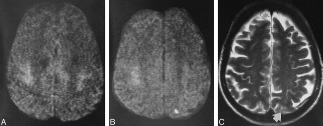
  • fig 3.
    • Download figure
    • Open in new tab
    • Download powerpoint
    fig 3.

    A–C, Pre- and postoperative images of a patient who had had an episode of amaurosis fugax. The preoperative DWI (A) shows no signal abnormality. The postoperative DWI (B) shows a small cortical lesion in the distribution of the posterior branches of the MCA; on the postoperative T2-weighted image (C), this lesion (arrow) can be seen only in light of the findings on the DWI

  • fig 4.
    • Download figure
    • Open in new tab
    • Download powerpoint
    fig 4.

    A–D, Pre- and postoperative images of a patient who underwent carotid endarterectomy 14 days after the clinical event. The preoperative T2-weighted (A) and DWI (B) studies show four small cortical lesions and one subcortical lesion (arrow, B). The cortical lesions cannot be detected on the corresponding postoperative DWI (D). The subcortical lesion is still present, but there is a new, smaller territorial infarction nearby as shown on the T2-weighted (C) and DWI (D) studies. These findings may suggest that cortical lesions disappear sooner than subcortical lesions, probably because of the better collateral blood flow in cortical regions as compared with subcortical regions

  • fig 5.
    • Download figure
    • Open in new tab
    • Download powerpoint
    fig 5.

    A–C, Preoperative CT scan (A) of a patient with an asymptomatic tight stenosis of the right internal carotid artery. In the right parasagittal area, the slight leukoencephalopathy is one aspect of the coincident microvascular disease, which is more clearly present on the other CT sections (not shown). Postoperatively, an infarct in the right precentral gyrus is seen on the axial T2-weighted (B) and DWI (C) studies

Tables

  • Figures
  • Table1

    Types of hyperintense signal distribution on postoperative diffusion-weighted images in patients who had endarterectomies performed without and with shunting

PreviousNext
Back to top

In this issue

American Journal of Neuroradiology
Vol. 21, Issue 1
1 Jan 2000
  • Table of Contents
  • Index by author
Advertisement
Print
Download PDF
Email Article

Thank you for your interest in spreading the word on American Journal of Neuroradiology.

NOTE: We only request your email address so that the person you are recommending the page to knows that you wanted them to see it, and that it is not junk mail. We do not capture any email address.

Enter multiple addresses on separate lines or separate them with commas.
Ischemia after Carotid Endarterectomy: Comparison between Transcranial Doppler Sonography and Diffusion-Weighted MR Imaging
(Your Name) has sent you a message from American Journal of Neuroradiology
(Your Name) thought you would like to see the American Journal of Neuroradiology web site.
CAPTCHA
This question is for testing whether or not you are a human visitor and to prevent automated spam submissions.
Cite this article
Martin Müller, Werner Reiche, Philipp Langenscheidt, Jens Haßfeld, Thomas Hagen
Ischemia after Carotid Endarterectomy: Comparison between Transcranial Doppler Sonography and Diffusion-Weighted MR Imaging
American Journal of Neuroradiology Jan 2000, 21 (1) 47-54;

Citation Manager Formats

  • BibTeX
  • Bookends
  • EasyBib
  • EndNote (tagged)
  • EndNote 8 (xml)
  • Medlars
  • Mendeley
  • Papers
  • RefWorks Tagged
  • Ref Manager
  • RIS
  • Zotero
0 Responses
Respond to this article
Share
Bookmark this article
Ischemia after Carotid Endarterectomy: Comparison between Transcranial Doppler Sonography and Diffusion-Weighted MR Imaging
Martin Müller, Werner Reiche, Philipp Langenscheidt, Jens Haßfeld, Thomas Hagen
American Journal of Neuroradiology Jan 2000, 21 (1) 47-54;
del.icio.us logo Twitter logo Facebook logo Mendeley logo
  • Tweet Widget
  • Facebook Like
  • Google Plus One

Jump to section

  • Article
    • Abstract
    • Methods
    • Results
    • Discussion
    • Conclusion
    • Footnotes
    • References
  • Figures & Data
  • Info & Metrics
  • Responses
  • References
  • PDF

Related Articles

  • No related articles found.
  • PubMed
  • Google Scholar

Cited By...

  • Symptomatic Carotid Occlusion Is Frequently Associated With Microembolization
  • Intracranial Hemodynamics Is Altered by Carotid Artery Disease and After Endarterectomy: A Dynamic Magnetic Resonance Angiography Study
  • Cerebral Microemboli and Brain Injury During Carotid Artery Endarterectomy and Stenting
  • Assessing Carotid Revascularization: Should We Abandon the Neurological Examination?
  • New Brain Lesions After Carotid Stenting Versus Carotid Endarterectomy: A Systematic Review of the Literature
  • Internal and Cortical Border-Zone Infarction: Clinical and Diffusion-Weighted Imaging Features
  • Pathogenesis of deep white matter medullary infarcts: a diffusion weighted magnetic resonance imaging study
  • The Pathophysiology of Watershed Infarction in Internal Carotid Artery Disease: Review of Cerebral Perfusion Studies
  • Subcortical White Matter Infarcts: Comparison of Superficial Perforating Artery and Internal Border-Zone Infarcts Using Diffusion-Weighted Magnetic Resonance Imaging
  • Brain diffusion changes in carotid occlusive disease treated with endarterectomy
  • Protecting the brain:how do we measure success?
  • Assessment of Silent Embolism from Carotid Endarterectomy by Use of Diffusion-weighted Imaging: Work in Progress
  • Detection of Clinically Silent Infarcts after Carotid Endarterectomy by Use of Diffusion-weighted Imaging
  • Systematic Review of Diffusion and Perfusion Imaging in Acute Ischemic Stroke
  • Crossref
  • Google Scholar

This article has not yet been cited by articles in journals that are participating in Crossref Cited-by Linking.

More in this TOC Section

  • Multimodal CT Provides Improved Performance for Lacunar Infarct Detection
  • Statin Therapy Does Not Affect the Radiographic and Clinical Profile of Patients with TIA and Minor Stroke
  • Optimal MRI Sequence for Identifying Occlusion Location in Acute Stroke: Which Value of Time-Resolved Contrast-Enhanced MRA?
Show more Brain

Similar Articles

Advertisement

Indexed Content

  • Current Issue
  • Accepted Manuscripts
  • Article Preview
  • Past Issues
  • Editorials
  • Editors Choice
  • Fellow Journal Club
  • Letters to the Editor

Cases

  • Case Collection
  • Archive - Case of the Week
  • Archive - Case of the Month
  • Archive - Classic Case

Special Collections

  • Special Collections

Resources

  • News and Updates
  • Turn around Times
  • Submit a Manuscript
  • Author Policies
  • Manuscript Submission Guidelines
  • Evidence-Based Medicine Level Guide
  • Publishing Checklists
  • Graphical Abstract Preparation
  • Imaging Protocol Submission
  • Submit a Case
  • Become a Reviewer/Academy of Reviewers
  • Get Peer Review Credit from Publons

Multimedia

  • AJNR Podcast
  • AJNR SCANtastic
  • Video Articles

About Us

  • About AJNR
  • Editorial Board
  • Not an AJNR Subscriber? Join Now
  • Alerts
  • Feedback
  • Advertise with us
  • Librarian Resources
  • Permissions
  • Terms and Conditions

American Society of Neuroradiology

  • Not an ASNR Member? Join Now

© 2025 by the American Society of Neuroradiology All rights, including for text and data mining, AI training, and similar technologies, are reserved.
Print ISSN: 0195-6108 Online ISSN: 1936-959X

Powered by HighWire