Advertisement

Main menu

  • Home
  • Content
    • Current Issue
    • Accepted Manuscripts
    • Article Preview
    • Past Issue Archive
    • AJNR Case Collection
    • Case of the Week Archive
    • Classic Case Archive
    • Case of the Month Archive
  • Special Collections
    • Spinal CSF Leak Articles (Jan 2020-June 2024)
    • 2024 AJNR Journal Awards
    • Most Impactful AJNR Articles
  • Multimedia
    • AJNR Podcast
    • AJNR Scantastics
    • Video Articles
  • For Authors
    • Submit a Manuscript
    • Author Policies
    • Fast publishing of Accepted Manuscripts
    • Graphical Abstract Preparation
    • Manuscript Submission Guidelines
    • Imaging Protocol Submission
    • Submit a Case for the Case Collection
  • About Us
    • About AJNR
    • Editorial Board
  • More
    • Become a Reviewer/Academy of Reviewers
    • Subscribers
    • Permissions
    • Alerts
    • Feedback
    • Advertisers
    • ASNR Home
  • Other Publications
    • ajnr

User menu

  • Alerts
  • Log in

Search

  • Advanced search
American Journal of Neuroradiology

American Journal of Neuroradiology

ASHNR American Society of Functional Neuroradiology ASHNR American Society of Pediatric Neuroradiology ASSR
  • Alerts
  • Log in

Advanced Search

  • Home
  • Content
    • Current Issue
    • Accepted Manuscripts
    • Article Preview
    • Past Issue Archive
    • AJNR Case Collection
    • Case of the Week Archive
    • Classic Case Archive
    • Case of the Month Archive
  • Special Collections
    • Spinal CSF Leak Articles (Jan 2020-June 2024)
    • 2024 AJNR Journal Awards
    • Most Impactful AJNR Articles
  • Multimedia
    • AJNR Podcast
    • AJNR Scantastics
    • Video Articles
  • For Authors
    • Submit a Manuscript
    • Author Policies
    • Fast publishing of Accepted Manuscripts
    • Graphical Abstract Preparation
    • Manuscript Submission Guidelines
    • Imaging Protocol Submission
    • Submit a Case for the Case Collection
  • About Us
    • About AJNR
    • Editorial Board
  • More
    • Become a Reviewer/Academy of Reviewers
    • Subscribers
    • Permissions
    • Alerts
    • Feedback
    • Advertisers
    • ASNR Home
  • Follow AJNR on Twitter
  • Visit AJNR on Facebook
  • Follow AJNR on Instagram
  • Join AJNR on LinkedIn
  • RSS Feeds

Welcome to the new AJNR, Updated Hall of Fame, and more. Read the full announcements.


AJNR is seeking candidates for the position of Associate Section Editor, AJNR Case Collection. Read the full announcement.


Research ArticleBRAIN

Increased Conspicuity of Intraventricular Lesions Revealed by Three-dimensional Constructive Interference in Steady State Sequences

Dewen Yang, Yukunori Korogi, Yukitaka Ushio and Mutsumasa Takahashi
American Journal of Neuroradiology June 2000, 21 (6) 1070-1072;
Dewen Yang
  • Find this author on Google Scholar
  • Find this author on PubMed
  • Search for this author on this site
Yukunori Korogi
  • Find this author on Google Scholar
  • Find this author on PubMed
  • Search for this author on this site
Yukitaka Ushio
  • Find this author on Google Scholar
  • Find this author on PubMed
  • Search for this author on this site
Mutsumasa Takahashi
  • Find this author on Google Scholar
  • Find this author on PubMed
  • Search for this author on this site
  • Article
  • Figures & Data
  • Info & Metrics
  • Responses
  • References
  • PDF
Loading

Abstract

Summary: We describe our preliminary experience with the three-dimensional constructive interference in steady state (3D-CISS) sequence for the evaluation of intraventricular lesions. Cyst walls, extent and margins of tumors, and intratumoral cystic structures were clearly depicted on 3D-CISS images. The 3D-CISS sequence can offer additional information to conventional MR studies to define intraventricular lesions better.

Although MR imaging is useful for the evaluation of intraventricular lesions, it may sometimes be difficult to determine lesion location, margin, and extent when using conventional MR techniques (1, 2). The three-dimensional constructive interference in steady state (3D-CISS) sequence can provide high-resolution images with excellent contrast between cerebrospinal fluid (CSF) and solid structures. Several investigators have used the 3D-CISS sequence to show the fine anatomic structures of the inner ear and the cerebellopontine angle (3–5). In this study, we describe our preliminary experience with the 3D-CISS sequence for imaging intraventricular lesions.

Methods

Twenty-two patients with intraventricular lesions (18 patients with intraventricular masses and four with intraventricular cysts) were studied on a 1.5-T superconductive unit with a 25-mT/m maximum-gradient capability (Magnetom Vision; Siemens, Erlangen, Germany) and a standard head coil. For the 3D-CISS images, two 3D Fourier transform data sets were acquired with true fast imaging with steady-state precession (FISP) sequences, which included flow compensation applied to each gradient over each repetition-time cycle. One data set was acquired with an alternating radiofrequency pulse, and one was acquired with a nonalternating radiofrequency pulse. After conventional T1- and T2-weighted spin-echo (SE) imaging was completed, the 3D-CISS sequence was performed (parameters: 12.2/5.9/1 [TR/ TE/excitations]; flip angle, 70°; matrix, 192—256 × 512; field of view, 150—210 × 200–210 mm; in-plane resolution, 0.78 × 0.39 mm). An axial or sagittal 50-mm-thick slab with 70 partitions was used to cover the intraventricular lesions based on conventional T1- or T2-weighted images or both. Image acquisition time ranged from 5 minutes 2 seconds to 6 minutes 43 seconds. The phase-encoding direction was right to left for all patients.

Results

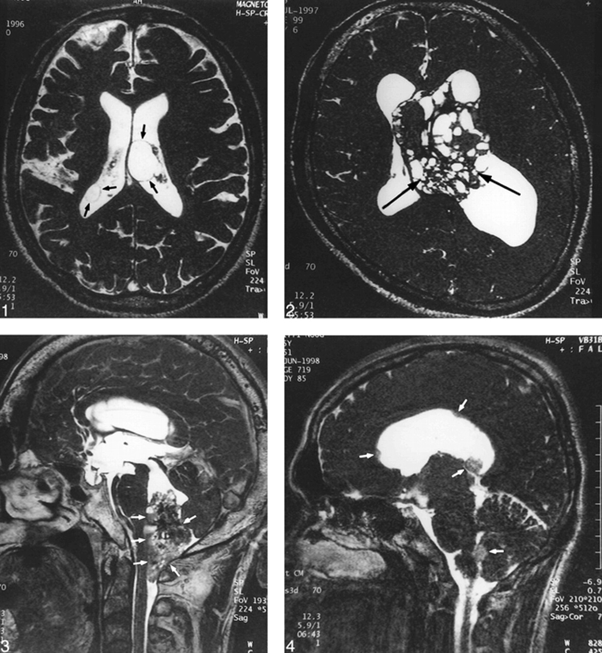
In all cases, 3D-CISS sequences could produce homogeneous images over the entire field of view and high contrast between the CSF and solid structures. The thin wall of the cysts and small cystic components in the tumors were clearly shown on 3D-CISS images (Figs 1 and 2). The tumor margin and dissemination of the tumor were also clearly visible on images obtained from 3D-CISS sequences (Figs 3 and 4).

fig 1.
  • Download figure
  • Open in new tab
  • Download powerpoint
fig 1.

A 70-year-old man with intraventricular cysts in both lateral ventricles. The cyst walls (arrows) are clearly depicted on this 3D-CISS image.

fig 2. A 28-year-old man with central neurocytoma. Many small cystic components (arrows) are clearly visible within the tumor on this 3D-CISS image.

fig 3. A 51-year-old man with ependymoma of the fourth ventricle. A 3D-CISS sequence clearly shows the relationship between the tumor and adjacent structures (arrows), making determination of tumor origin easy.

fig 4. A 25-year-old man with anaplastic astrocytoma. A 3D-CISS sequence depicts multiple dissemination (arrows) in the ventricles.

Discussion

To evaluate the anatomic location of an intraventricular mass lesion accurately, an image with high resolution and high contrast between CSF and solid structures is necessary (2). In general, it is impossible to acquire sections thinner than 3 mm with the 2D-SE technique. Recently, improvements in gradient strength and profile have allowed the capability to obtain much thinner sections, but it sometimes causes prominent signal loss in the CSF due to a washout phenomenon (6). For this reason, the thinner-section 2D-SE scans would not be favorable for imaging the ventricular system.

The 3D-CISS MR sequence is specifically designed for MR cisternography. The 3D-CISS sequence has two data sets with a true FISP sequence of alternating and nonalternating radiofrequency pulses. Although the FISP sequence produces artifacts of low signal intensity bands that are caused by very small magnetic field inhomogeneities and local field distortions (susceptibility changes), the position of the dark bands is shifted in the second data set to the position of high intensity of the first data set. Thus, the 3D-CISS sequence is an effective method of reducing these dark bands by the summation of two flow-compensation 3D-FISP sequences, and produces an image with homogeneous intensity distribution over the whole image and excellent contrast between CSF and solid tissue (3).

The 3D-CISS sequence has several advantages over conventional MR imaging. First, the thin contiguous sections and high in-plane resolution make it possible to depict small structures or lesions, such as intraventricular CSF dissemination and the thin wall of a cyst. Second, any desired imaging plane could be obtained by multiplanar reconstructive technique. For example, the reconstruction images can easily depict a relationship between the lesion and important structures such as the foramen of Monro or aqueduct. Third, the 3D-CISS sequence may be used to evaluate intraventricular solid masses after contrast enhancement. A previous study has reported that the contrast between the cranial nerves and acoustic tumors increases after gadolinium administration, whereas the contrast between CSF and tumor remains (7). Finally, we believe that the 3D-CISS sequence could be added to the MR examination of the intraventricular system for patients with a contraindication to receiving contrast material.

Conclusion

The 3D-CISS sequence enables the examiner to obtain high-resolution and heavily T2-weighted 3D images in a relatively short imaging time for diagnosis of intraventricular lesions.

Footnotes

  • ↵1 Address reprint requests to Yukunori Korogi, MD, Department of Radiology, Kumamoto University School of Medicine. 1-1-1 Honjo, Kumamoto 860-8556, Japan.

References

  1. ↵
    Jelinek J, Smirniotopoulos JG, Parisi JE, Kanzer M. Lateral ventricular neoplasms of the brain: differential diagnosis based on clinical, CT, and MR findings. AJNR Am J Neuroradiol 1990;11:567-574
    Abstract/FREE Full Text
  2. ↵
    Tien RD. Intraventricular mass lesions of the brain: CT and MRI findings. AJR Am J Roentgenol 1991;157:1283-1290
    PubMed
  3. ↵
    Casselman JW, Kuhweide R, Deimling M, et al. Constructive interference in steady state-3DFT MR imaging of the inner ear and cerebellopontine angle. AJNR Am J Neuroradiol 1993;14:47-57
    Abstract/FREE Full Text
  4. Casselman JW, Kuhweide R, Ampe W, et al. Inner ear malformations in patients with sensorineural hearing loss: detection with gradient-echo (3DFT-CISS) MRI. Neuroradiology 1996;38:278-286
    CrossRefPubMed
  5. Stuckey SL, Harris AJ, Mannolini SM. Detection of acoustic schwannoma: use of constructive inteference in the steady state three-dimensional MR. AJNR Am J Neuroradiol 1996;17:1219-1225
    Abstract
  6. ↵
    Iwayama E, Naganawa S, Ito T, et al. High-resolution MR cisternography of the cerebellopontine angle: 2D versus 3D fast spin-echo sequences. AJNR Am J Neuroradiol 1999;20:889-895
    Abstract/FREE Full Text
  7. ↵
    Sigemutsu Y, Korogi Y, Hirai T, et al. Contrast-enhanced CISS MR Imaging of vestibular schwannomas: phantom and clinical studies. J Comput Assist Tomogr 1999;23:224-231
    CrossRefPubMed
  • Received June 21, 1999.
  • Accepted after revision January 12, 2000.
  • Copyright © American Society of Neuroradiology
View Abstract
PreviousNext
Back to top

In this issue

American Journal of Neuroradiology
Vol. 21, Issue 6
1 Jun 2000
  • Table of Contents
  • Index by author
Advertisement
Print
Download PDF
Email Article

Thank you for your interest in spreading the word on American Journal of Neuroradiology.

NOTE: We only request your email address so that the person you are recommending the page to knows that you wanted them to see it, and that it is not junk mail. We do not capture any email address.

Enter multiple addresses on separate lines or separate them with commas.
Increased Conspicuity of Intraventricular Lesions Revealed by Three-dimensional Constructive Interference in Steady State Sequences
(Your Name) has sent you a message from American Journal of Neuroradiology
(Your Name) thought you would like to see the American Journal of Neuroradiology web site.
CAPTCHA
This question is for testing whether or not you are a human visitor and to prevent automated spam submissions.
Cite this article
Dewen Yang, Yukunori Korogi, Yukitaka Ushio, Mutsumasa Takahashi
Increased Conspicuity of Intraventricular Lesions Revealed by Three-dimensional Constructive Interference in Steady State Sequences
American Journal of Neuroradiology Jun 2000, 21 (6) 1070-1072;

Citation Manager Formats

  • BibTeX
  • Bookends
  • EasyBib
  • EndNote (tagged)
  • EndNote 8 (xml)
  • Medlars
  • Mendeley
  • Papers
  • RefWorks Tagged
  • Ref Manager
  • RIS
  • Zotero
0 Responses
Respond to this article
Share
Bookmark this article
Increased Conspicuity of Intraventricular Lesions Revealed by Three-dimensional Constructive Interference in Steady State Sequences
Dewen Yang, Yukunori Korogi, Yukitaka Ushio, Mutsumasa Takahashi
American Journal of Neuroradiology Jun 2000, 21 (6) 1070-1072;
del.icio.us logo Twitter logo Facebook logo Mendeley logo
  • Tweet Widget
  • Facebook Like
  • Google Plus One

Jump to section

  • Article
    • Abstract
    • Methods
    • Results
    • Discussion
    • Conclusion
    • Footnotes
    • References
  • Figures & Data
  • Info & Metrics
  • Responses
  • References
  • PDF

Related Articles

  • No related articles found.
  • PubMed
  • Google Scholar

Cited By...

  • No citing articles found.
  • Crossref
  • Google Scholar

This article has not yet been cited by articles in journals that are participating in Crossref Cited-by Linking.

More in this TOC Section

  • Multimodal CT Provides Improved Performance for Lacunar Infarct Detection
  • Statin Therapy Does Not Affect the Radiographic and Clinical Profile of Patients with TIA and Minor Stroke
  • Optimal MRI Sequence for Identifying Occlusion Location in Acute Stroke: Which Value of Time-Resolved Contrast-Enhanced MRA?
Show more Brain

Similar Articles

Advertisement

Indexed Content

  • Current Issue
  • Accepted Manuscripts
  • Article Preview
  • Past Issues
  • Editorials
  • Editors Choice
  • Fellow Journal Club
  • Letters to the Editor

Cases

  • Case Collection
  • Archive - Case of the Week
  • Archive - Case of the Month
  • Archive - Classic Case

Special Collections

  • Special Collections

Resources

  • News and Updates
  • Turn around Times
  • Submit a Manuscript
  • Author Policies
  • Manuscript Submission Guidelines
  • Evidence-Based Medicine Level Guide
  • Publishing Checklists
  • Graphical Abstract Preparation
  • Imaging Protocol Submission
  • Submit a Case
  • Become a Reviewer/Academy of Reviewers
  • Get Peer Review Credit from Publons

Multimedia

  • AJNR Podcast
  • AJNR SCANtastic
  • Video Articles

About Us

  • About AJNR
  • Editorial Board
  • Not an AJNR Subscriber? Join Now
  • Alerts
  • Feedback
  • Advertise with us
  • Librarian Resources
  • Permissions
  • Terms and Conditions

American Society of Neuroradiology

  • Not an ASNR Member? Join Now

© 2025 by the American Society of Neuroradiology All rights, including for text and data mining, AI training, and similar technologies, are reserved.
Print ISSN: 0195-6108 Online ISSN: 1936-959X

Powered by HighWire