
Article Figures & Data
Figures
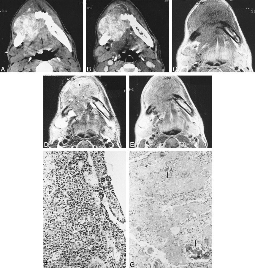
- fig 1.
32-year-old man with aggressive epithelial odontogenic ghost cell tumor in the mandible.
A, Axial unenhanced CT scan shows a large, poorly marginated, lobulated soft-tissue mass causing irregular destruction of the body of the mandible, predominantly in the right. There are numerous calcifications varying in size within the mass. Compared with adjacent muscle, the majority of soft-tissue components of the mass show higher attenuation, while other components, mainly in the anterior aspect of the mass, show similar or slightly lower attenuation. Note lymph node enlargement (arrow) anterior to the right submandibular gland.
B, Corresponding axial contrast-enhanced CT scan more clearly shows the margin of the mass, which invades the mouth floor and tongue base. Moderate, nonhomogeneous enhancement is seen throughout the mass, with several cystic areas (arrows) remaining un[chen[chhanced. An enlarged right submandibular lymph node is also enhanced moderately, with central portions enhanced to a lesser degree.
C, Axial T1-weighted MR image shows a large soft-tissue mass destroying the mandible. Signal intensity of the mass is grossly the same as that of adjacent muscle. Also note right submandibular lymphadenopathy.
D, Corresponding axial T2-weighted MR image more clearly shows the lobulated contour and internal heterogeneity of the mass. The majority of soft-tissue components of the mass show significantly lower signal intensity than other portions, located mainly in the anterior aspect of the mass. These variations in signal intensity correlate well with density variations on corresponding unenhanced CT scan (A). Note bright signals (large arrows) from cystic components of the mass. Tiny hyperintensities (small arrows) within the mass may reflect necrotic foci or small cysts. Also note hyperintense right submandibular lymph node. Although there are several foci of small, dark signal intensities (arrowheads), calcifications are poorly seen on this MR image.
E, Axial contrast-enhanced T1-weighted MR image shows moderate, homogeneous enhancement of the mass, with cystic portions (arrow) remaining unenhanced. There is also significant enhancement of right submandibular lymphadenopathy.
F, Photomicrograph of tissue obtained from larger, posterior portion of the mass (which was hyperdense on unenhanced CT scans and hypointense on T2-weighted MR images) shows irregular islands of highly compact epithelial cells, composed primarily of small basaloid cells with hyperchromatic nuclei and scanty cytoplasm. There is prominent cellular and nuclear pleomorphism as well as evidence of frequent mitoses (hematoxylin-eosin, original magnification ×200).
G, Photomicrograph of tissue obtained from smaller, anterior portion of the mass (which was isodense or slightly hypodense on unenhanced CT scans and hyperintense on T2-weighted MR images) shows abundant ghost cell nests and eosinophilic materials containing highly basophilic foci of calcification (arrows). Epithelial cells are remarkably lacking in this area (hematoxylin-eosin, original magnification ×200).

In this issue


Jump to section
Related Articles
Cited By...
- No citing articles found.