Advertisement

Main menu

  • Home
  • Content
    • Current Issue
    • Accepted Manuscripts
    • Article Preview
    • Past Issue Archive
    • AJNR Case Collection
    • Case of the Week Archive
    • Classic Case Archive
    • Case of the Month Archive
  • Special Collections
    • Spinal CSF Leak Articles (Jan 2020-June 2024)
    • 2024 AJNR Journal Awards
    • Most Impactful AJNR Articles
  • Multimedia
    • AJNR Podcast
    • AJNR Scantastics
    • Video Articles
  • For Authors
    • Submit a Manuscript
    • Author Policies
    • Fast publishing of Accepted Manuscripts
    • Graphical Abstract Preparation
    • Manuscript Submission Guidelines
    • Imaging Protocol Submission
    • Submit a Case for the Case Collection
  • About Us
    • About AJNR
    • Editorial Board
  • More
    • Become a Reviewer/Academy of Reviewers
    • Subscribers
    • Permissions
    • Alerts
    • Feedback
    • Advertisers
    • ASNR Home
  • Other Publications
    • ajnr

User menu

  • Alerts
  • Log in

Search

  • Advanced search
American Journal of Neuroradiology

American Journal of Neuroradiology

ASHNR American Society of Functional Neuroradiology ASHNR American Society of Pediatric Neuroradiology ASSR
  • Alerts
  • Log in

Advanced Search

  • Home
  • Content
    • Current Issue
    • Accepted Manuscripts
    • Article Preview
    • Past Issue Archive
    • AJNR Case Collection
    • Case of the Week Archive
    • Classic Case Archive
    • Case of the Month Archive
  • Special Collections
    • Spinal CSF Leak Articles (Jan 2020-June 2024)
    • 2024 AJNR Journal Awards
    • Most Impactful AJNR Articles
  • Multimedia
    • AJNR Podcast
    • AJNR Scantastics
    • Video Articles
  • For Authors
    • Submit a Manuscript
    • Author Policies
    • Fast publishing of Accepted Manuscripts
    • Graphical Abstract Preparation
    • Manuscript Submission Guidelines
    • Imaging Protocol Submission
    • Submit a Case for the Case Collection
  • About Us
    • About AJNR
    • Editorial Board
  • More
    • Become a Reviewer/Academy of Reviewers
    • Subscribers
    • Permissions
    • Alerts
    • Feedback
    • Advertisers
    • ASNR Home
  • Follow AJNR on Twitter
  • Visit AJNR on Facebook
  • Follow AJNR on Instagram
  • Join AJNR on LinkedIn
  • RSS Feeds

Welcome to the new AJNR, Updated Hall of Fame, and more. Read the full announcements.


AJNR is seeking candidates for the position of Associate Section Editor, AJNR Case Collection. Read the full announcement.


Research ArticleINTERVENTIONAL

Balloon-assisted Coil Embolization of Wide-necked Aneurysms of the Internal Carotid Artery: Medium-term Angiographic and Clinical Follow-up in 22 Patients

Peter Kim Nelson and David I. Levy
American Journal of Neuroradiology January 2001, 22 (1) 19-26;
Peter Kim Nelson
  • Find this author on Google Scholar
  • Find this author on PubMed
  • Search for this author on this site
David I. Levy
  • Find this author on Google Scholar
  • Find this author on PubMed
  • Search for this author on this site
  • Article
  • Figures & Data
  • Info & Metrics
  • Responses
  • References
  • PDF
Loading

Article Figures & Data

Figures

  • Tables
  • fig 1.
    • Download figure
    • Open in new tab
    • Download powerpoint
    fig 1.

    A and B, Frontal (A) and lateral (B) arterial phase images from digital subtraction angiography (DSA) of the left ICA show a large wide-necked aneurysm of the ophthalmic segment.

    C and D, Road-mapped live subtraction images of the left ICA (lateral projection) show placement of the first coil. Note the herniation of a coil loop (arrow) through the neck of the aneurysm into the left ICA (C). In D, the balloon (thick arrow) has been inflated across the aneurysmal neck, permitting the framing coils to be deployed within the aneurysm. Arrowheads identify the indwelling 0.010-inch guidewire within the balloon microcatheter. An unextruded segment of a GDC (thin arrow) identi-fies the course of the coil-delivery microcatheter.

    E and F, Frontal (E) and lateral (F) unsubtracted radiographs show deployment of a subsequent GDC. The images serve to orient the viewer with respect to the course of the ICA in relation to the aneurysmal base. The balloon (slanted arrow, E and F) has been inflated within the paraclinoid segment of the ICA, which sweeps lateral to medial across the face of the aneurysmal neck. The supraclinoid segment courses medial to the lower portion of the aneurysmal fundus and is partially obscured by the aneurysm in lateral projection. A small niche of aneurysm lying posterior to the ICA could not be angiographically thrown off the ICA by any of the views attempted. In this respect, the inflated balloon defines the boundary of the ICA lumen and assists the operator in coiling the aneurysmal base. The final segment of coil 14 (thin arrow, E) has been deployed in F. Note the alignment of the delivery wire marker with the proximal microcatheter marker (curved arrow, F). An indwelling 0.010-inch guidewire (arrowhead, E and F) identifies the course of the balloon microcatheter.
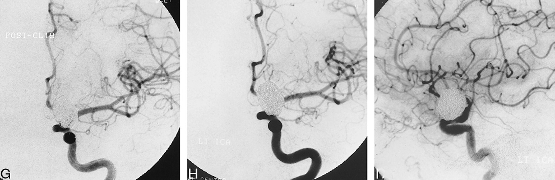
  • fig 1.
    • Download figure
    • Open in new tab
    • Download powerpoint
    fig 1.

    Continued.

    G, Mid-arterial phase image (frontal projection) from the immediate posttreatment left ICA DSA after deployment of the 18th GDC within the aneurysm. Coils are present throughout the aneurysmal base; however, minor opacification through the coil interstices is evident.

    H and I, Frontal (H) and lateral (I) arterial phase images from the follow-up angiogram 18 months later confirm stable occlusion of the aneurysm.

  • fig 2.
    • Download figure
    • Open in new tab
    • Download powerpoint
    fig 2.

    A, Arterial phase DSA image (lateral view) of the left ICA shows a small aneurysm of the superior hypophyseal segment.

    B, Several attempts were made to coil the aneurysm initially. Road-mapped live subtraction image of the left ICA (lateral projection) shows extrusion of a 4-mm × 8-cm soft 0.010-inch GDC (arrow). A subsequent attempt to place a 3-mm × 8-cm coil was likewise unsuccessful.

    C, Road-mapped live subtraction image of the left ICA shows deployment of a 4-mm × 8-cm soft GDC within the aneurysm during balloon inflation. The balloon (thick arrow) has been inflated across the proximal two thirds of the aneurysmal orifice, preventing coil herniation into the ICA and allowing coiling of the aneurysmal fundus. An indwelling 0.014-inch guidewire (arrowhead) and an unextruded coil segment (thin arrow) identify the balloon microcatheter and the GDC microcatheter, respectively.

    D, Posttreatment image from a left common carotid DSA, 3 days later, shows occlusion of the aneurysm.

    E, Mid-arterial phase image (lateral projection) from follow-up angiography of the left ICA at 22 months confirms stability of aneurysmal occlusion.

  • fig 3.
    • Download figure
    • Open in new tab
    • Download powerpoint
    fig 3.

    A, Arterial phase image from a DSA of the left ICA (lateral projection) shows a small dural ring aneurysm with a broad neck. The aneurysm was occluded with five GDCs under balloon protection.

    B, Follow-up angiogram 2 days after treatment confirms aneurysmal occlusion by GDCs.

    C, A subsequent study at 8 months' follow-up shows a small recurrence (arrow) at the aneurysmal neck.

    D, Repeat angiography 24 months after treatment shows negligible change in the size of the recurrence.

Tables

  • Figures
  • Table1

    Demographics and treatment outcome in 22 patients with ICA aneurysms treated by balloon-assisted coiling

PreviousNext
Back to top

In this issue

American Journal of Neuroradiology
Vol. 22, Issue 1
1 Jan 2001
  • Table of Contents
  • Index by author
Advertisement
Print
Download PDF
Email Article

Thank you for your interest in spreading the word on American Journal of Neuroradiology.

NOTE: We only request your email address so that the person you are recommending the page to knows that you wanted them to see it, and that it is not junk mail. We do not capture any email address.

Enter multiple addresses on separate lines or separate them with commas.
Balloon-assisted Coil Embolization of Wide-necked Aneurysms of the Internal Carotid Artery: Medium-term Angiographic and Clinical Follow-up in 22 Patients
(Your Name) has sent you a message from American Journal of Neuroradiology
(Your Name) thought you would like to see the American Journal of Neuroradiology web site.
CAPTCHA
This question is for testing whether or not you are a human visitor and to prevent automated spam submissions.
Cite this article
Peter Kim Nelson, David I. Levy
Balloon-assisted Coil Embolization of Wide-necked Aneurysms of the Internal Carotid Artery: Medium-term Angiographic and Clinical Follow-up in 22 Patients
American Journal of Neuroradiology Jan 2001, 22 (1) 19-26;

Citation Manager Formats

  • BibTeX
  • Bookends
  • EasyBib
  • EndNote (tagged)
  • EndNote 8 (xml)
  • Medlars
  • Mendeley
  • Papers
  • RefWorks Tagged
  • Ref Manager
  • RIS
  • Zotero
0 Responses
Respond to this article
Share
Bookmark this article
Balloon-assisted Coil Embolization of Wide-necked Aneurysms of the Internal Carotid Artery: Medium-term Angiographic and Clinical Follow-up in 22 Patients
Peter Kim Nelson, David I. Levy
American Journal of Neuroradiology Jan 2001, 22 (1) 19-26;
del.icio.us logo Twitter logo Facebook logo Mendeley logo
  • Tweet Widget
  • Facebook Like
  • Google Plus One

Jump to section

  • Article
    • Abstract
    • Methods
    • Results
    • Discussion
    • Conclusion
    • Footnotes
    • References
  • Figures & Data
  • Info & Metrics
  • Responses
  • References
  • PDF

Related Articles

  • No related articles found.
  • PubMed
  • Google Scholar

Cited By...

  • Assisted coiling of saccular wide-necked unruptured intracranial aneurysms: stent versus balloon
  • Safety and performance of the Penumbra Liberty stent system in a rabbit aneurysm model
  • Silent embolism after stent-assisted coiling of cerebral aneurysms: diffusion-weighted MRI study of 75 cases
  • Treatment of basilar tip aneurysms with horizontal PCA to PCA stent-assisted coiling: case series
  • Combined balloon stent technique with the Scepter C balloon and low-profile visualized intraluminal stent for the treatment of intracranial aneurysms
  • Balloon-assisted coil embolization of intracranial aneurysms is not associated with increased periprocedural complications
  • Immediate and Midterm Results following Treatment of Unruptured Intracranial Aneurysms with the Pipeline Embolization Device
  • Safety and Efficacy of Balloon Remodeling Technique during Endovascular Treatment of Intracranial Aneurysms: Critical Review of the Literature
  • Thromboembolic events associated with endovascular treatment of cerebral aneurysms
  • An Analysis of Inflation Times During Balloon-Assisted Aneurysm Coil Embolization and Ischemic Complications
  • Endovascular treatment of unruptured intracranial aneurysms in the elderly: analysis of procedure related complications
  • Acutely ruptured intracranial saccular aneurysms treated with stent assisted coiling: complications and outcomes in 42 consecutive patients
  • Balloon assisted treatment of intracranial aneurysms: the conglomerate coil mass technique
  • Treatment of Wide-Necked Intracranial Aneurysms with a Self-Expanding Stent System: Initial Clinical Experience
  • Crossref
  • Google Scholar

This article has not yet been cited by articles in journals that are participating in Crossref Cited-by Linking.

More in this TOC Section

  • SAVE vs. Solumbra Techniques for Thrombectomy
  • CT Perfusion&Reperfusion in Acute Ischemic Stroke
  • Delayed Reperfusion Post-Thrombectomy&Thrombolysis
Show more Interventional

Similar Articles

Advertisement

Indexed Content

  • Current Issue
  • Accepted Manuscripts
  • Article Preview
  • Past Issues
  • Editorials
  • Editors Choice
  • Fellow Journal Club
  • Letters to the Editor

Cases

  • Case Collection
  • Archive - Case of the Week
  • Archive - Case of the Month
  • Archive - Classic Case

Special Collections

  • Special Collections

Resources

  • News and Updates
  • Turn around Times
  • Submit a Manuscript
  • Author Policies
  • Manuscript Submission Guidelines
  • Evidence-Based Medicine Level Guide
  • Publishing Checklists
  • Graphical Abstract Preparation
  • Imaging Protocol Submission
  • Submit a Case
  • Become a Reviewer/Academy of Reviewers
  • Get Peer Review Credit from Publons

Multimedia

  • AJNR Podcast
  • AJNR SCANtastic
  • Video Articles

About Us

  • About AJNR
  • Editorial Board
  • Not an AJNR Subscriber? Join Now
  • Alerts
  • Feedback
  • Advertise with us
  • Librarian Resources
  • Permissions
  • Terms and Conditions

American Society of Neuroradiology

  • Not an ASNR Member? Join Now

© 2025 by the American Society of Neuroradiology All rights, including for text and data mining, AI training, and similar technologies, are reserved.
Print ISSN: 0195-6108 Online ISSN: 1936-959X

Powered by HighWire