
Article Figures & Data
Figures
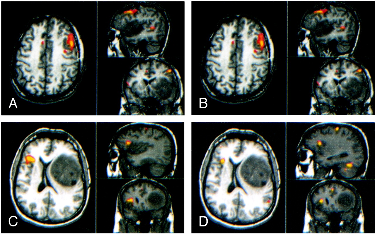
- fig 1.
Sagittal (top right), axial (left), and coronal (bottom right) fMR images in patients performing word-generation tasks.
A, Activation is evident in the pars triangularis and pars opercularis of the left inferior frontal gyrus (Brodmann areas 44 and 45) in a patient performing an LWG task.
B, Activation also is evident in Brodmann areas 44 and 45 in a patient with an arteriovenous malformation in the right hemisphere who is performing an LWG task.
C, Activation appears in Brodmann areas 9 and 46 in a patient with a tumor involving the inferior left frontal lobe who is performing a CWG task.
- fig 2.
Sagittal (top right), axial (left), and coronal (bottom right) fMR images in a patient with a large left frontal tumor who is performing an LWG task. Activation is identified primarily in the right inferior frontal gyrus (Brodmann area 45)
- fig 3.
Sagittal (top right), axial (left), and coronal (bottom right) activation images in two iterations of an LWG task.
A and B, Patient with a left frontal tumor. Activation occurs in the same gyri between iterations, although with slightly different patterns.
C and D, Patient with a left frontal tumor and right hemispheric activation.
- fig 4.
Coronal fMR imaging images of two iterations of an LWG task in a patient with a left middle cerebral arteriovenous malformation.
A, Activation in the first iteration.
B, Activation in the second iteration, showing similar activation compared with A.
C, Intersect map shows the voxels activated in the two iterations. The proportion of voxels in the intersect map compared with the average number of voxels in A and B (the CR) is 45%.
- fig 5.
Sagittal (top right), axial (left), and coronal (bottom right) projections in a patient with a right frontal tumor.
A, Activation from a CWG task.
B, Activation from an LWG task.
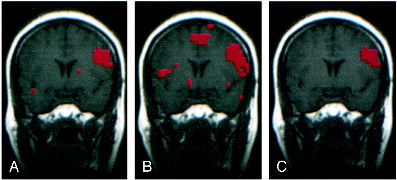
- fig 6.
Coronal images in a patient with a right frontoparietal arteriovenous malformation.
A, Activation from an AWG task
B, Activation from a CWG task. The activation patterns from the two variations of the word-generation task are similar.
C, Intersect map shows the voxels activated by both versions of the task. The CR was 52%.
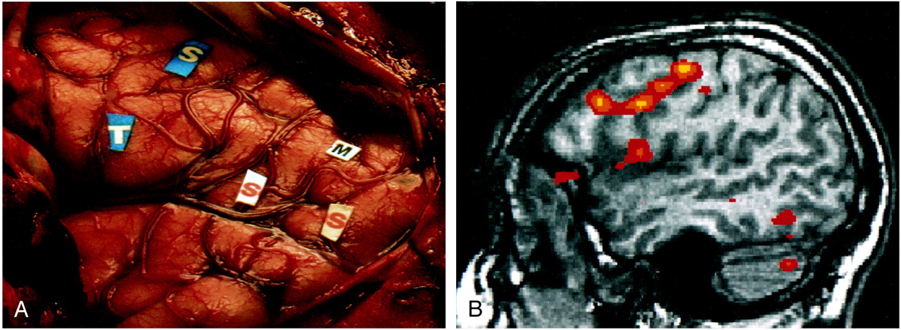
- fig 7.
Comparison of intraoperative localization of speech function (ECS) and activation from the word-generation task (fMR imaging) in a patient with a left frontal-lobe glioma. Both activation and intraoperative localization of the speech mapping were classified as belonging to Brodmann areas 44 and 45. M indicates motor cortex; S, speech areas; T, tumor


Tables
 TABLE 1:
Location of activation, threshold for fMR images, word-generation paradigm, and clinical diagnosis in the 31 patients with activation
- TABLE 2:
CRs for five-section comparisons in patients performing two iterations of an LWG task
- TABLE 3:
CRs for single-section comparisons in patients performing two iterations of an LWG task
- TABLE 4:
CRs for five-section comparisons in patients performing two or more versions of the language task
- TABLE 5:
CRs for single-section comparisons in patients performing two iterations of the language task
- TABLE 6:
Brodmann area of speech function in patients undergoing both ECS and fMR imaging


In this issue


Jump to section
Related Articles
Cited By...
- Reliability of single-subject neural activation patterns in speech production tasks
- American Society of Functional Neuroradiology-Recommended fMRI Paradigm Algorithms for Presurgical Language Assessment
- Reproducibility of Activation Maps for Longitudinal Studies of Visual Function by Functional Magnetic Resonance Imaging
- Impact of cognitive performance on the reproducibility of fMRI activation in schizophrenia
- Intrasubject reproducibility of presurgical language lateralization and mapping using fMRI