
Article Figures & Data
Figures
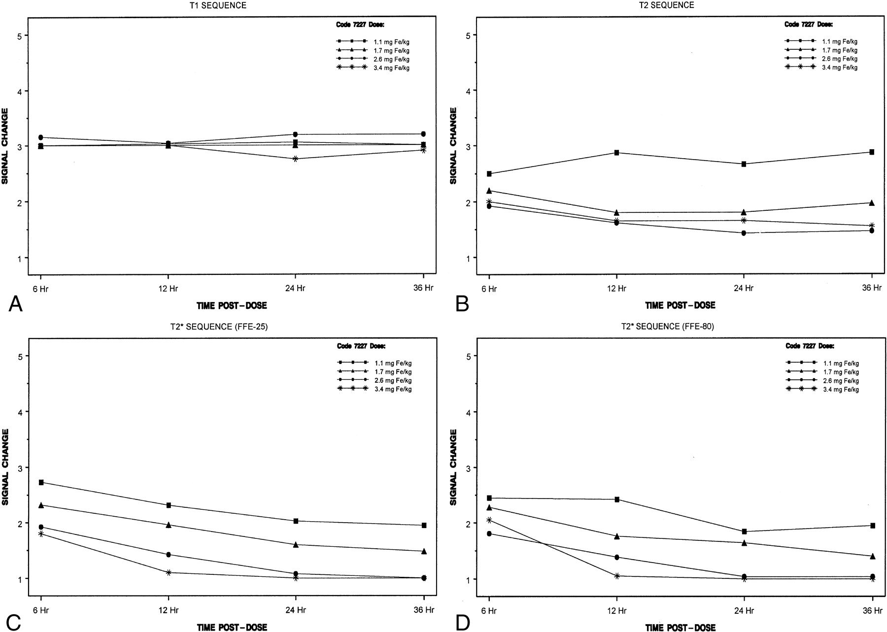
- Fig 1.
Mean change in signal intensity over time in the blinded evaluation
A, With T1-weighted imaging.
B, With T2-weighted imaging.
C, With T2*-weighted (FFE-25) imaging.
D, With T2*-weighted (FFE-80) imaging.
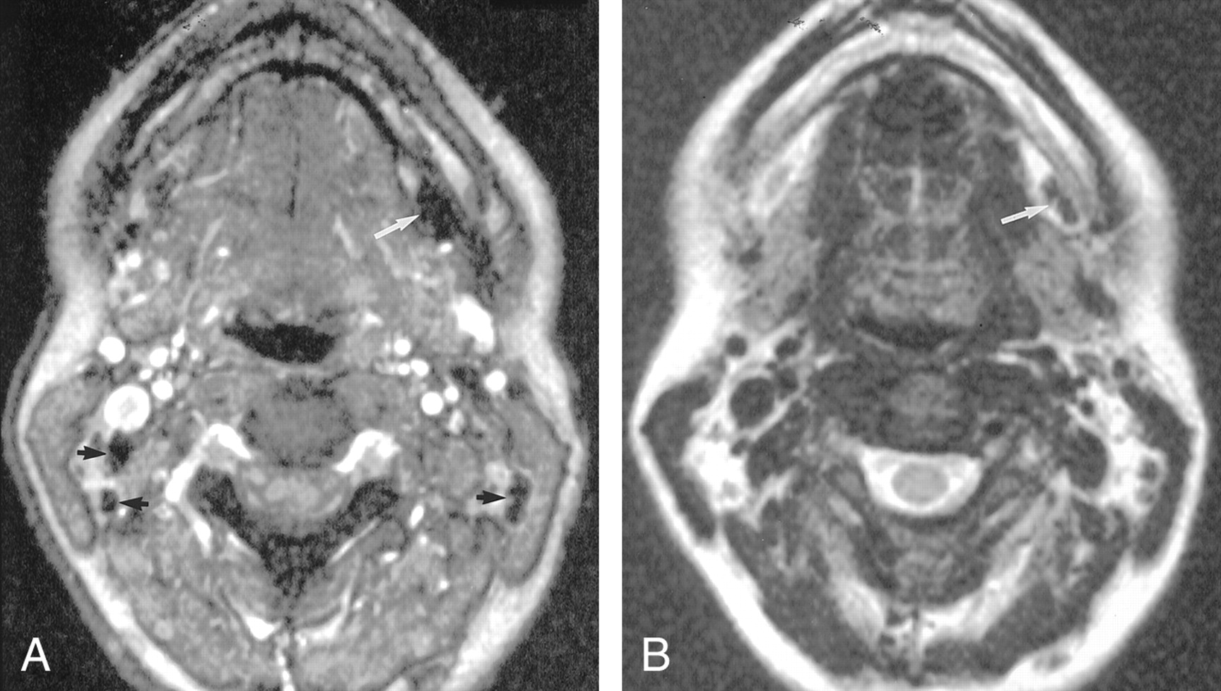
- Fig 2.
Axial nonenhanced MR images in a healthy volunteer
A, Gradient-echo (FFE-80) (120/7.8/1, 80° flip angle) image shows a small left submandibular node (arrow).
B, On the fast spin-echo T2-weighted (3500/120/1) image, the node (arrow) is difficult to visualize because it is nearly isointense to fat.
- Fig 3.
Axial MR images obtained 36 hours after the intravenous administration of 2.6 mg. Fe/kg ferumoxtran-10 in the same volunteer as in Figure 2.
A, Gradient-echo (FFE-80) (120/7.8/1, 80° flip angle) image shows that the benign node is now homogeneously hypointense (white arrow). Note the multiple additional nodes, some of which are delineated by black arrows, all of which are better visualized now than before contrast enhancement.
B,T2-weighted (3500/120/1) image obtained at the same level as Figure 1B shows excellent contrast between the enhanced left submandibular node (arrow) and the subcutaneous fat.


Tables
Dose Group and Postdose Time Point Total Lymph Nodes Sequence Specificity (%) T2-Weighted FFE-25 FFE-80 1.1 mg Fe/kg 6 h 41 78 12 20 12 h 41 90 56* 74* 24 h 41 80 62* 87* 36 h 41 56 62* 65† 1.7 mg Fe/kg 6 h 25 88 32 44 12 h 25 92 64 84 24 h 25 100 72 84 36 h 25 92 84 76 2.6 mg Fe/kg 6 h 25 92 56 68 12 h 25 96 84 96 24 h 25 100 96 100 36 h 25 100 96 92 3.4 mg Fe/kg 6 h 19 95 63 89 12 h 19 100 83‡ 95 24 h 19 89 84 95 36 h 19 79 79 79 Note.—The specificity was based on the signal intensity and lymph node architecture. A decrease in the signal intensity of a heterogeneous lymph node indicated a false-positive result.
* The total number of lymph nodes was 39.
† The total number of lymph nodes was 40.
‡ The total number of lymph nodes was 18.
Dose Group and Postdose Time Point Total Lymph Nodes Sequence Specificity (%) T2-Weighted FFE-25 FFE-80 1.1 mg Fe/kg 6 h 38 45 16* 37 12 h 38 11 40† 47 24 h 38 29 53 66 36 h 38 14* 50 50 1.7 mg Fe/kg 6 h 25 60 52 52 12 h 25 80 32 68 24 h 25 88 48 64 36 h 25 80 48 64 2.6 mg Fe/kg 6 h 26 88 42 50 12 h 26 81 58 54 24 h 26 88 73 85 36 h 26 88 85 88 3.4 mg Fe/kg 6 h 20 60 55 35 12 h 20 70 70 80 24 h 20 75 95 95 36 h 20 75 100 80 Note.— The specificity was based on the signal intensity and lymph node architecture. A decrease in the signal intensity of a heterogeneous lymph node indicated a false-positive result.
* The total number of lymph nodes was 37.
† The total number of lymph nodes was 35.
- TABLE 3:
Descriptive statistics for quantitative signal intensity measurements of lymph nodes
Dose Group and Time Point Total Lymph Nodes Quantitative Signal Intensity Measurements by Sequence* T1-Weighted T2-Weighted FFE-25 FFE-80 1.1 mg Fe/kg Predose 18 486.1 + 45.8 536.0 + 64.6 1170.6 + 179.3 784.9 + 98.8 6 h 18 626.6 + 70.3 277.1 + 45.4 995.6 + 129.4 510.6 + 69.7 12 h 18 587.9 + 53.7 297.3 + 55.1 906.4 + 197.1 524.3 + 143.6 24 h 18 596.9 + 98.2 280.1 + 34.7 904.7 + 178.9 442.4 + 117.7 36 h 18 570.3 + 92.2 323.0 + 40.5 852.9 + 231.4 447.3 + 137.9 1.7 mg Fe/kg Predose 18 498.5 + 59.0 505.5 + 48.6 979.7 + 283.4 669.5 + 139.8 6 h 15 618.7 + 89.1 256.1 + 67.0 901.3 + 257.9 473.8 + 167.6 12 h 18 555.2 + 81.3 275.8 + 35.4 725.7 + 276.5 372.5 + 150.3 24 h 18 567.9 + 101.0 279.9 + 74.6 676.7 + 266.4 341.7 + 135.5 36 h 18 545.0 + 93.0 291.5 + 46.6 491.3 + 171.6 311.7 + 137.6 2.6 mg Fe/kg Predose 18 470.3 + 55.4 561.2 + 71.6 1150.0 + 125.2 777.1 + 95.2 6 h 18 568.1 + 80.0 293.8 + 54.5 754.2 + 148.1 493.3 + 116.5 12 h 18 534.7 + 109.7 225.7 + 50.4 535.6 + 93.6 328.1 + 64.1 24 h 18 511.3 + 85.4 196.6 + 42.7 335.2 + 85.9 243.2 + 66.5 36 h 18 494.6 + 119.3 209.2 + 41.9 316.3 + 79.4 188.4 + 48.6 3.4 mg Fe/kg Predose 18 515.0 + 76.9 583.6 + 140.7† 1132.2 + 164.7 827.2 + 129.3 6 h 18 608.3 + 68.8 276.8 + 110.9† 710.8 + 166.4 550.6 + 117.9 12 h 18 559.9 + 74.9 219.8 + 63.9† 452.5 + 125.3 346.5 + 72.2 24 h 18 513.1 + 68.6 178.3 + 58.7† 326.2 + 98.4 255.4 + 76.9 36 h 18 459.4 + 108.4 206.8 + 69.8† 300.6 + 72.6 221.9 + 35.5 10 d 6 487.7 + 58.2 570.9 + 113.2‡ 594.1 + 80.6 530.8 + 103.8 1 mo 6 457.8 + 81.2 561.3 + 115.4‡ 726.9 + 450.8 617.3 + 185.4 3 mo 6 502.6 + 14.0 595.3 + 27.7‡ 965.2 + 272.3 895.8 + 181.5 * Data are the mean + the SD.
† The total number of lymph nodes was 17.
‡ The total number of lymph nodes was 5.


