
Article Figures & Data
Figures
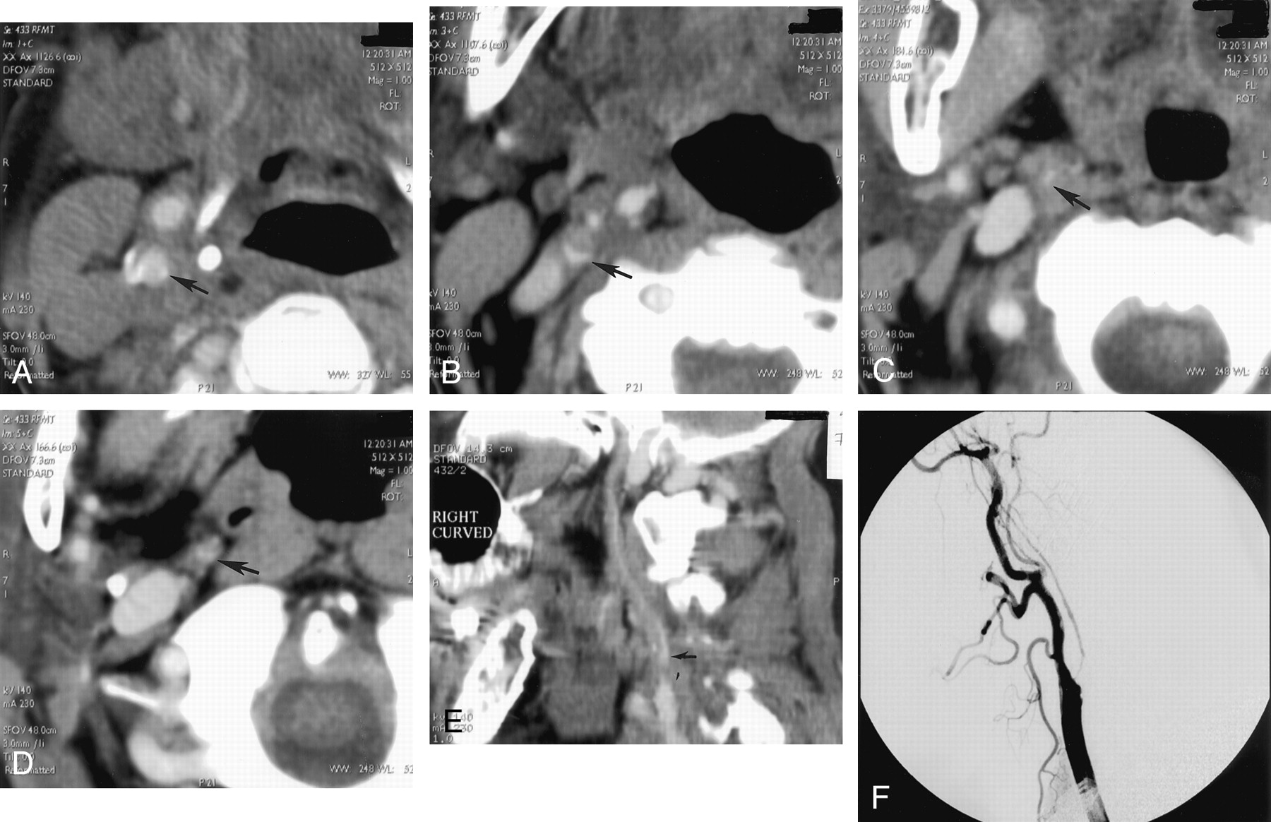
- Fig 1.
A−D, Selected axial view source scans from a CT angiogram of a 74-year-old female patient show continuity of the contrast-filled lumen (arrows) in consecutive axial views of the right ICA, reflecting a patent vascular lumen. Scans progress from inferior to superior levels, beginning at the patent carotid bifurcation. Note that in A, the jugular vein, posterior and lateral to the carotid artery, has not yet opacified.
E, Sagittal view curved reformatted projection of this CT angiography dataset shows a hairline residual right ICA lumen with a slim sign extending into the petrous canal at the skull base. Note that the vessel appears falsely discontinuous along portions of the scan, which is a potential pitfall of this postprocessing technique, emphasizing the need for review of the axial view source scans. ICA origin is seen just superior to a discontinuous segment (arrow).
F, Lateral view digital subtraction arteriogram obtained during injection of the right common carotid artery confirms critical stenosis of the right ICA origin, with diffuse narrowing (hairline residual lumen) of the more distal right ICA.
- Fig 2.
A−D, Selected source scans from a CT angiogram of a 45-year-old man. Unlike those shown in Figure 1, these scans reveal discontinuity of the intraluminal left ICA contrast column, reflecting an occluded vessel (arrow in A indicates residual ICA lumen; arrows in B−D indicate unopacified ICA lumen/enhancing vasa vasorum). Scans progress from inferior to superior levels.
E, Curved reformatted projection constructed from this CT angiography dataset shows left ICA occlusion, with absent intraluminal contrast opacification. An opacified ascending pharyngeal artery (arrow) mimics a patent left ICA but cannot be followed into the petrous canal.
F, Lateral view digital subtraction arteriogram obtained during injection of the left common carotid artery confirms total occlusion of the proximal left ICA.
- Fig 3.
Receiver operating characteristic curves for detection of hairline residual lumen by readers 1 and 2. Area under the receiver operating characteristic curve for reader 1 (which correlates with accuracy) was 0.98 ± 0.03; for reader 2, this area was 0.91 ± 0.06. No significant difference was observed between the accuracy of the two readers (P = .28, two-tailed t test). Of note, at an operating point corresponding to 90% sensitivity, reader 1 achieved 95% specificity and reader 2 achieved 80% specificity. At an operating point corresponding to a sensitivity of 95%, specificity was reduced to approximately 90% for reader 1 and 75% for reader 2.


Tables
Patient No. Vessel Radiographic Total Occlusion Rating (Reader 1) Rating (Reader 2) 1 LIC1 Y 1 1 2 RIC2 Y 1 3 3 LIC Y 1 5† 4 RIC Y 1 1 5 RIC Y 1 2 6 RIC Y 2 2 7 LIC Y 1 1 8 LIC Y 1 1 9 RIC Y 1 1 10 RIC Y 1 2 11 RIC Y 1 2 12 RIC Y 1 2 13 LIC Y 1 1 14 LIC Y 1 1 15 LIC N 5 5 16 LIC N 5 5 17 RIC N 4 4 18 LIC N 4 4 19 LIC N 2 5 20 RIC N 4 3 21 RIC N 5 5 Note.—LIC indicates left internal carotid artery; RIC, right internal carotid artery; Y, yes; N, no;
† false positive for hairline residual lumen (occluded internal carotid artery confused with patent ascending pharyngeal artery).
* Chart shows the vessel patency ratings for both readers. Note that for patient 3, with an internal carotid artery occlusion, one reader mistook the ascending pharyngeal artery for a patent hairline internal carotid artery. 1 = definitely occluded; 2 = probably occluded; 3 = indeterminate; 4 = probably patent; and 5 = definitely patent. During data analysis, vessels with rating scores of 1 or 2 were categorized as “occluded,” whereas those with rating scores of 4 or 5 were categorized as “hairline.”
Occlusions Hairline Lumina Total Agreed 7 of 14 = 50% 5 of 7 = 71% 12 of 21 = 57% Disagree 1 level 5 of 14 = 36% 0 of 7 = 0% 05 of 21 = 24% Disagree 2 levels 1 of 14 = 7% 1 of 7 = 14% 02 of 21 = 10% Disagree 3 levels 0 of 14 = 0% 0 of 7 = 0% 0 of 21 = 0% Disagree 4 levels 1 of 14 = 7% 1 of 7 = 14% 02 of 21 = 10% * Interobserver variability of reader ratings. No or insignificant disagreements (same diagnosis) in 19 (90.4%) of 21 cases and significant disagreements (different diagnosis) in two (9.5%) of 21 cases.
In this issue


Jump to section
Related Articles
Cited By...
- Acute Ischemic Stroke Therapy Overview
- Carotid Near-Occlusion: A Comprehensive Review, Part 1--Definition, Terminology, and Diagnosis
- Imaging Recommendations for Acute Stroke and Transient Ischemic Attack Patients: A Joint Statement by the American Society of Neuroradiology, the American College of Radiology, and the Society of NeuroInterventional Surgery
- Acute Stroke Imaging Research Roadmap II
- Guidelines for the Early Management of Patients With Acute Ischemic Stroke: A Guideline for Healthcare Professionals From the American Heart Association/American Stroke Association
- Recommendations for Imaging of Acute Ischemic Stroke: A Scientific Statement From the American Heart Association
- Contrast-Enhanced MR Angiography Is Not More Accurate Than Unenhanced 2D Time-of-Flight MR Angiography for Determining >=70% Internal Carotid Artery Stenosis
- Complete occlusion of extracranial internal carotid artery: clinical features, pathophysiology, diagnosis and management
- Systematic Review of Computed Tomographic Angiography for Assessment of Carotid Artery Disease