
Article Figures & Data
Figures
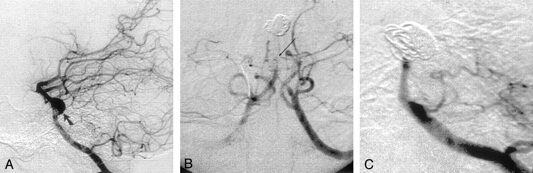
- Fig 1.
Anteroposterior digital angiography, right VA injection, performed after attempted wrapping of the aneurysm, immediately prior to coiling (A). There is no filling of the PCAs via the BA. The RSCA is patent (small arrows), but the LSCA is occluded at its origin. An extensive network of collateral vessels is already evident, arising from the left AICA and PICA (arrowheads). Following the first coiling, no residual filling of the aneurysm is seen (B). The coil mass (curved arrows) occludes the distal BA trunk, and flow in the RSCA is no longer evident. Several collateral vessels (small arrows) from the right AICA and PICA are present. Angiography 3 years following the last procedure (C), shows continued near-total aneurysm occlusion, with only a miniscule amount of body filling (small arrows). Both SCAs have recanalised, but the BA tip remains occluded between the SCA origins and the BA bifurcation (open, curved arrow).
- Fig 2.
Lateral digital subtraction angiography, left VA injection, performed prior to endovascular treatment (A). A fusiform aneurysm affecting the middle of the BA is shown (solid arrow). Angiography, AP projection, left VA injection, performed on day 7 after coiling (B), shows tapering of the proximal BA leading to a short segment of occlusion (long arrow) between the AICA and SCA origins. Some collateral vessels are seen arising from the PICAs and AICAs.
Angiography performed 4 months after coiling (C) shows persistent occlusion of both the BA and the aneurysm.
- Fig 3.
Oblique digital subtraction angiography, right VA injection, performed at the time of presentation (A). A giant fusiform aneurysm of the BA is shown. Although the AICA origins are apparently separate from the bulk of the aneurysm, the left AICA originates from the junction of normal and ectatic vessel (small arrow). The straight configuration of the inferior aspect of the aneurysm sac suggests the presence of intra-aneurysmal thrombus (arrowheads). Note the large left PCoA (short curved arrow). Following deployment of 18 coils (B), near-total occlusion of the aneurysm sac is shown. A small amount of residual inferior body filling was accepted due to the proximity of the left AICA origin to this loculus (curved arrow). The BA trunk is occluded, and continued filling of the distal BA and PCAs via the PCoAs was confirmed on bilateral ICA angiography (not shown). Angiography performed prior to the second coiling procedure, 4 months after initial presentation (C). There has been aneurysm recanalisation and coil compaction. The BA has recanalised. Angiography performed after the second coiling procedure (D). The BA trunk has been reoccluded. No residual aneurysm filling is seen, and the AICAs have been preserved (arrowheads). Lateral right ICA angiograms (E) confirms good filling of the PCAs and the distal BA via a large PcoA (long arrow).


