
Article Figures & Data
Figures
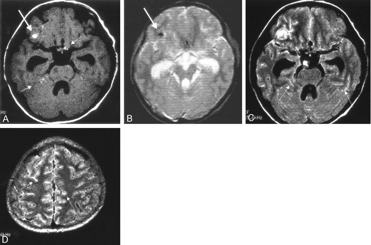
- Fig 1.
Initial MR imaging 3 days after the CT study.
A, T1-weighted image (616/14) shows a high signal intensity focus in the area of the right frontal lobe (arrow). Other high signal intensity foci are seen at multiple sites (small arrows).
B, T2*-weighted image (GRE, 400/20/FA 20) shows a low-signal intensity focus in the corresponding area of the right frontal lobe (arrow), which was confirmed as hemorrhage at surgery. No other low signal intensity lesions are seen.
C and D, FLAIR images (8002/133/2000) show diffuse leptomeningeal hyperintensity in the sulci (small arrows).
- Fig 2.
Follow-up MR imaging 3 weeks after the initial MR study.
A, T1-weighted image (616/14) shows high signal intensity in both trigeminal nerves (arrows). High signal intensity foci are also seen in the cerebellar sulci (small arrow). Note that hydrocephalus has progressed.
B, FLAIR image (8002/133/2000) shows diffuse leptomeningeal hyperintensity (small arrows). Both trigeminal nerves are thickened and hyperintense (arrows).
C, Contrast-enhanced T1-weighted image (616/14) shows diffuse leptomeningeal enhancement corresponding to the findings of FLAIR imaging.
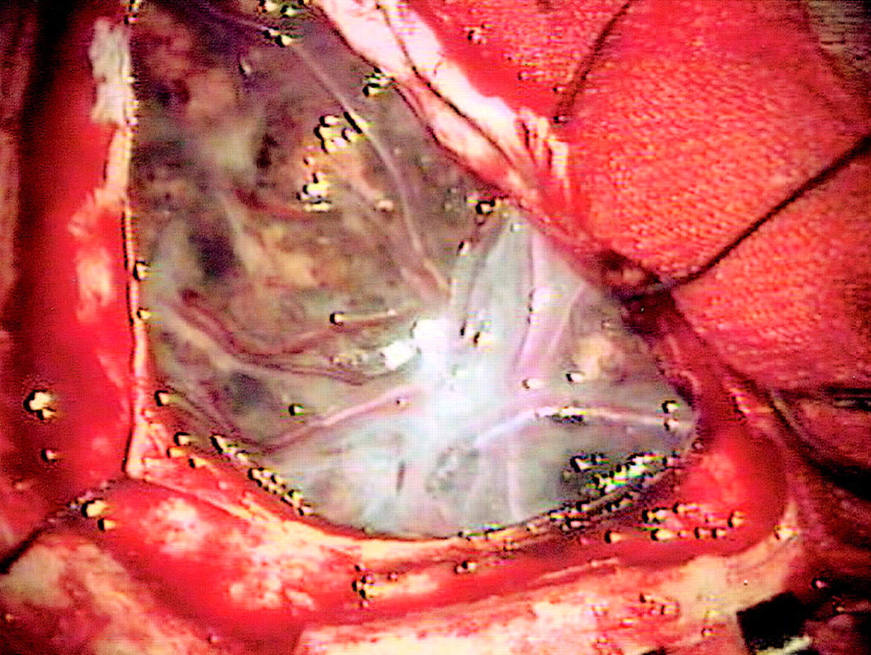
- Fig 3.
Appearance at surgery. Diffuse black pigmentation of the leptomeninges is noted.
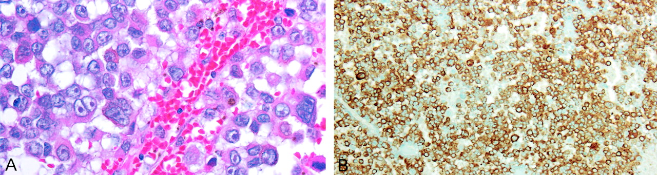
- Fig 4.
A, Photomicrograph (hematoxylin and eosin stain, × 400) shows the atypical cells with dark melanin pigment. The mitotic figures were frequently observed in the neoplasm.
B, Photomicrograph shows a strongly positive immunohistochemical reaction to antimelanoma antibody (HMB-45).


