
Article Figures & Data
Figures
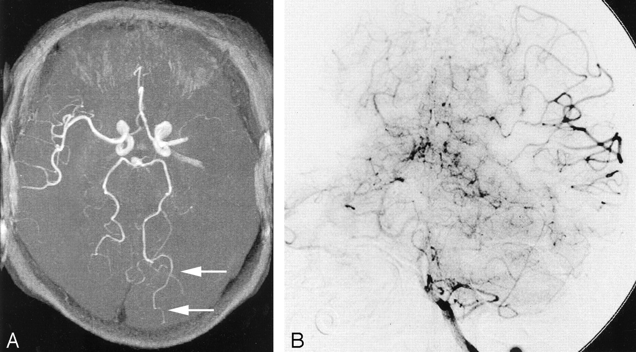
- Fig 1.
Ipsilateral P4 sign in a 73-year-old female patient who presented with acute-onset right hemiplegia.
A, 3D TOF MRA obtained 3 hours after onset showed left M1 occlusion. Ipsilateral P4 (parieto-occipital artery) is observable over almost half of the segment (white arrows).
B, Anteroposterior view of left vertebral arteriogram shows prominent collateral flow from the PCA to the ipsilateral MCA during the capillary phase (black arrows).

Tables
- TABLE 1:
DSA scoring system of collateral flow from the PCA to the ipsilateral MCA territory via the LMA:
Observable MCA Branches (A) Score Extent of Retrograde Filling (B) Score 0 0 None 0 1–2 1 To M4 1 3–4 2 To M3 2 >5 3 To M2 3 Ipsilateral PCA Score Two distal segments 2 One distal segment 1 Bilaterally equal 0 Contralaterally distal −1 - TABLE 3:
Relationship between degree of collateral flow and observable laterality of PCA at MRA
Degree of Collateral Flow (Ipsilateral-Contralateral) Score of PCA Laterality 2 1 0 P4–P2p P4–P3 P3–P2p P2p–P2a P4–P4 P3–P3 P2a–P2a Prominent 1 8 1 0 1 0 0 Mild 0 0 1 1 1 7 0 Absent 0 0 0 0 0 3 1 - TABLE 4:
Frequency of observable PCA laterality at MRA in patients with unilateral M1 occlusion and that in control subjects
PCA Laterality Positive Negative Patients (n = 25) 13 12 Controls (n = 56) 5 51 PCA Origin PCA Laterality Positive Negative Bilateral normal type (n = 28) 1 27 Unilateral fetal type (n = 28) 3 25
In this issue


Jump to section
Related Articles
Cited By...
- The Association between FLAIR Vascular Hyperintensity and Stroke Outcome Varies with Time from Onset
- In Vivo Assessment of the Impact of Regional Intracranial Atherosclerotic Lesions on Brain Arterial 3D Hemodynamics
- Significance of Development and Reversion of Collaterals on MRI in Early Neurologic Improvement and Long-Term Functional Outcome after Intravenous Thrombolysis for Ischemic Stroke
- Letter by Gomez-Choco and Valdueza Regarding Article, "Posterior Cerebral Artery Laterality on Magnetic Resonance Angiography Predicts Functional Outcome"
- Posterior Cerebral Artery Laterality on Magnetic Resonance Angiography Predicts Long-Term Functional Outcome in Middle Cerebral Artery Occlusion
- Systematic Review of Methods for Assessing Leptomeningeal Collateral Flow