Advertisement

Main menu

  • Home
  • Content
    • Current Issue
    • Accepted Manuscripts
    • Article Preview
    • Past Issue Archive
    • AJNR Case Collection
    • Case of the Week Archive
    • Classic Case Archive
    • Case of the Month Archive
  • Special Collections
    • Spinal CSF Leak Articles (Jan 2020-June 2024)
    • 2024 AJNR Journal Awards
    • Most Impactful AJNR Articles
  • Multimedia
    • AJNR Podcast
    • AJNR Scantastics
    • Video Articles
  • For Authors
    • Submit a Manuscript
    • Author Policies
    • Fast publishing of Accepted Manuscripts
    • Graphical Abstract Preparation
    • Manuscript Submission Guidelines
    • Imaging Protocol Submission
    • Submit a Case for the Case Collection
  • About Us
    • About AJNR
    • Editorial Board
  • More
    • Become a Reviewer/Academy of Reviewers
    • Subscribers
    • Permissions
    • Alerts
    • Feedback
    • Advertisers
    • ASNR Home
  • Other Publications
    • ajnr

User menu

  • Alerts
  • Log in

Search

  • Advanced search
American Journal of Neuroradiology

American Journal of Neuroradiology

ASHNR American Society of Functional Neuroradiology ASHNR American Society of Pediatric Neuroradiology ASSR
  • Alerts
  • Log in

Advanced Search

  • Home
  • Content
    • Current Issue
    • Accepted Manuscripts
    • Article Preview
    • Past Issue Archive
    • AJNR Case Collection
    • Case of the Week Archive
    • Classic Case Archive
    • Case of the Month Archive
  • Special Collections
    • Spinal CSF Leak Articles (Jan 2020-June 2024)
    • 2024 AJNR Journal Awards
    • Most Impactful AJNR Articles
  • Multimedia
    • AJNR Podcast
    • AJNR Scantastics
    • Video Articles
  • For Authors
    • Submit a Manuscript
    • Author Policies
    • Fast publishing of Accepted Manuscripts
    • Graphical Abstract Preparation
    • Manuscript Submission Guidelines
    • Imaging Protocol Submission
    • Submit a Case for the Case Collection
  • About Us
    • About AJNR
    • Editorial Board
  • More
    • Become a Reviewer/Academy of Reviewers
    • Subscribers
    • Permissions
    • Alerts
    • Feedback
    • Advertisers
    • ASNR Home
  • Follow AJNR on Twitter
  • Visit AJNR on Facebook
  • Follow AJNR on Instagram
  • Join AJNR on LinkedIn
  • RSS Feeds

Welcome to the new AJNR, Updated Hall of Fame, and more. Read the full announcements.


AJNR is seeking candidates for the position of Associate Section Editor, AJNR Case Collection. Read the full announcement.


Research ArticleHEAD AND NECK

Combined Use of Color Duplex Ultrasonography and B-Flow Imaging for Evaluation of Patients with Carotid Artery Stenosis

Muharrem Tola, Mehmet Yurdakul and Turhan Cumhur
American Journal of Neuroradiology November 2004, 25 (10) 1856-1860;
Muharrem Tola
  • Find this author on Google Scholar
  • Find this author on PubMed
  • Search for this author on this site
Mehmet Yurdakul
  • Find this author on Google Scholar
  • Find this author on PubMed
  • Search for this author on this site
Turhan Cumhur
  • Find this author on Google Scholar
  • Find this author on PubMed
  • Search for this author on this site
  • Article
  • Figures & Data
  • Info & Metrics
  • Responses
  • References
  • PDF
Loading

Abstract

BACKGROUND AND PURPOSE: Color duplex ultrasonography (CDU) is a standard method of noninvasive evaluation of internal carotid artery stenosis (ICAS). B-flow imaging (BFI), on the other hand, is a newer method. We investigated the accuracy of the two noninvasive tests—CDU and BFI—each separately and as a combination of the two tests by comparing with digital subtraction angiography as a reference standard.

METHODS: We performed CDU, BFI, and digital subtraction angiography on 95 consecutive patients with ICAS. Separate and combined test results of CDU and BFI were compared with digital subtraction angiography results.

RESULTS: For identifying 70% to 99% ICAS, as CDU criterion, the ratio of internal carotid artery to common carotid artery peak systolic velocity had the highest diagnostic accuracy (sensitivity, 94%; specificity, 96%). The sensitivity and specificity of BFI were 65% and 98%, respectively. With CDU and BFI, results were concordant in 144 (89%) cases for 70% to 99% ICAS. Sensitivity and specificity of combined CDU and BFI results for identification of ICAS were 95% and 99%, respectively. The misclassification rates of CDU and BFI were 4.7% and 8.1%, respectively. When combined test results were concordant, the misclassification rate decreased to 1.4%.

CONCLUSION: CDU showed a slightly better accuracy than did BFI in the diagnosis of carotid artery stenosis. Combined use of CDU and BFI is more accurate than use of either test alone.

The results of the North American Symptomatic Carotid Endarterectomy Trial, the European Carotid Surgery Trial, and the Asymptomatic Carotid Atherosclerosis Study trials have shown the importance of detecting and grading symptomatic and asymptomatic internal carotid artery stenosis (ICAS) (1–4). Those studies, which showed that severity of carotid stenosis is a fundamental criterion for surgical intervention, were based on the use of angiography. Angiography has associated neurologic and systemic risks, although less so in experienced hands (5), which decreases the benefit obtained from surgical operation (5–9). Noninvasive tests, such as color duplex ultrasonography (CDU), MR angiography, and, most recently, CT angiography have been used without the use of digital subtraction angiography in screening for carotid disease and in indicating carotid endarterectomy (10–19). Invasive diagnostic methods are necessary in only a small proportion of patients, mainly because of poor quality or inadequacy of noninvasive tests (19–23). The most widely used method among these is CDU. Although the accuracy of CDU in diagnosis of carotid stenosis is satisfactory (13–15, 24–33), benefit could be gained by pursuing further improvement.

A novel imaging technique for the detection of blood flow by using sonography (B-flow) has been developed (34–37). B-flow uses digitally encoded sonography techniques to boost blood echoes and to preferentially suppress non-moving tissue signals. B-flow imaging (BFI) provides real-time visualization of blood flow by directly visualizing blood reflectors and presenting this information in grayscale. BFI has higher spatial and temporal resolution than does Doppler imaging because of the clearer definition of the vessel lumen. The aim of this study was to assess the accuracy of the two noninvasive tests—CDU and BFI—each alone and as a combination of the two tests, by comparing their results with the results of digital subtraction angiography as a reference standard in the diagnosis of ICAS.

Methods

From April 2002 to July 2003, before undergoing coronary artery bypass grafting, 95 consecutive patients who were screened for carotid artery stenosis with CDU and who were sent for digital subtraction angiography after being diagnosed as having carotid artery stenosis were examined with BFI before digital subtraction angiography. The study group included 67 men and 28 women with a mean age of 66 years (age range, 46–85 years). Patients were excluded from this study if they had any of the following: 1) previous ipsilateral carotid endarterectomy; 2) calcification extensive enough to obscure sonography signal intensity in the stenotic area; 3) high bifurcation of the carotid artery or a short neck, which would mean that the more normal distal portion of the ICA beyond the post-stenosis dilation could not be observed; or 4) a situation that had no role in duplex velocity measurement, such as occlusion or string sign. All patients provided oral informed consent. The institutional review board approved the study.

All CDU and BFI examinations were performed by the same radiologists (M.T. and M.Y., respectively). The radiologists were unaware of each other’s test results. CDU and BFI were performed on a longitudinal plane by using a GE Logiq 700 (General Electric Company, Milwaukee, WI) equipped with a 5- to 10-MHz linear array transducer. The common carotid artery (CCA) and internal carotid artery (ICA) were scanned in transverse and longitudinal planes by using B-mode and color mode. Velocity waveforms were obtained routinely from the CCA in the center stream approximately 2 cm below the bifurcation. The ICA was sampled proximally just beyond the bulb widening. When color flow imaging showed areas of abnormal flow that appeared as heterogeneous color patterns, luminal narrowing, or both, the sample volume was moved slowly, from proximal to distal in the ICAS to obtain the highest flow velocity. The measured angle of insonation was kept at the 60-degree standard. The highest peak systolic velocity (PSV) and the end diastolic velocity of blood flow in the CCA and ICA were recorded. On the basis of these values, the ratio of the ICA to the CCA PSV (PSVICA/CCA) was calculated and recorded. Criteria for identification of 70% to 99% ICAS by CDU was based on an unpublished study of 147 patients at our laboratory who had undergone both CDU and digital subtraction angiography (Table 1). With BFI, time gain compensation was fixed in a medium position for all patients, gain was adapted for optimized image quality at approximately 50%, and the dynamic range was 60 dB, with linear grayscale calibration.

View this table:
  • View inline
  • View popup
Table 1:

Color duplex ultrasonography criteria for identification of 70% to 99% internal carotid artery stensosis

Thirty-six patients with 40 ICAS were examined for interobserver variability (25 men and 11 women; mean age, 65 years; age range, 48–85 years) with mean angiographic diameter stenosis of 58 ± 17% (range, 24–84%). Each patient was examined with BFI on the same day. Two radiologists (M.Y. and M.T.) measured carotid stenosis. The radiologists were unaware of each other’s test results.

Digital subtraction angiography was performed with a Polytron V 1000 angiographic unit (Siemens, Germany) by using a Seldinger technique. The tip of a 5-French Siomons Sidewinder (Cordis, The Netherlands) catheter was positioned in the right and left CCA. From each carotid bifurcation, at least two projections were acquired. For each projection, 8 mL of contrast medium (320 mg Iodium/mL Iodixanol; Nycomed, Ireland) was injected with a flow rate of 4 mL/s.

In BFI and digital subtraction angiography studies, the degree of stenosis was assessed by comparing the maximum stenotic area of the ICA with a more normal distal portion of the depicted ICA by using North American Symptomatic Carotid Endarterectomy Trial methodology. Stenosis was calculated as [1 − (s / n)] × 100, where s is the diameter of the maximum stenotic lumen and n is the diameter of the normal vessel. The radiologist who assessed the angiograms (T.C.) was blinded for the results of ultrasonography examinations.

Test results of CDU and BFI were first analyzed separately and each compared with the reference standard digital subtraction angiography. Results were interpreted by calculating sensitivity, specificity, positive predictive value (PPV), negative predictive value, and accuracy. Secondly, CDU and BFI results were combined and considered as a single test. The combined results were again compared with digital subtraction angiography.

Interobserver agreement was analyzed by using the statistical method described by Bland and Altman (38). We calculated the mean difference of each data point as an estimate of the average bias of one observer relative to the other. Additionally, we defined the 95% confidence limits.

Results

In our study, 29 carotid arteries were excluded (11 extensive calcifications, six high bifurcation of carotid artery or short neck, 10 occlusion, and two string sign). All the occlusions were diagnosed correctly with both CDU and BFI. A carotid artery in one patient that showed string sign was incorrectly evaluated as occlusion by both methods. The remaining 161 ICAs with 0–99% stenosis were included in the statistical evaluation. Seventy percent to 99% ICAS was present in 33 (17%) of nonoccluded imaged vessels. The overall distribution of carotid arteries with respect to degree of stenosis is shown in Figure 1. Images from one patient are shown in Figure 2.

Fig 1.
  • Download figure
  • Open in new tab
  • Download powerpoint
Fig 1.

Graph shows distribution of ICAS according to angiographic interpretation.

Fig 2.
  • Download figure
  • Open in new tab
  • Download powerpoint
Fig 2.

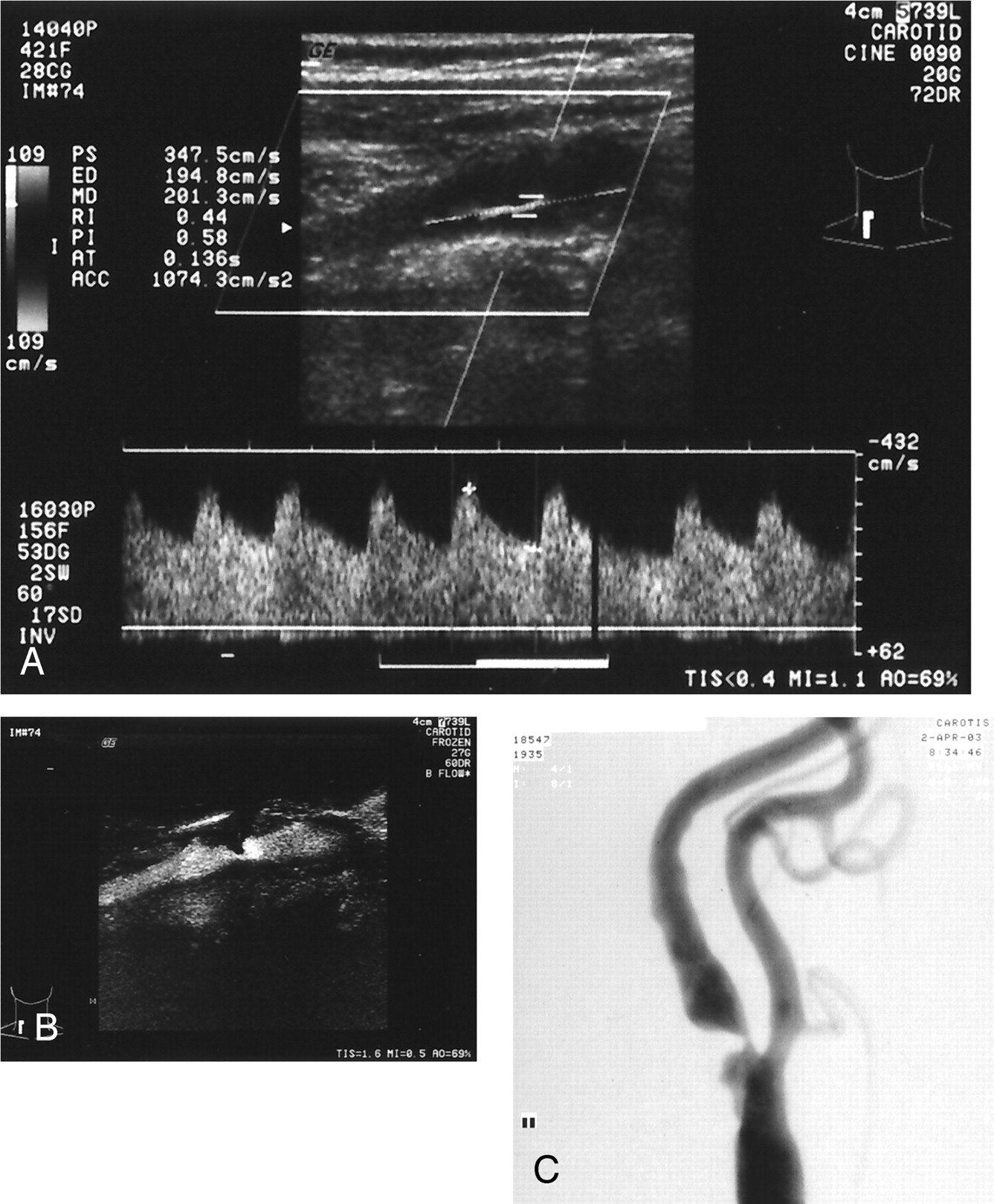
Severe left ICAS in a 68-year-old woman.

A, CDU shows >70% ICAS with PSV of 347.5 cm/s, end diastolic velocity of 194.8 cm/s, and ICA to CCA ratio of 11.6.

B, 82% of ICAS is measured with BFI.

C, Digital subtraction angiography assesses 83% stenosis.

Interobserver difference of BFI measurement was 0.1% (95% confidence limits: −16.3%, 16.5%). When PSV, end diastolic velocity, and PSV ratio each was used as the CDU criterion, the results of CDU and BFI were concordant in 143 of 161 (89%), 138 of 161 (86%), and 144 of 161 (89%) of the cases, respectively.

Table 2 summarizes the sensitivity, specificity, PPV, negative predictive value, accuracy, and misclassification rate results obtained with CDU, BFI, and a combination of the two tests. Compared with digital subtraction angiography, BFI underestimated ICAS so that the sensitivity and specificity values were 65% and 98%, respectively. CDU results had slightly better accuracy compared with BFI for detection of ICAS. For identifying 70% to 99% ICAS, CDU criterion PSVICA/CCA had the highest diagnostic accuracy (its sensitivity was 94% and its specificity was 96%). The PPVs of CDU for detection of 70% to 99% ICAS were 74% for PSV, 81% for end diastolic velocity, and 83% for PSVICA/CCA, respectively. These values increased to 90%, 95%, and 95%, respectively, when combined with BFI finding. The misclassification rate of the combined test is significantly decreased in comparison with each test used alone. This decrease is more apparent when end diastolic velocity and PSVICA/CCA is used as CDU criterion.

View this table:
  • View inline
  • View popup
Table 2:

Diagnostic performance color duplex ultrasonography, B-flow imaging, and a combination of these tests in detecting 70% of 99% internal carotid artery stensosis

Discussion

CDU has now become the principal method of investigation for carotid artery stenosis. CDU is used as the sole imaging test before carotid endarterectomy in some centers. Although the accuracy of CDU for categorizing disease severity approaches 90% in accredited vascular laboratories (13–15, 24–33), concern exists regarding its reliability. A reliable confirmatory noninvasive test is needed to improve confidence in correct patient selection for carotid endarterectomy.

In deciding whether carotid endarterectomy is necessary in cases of ICAS, the use of an MR angiography and CDU combination has been proposed and has received wide acceptance (16, 21, 23, 33, 39–44). MR angiography also provides assessments of the carotid artery high in the neck, in the head and the intracranial branches.

In this study, we assessed the accuracy of CDU and BFI separately and as a combination of the two tests for determining carotid artery stenosis. BFI is a method that has been used recently in the evaluation of carotid artery stenosis. This method uses Digitally Encoded Sonography Technology (34–37). Coded sound waves are transmitted into the body and vasculature and then the returning signals to enhance sensitivity of weak signals and to suppress nonmoving tissue signals. The remainder of the data processing is essentially the same as with the conventional B-mode. Consequently, B-flow can visualize real-time hemodynamic flow in relation to stationary tissue. Advantages of this technique are simultaneous imaging of tissue and blood-echo information, so that blooming artifacts are not possible. A high frame rate is possible, as is high spatial and transverse resolution, so that imaging of complex flow phenomena becomes possible. BFI also has some limitations (34, 35). A significant technical limitation of direct BFI measurement of carotid stenoses arises in the presence of extensive plaque calcification in the carotid artery. Calcification interferes with the ability to achieve a clear sonographic window to the carotid artery. In cases in which BFI measurement cannot be made because of calcification, changing the angle and position of the probe on the patient’s neck usually can provide a sonographic window that is clear enough to measure velocities. Although BFI measurement cannot be made because of extensive calcification, it was possible to make duplex velocity measurement in nine of 11 carotid arteries in our patient group. A limitation of BFI is that excessive pulsation of the vessel leads to movement of the surrounding structures, so that the vessel wall is sometimes ill defined. Another is that sensitivity in BFI is decreased with increasing depth because of strong dependence on signal intensity strength. This limitation is especially significant in evaluation of post-bulbar ICA, because ICA toward distal is more deeply rooted. Finally, the remaining two limitations are background flash and difficulty in showing slow flow. Slow flow limitation, especially in high degree stenosis, may reduce flow velocity at distal normal ICA and may cause difficulty in imaging of lumen and measurement of diameter.

CDU also has some limitations. With CDU, duplex velocity measurement at the site of stenosis is a primary diagnostic criterion. Velocity criteria may be inaccurate in a number of clinical conditions. Although cardiac arrhythmia, aortic valve insufficiency, and tandem plaques may result in underestimation of the degree of stenosis, carotid arterial coiling or kinking, arteriovenous malformations, carotid arterial body tumors, and contralateral severe stenosis or occlusion may promote overestimation of luminal narrowing (45). Many of the limitations of duplex Doppler can be overcome by performing velocity ratio measurement (46). This ratio is not clearly superior to absolute flow velocity (24), probably because of CCA diameter variation and variable collateral flow.

Our data show that CDU is more sensitive but that BFI is more specific for the detection of 70% to 99% ICAS. Concordant results of both tests had similar sensitivity, slightly better specificity, moderately high accuracy, and markedly high PPV, compared with CDU.

When a noninvasive test is to be used as a definitive diagnostic tool for an individual patient, however, the most important parameter is the PPV of the test. In the study, combining concordant data from both tests markedly increased PPV compared with CDU. By combining CDU with BFI, the PPV increased on average by 14%. CDU provides an anatomic picture of the carotid artery and 3D physiological assessment, whereas BFI provides an anatomic 2D picture of the carotid arteries. The reason for the improved performance of a combination of these tests may be, at least in part, the use of complementary methods for determining the degree of carotid stenosis.

BFI examination does not require separate equipment and can be implemented by using the same Doppler equipment that has the necessary hardware by adding some software. Both examinations can be performed by using single equipment, which is advantageous in terms of time and cost compared with the combination of CDU and MR angiography or CT angiography. However, as is the case in our study, calcification and high bifurcation or short neck anatomy causes limitation in evaluation of carotid bifurcation with ultrasonography methods. Additionally, it is not possible to evaluate distal ICA with ultrasonography methods. The results of BFI in determining ICA stenosis are less accurate compared with the results of CDU. Despite the several disadvantages of BFI examination mentioned above, it reaches a significant value when it is used in combination with CDU.

Conclusion

CDU showed slightly better accuracy than did BFI in the diagnosis of carotid artery stenosis. Combined use of CDU and BFI provides better diagnostic accuracy than either method alone. The combined use of CDU and BFI affords accurate noninvasive evaluation of carotid artery stenosis that is sufficient for surgical planning in most cases. Digital subtraction angiography remains indicated in cases in which both tests are discordant. Because of its high specificity and good interobserver variability, BFI can be used in combination with CDU as a confirmatory test for assessment of ICAS.

References

  1. ↵
    North American Symptomatic Carotid Endarterectomy Trial Collaborators. Beneficial effect of carotid endarterectomy in symptomatic patients with high-grade carotid stenosis. N Engl J Med 1991;325:445–453
    CrossRefPubMed
  2. European Carotid Surgery Trialist Collaborative Group. MRC European Carotid Surgery Trial: interim results for symptomatic patients with severe (70–99%) or with mild (0–29%) carotid stenosis. Lancet 1991;337:1235–1243
    CrossRefPubMed
  3. North American Symptomatic Carotid Endarterectomy Trial Collaborators. Benefit of carotid endarterectomy in patients with symptomatic moderate or severe stenosis. N Engl J Med 1998;339:1415–1425
    CrossRefPubMed
  4. ↵
    Asymptomatic Carotid Atherosclerosis Study Collaborators. Endarterectomy in asymptomatic carotid artery stenosis. JAMA 1995;273:1421–1428
    CrossRefPubMed
  5. ↵
    Willinsky RA, Taylor SM, TerBrugge K, Farb RI, Tomlinson G, Montanera W. Neurologic complication of cerebral angiography: prospective analysis of 2,899 procedures and review of the literature. Radiology 2003;227:522–528
    CrossRefPubMed
  6. Hankey GJ, Warlow CP, Molyneux AJ. Complications of cerebral angiography for patients with mild carotid territory ischaemia being considered for carotid endarterectomy. J Neurol Neurosurg Psychiatry 1990;53:542–548
    Abstract/FREE Full Text
  7. Davies KN, Humphrey PR. Complications of cerebral angiography in patients with symptomatic carotid territory ischaemia screened by carotid ultrasound. J Neurol Neurosurg Psychiatry 1993;56:967–972
    Abstract/FREE Full Text
  8. AbuRahma AF, Robinson PA, Boland JP, et al. Complications of arteriography in a recent series of 707 cases: factors affecting outcome. Ann Vasc Surg 1993;7:122–129
    CrossRefPubMed
  9. ↵
    Waugh JR, Sacharias N. Arteriographic complications in DSA era. Radiology 1992;182:243–246
    PubMed
  10. ↵
    Fontenelle LJ, Simper SC, Hanson TL. Carotid duplex scan versus angiography in evaluation of carotid artery disease. Am Surg 1994;60:864–868
    PubMed
  11. Golledge J, Ellis M, Sabharwal T, Sikdar T, Davies AH, Greenhalgh RM. Selection of patients for carotid endarterectomy. J Vasc Surg 1999;30:122–130
    CrossRefPubMed
  12. Collier PE. Changing trends in the use of preoperative carotid arteriography: the community experience. Cardiovasc Surg 1998;6:485–489
    CrossRefPubMed
  13. ↵
    Winkelaar GB, Chen JC, Salvian AJ, Taylor DC, Teal PA, Hsiang YN. New duplex ultrasound scan criteria for managing symptomatic 50% or greater carotid stenosis. J Vasc Surg 1999;29:986–994
    CrossRefPubMed
  14. Filis KA, Arko FR, Johnson BL, et al. Duplex ultrasound criteria for defining the severity of carotid stenosis. Ann Vasc Surg 2002;16:413–421
    CrossRefPubMed
  15. ↵
    Dinkel HP, Moll R, Debus S. Colour flow Doppler ultrasound of carotid bifurcation: can it replace routine angiography before carotid endarterectomy? Br J Radiol 2001;74:590–594
    Abstract/FREE Full Text
  16. ↵
    Borisch I, Horn M, Butz B, et al. Preoperative evaluation of carotid artery stenosis: comparison of contrast-enhanced MR angiography and duplex sonography with digital subtraction angiography. AJNR Am J Neuroradiol 2003;24:1117–1122
    Abstract/FREE Full Text
  17. Moll R, Dinkel HP. Value of the CT angiography in the diagnosis of common carotid artery bifurcation disease: CT angiography versus digital subtraction angiography and color flow Doppler. Eur J Radiol 2001;39:155–162
    CrossRefPubMed
  18. Patel SG, Collie DA, Wardlaw JM, et al. Outcome, observer reliability, and patient preferences if CTA, MRA, or Doppler ultrasound were used, individually or together, instead of digital subtraction angiography before carotid endarterectomy. J Neurol Neurosurg Psychiatry 2002;73:21–28
    Abstract/FREE Full Text
  19. ↵
    Friese S, Krapf H, Fetter M, Klose U, Skalej M, Kuker W. Ultrasonography and contrast-enhanced MRA in ICA-stenosis: is conventional angiography obsolete? J Neurol 2001;248:506–513
    CrossRefPubMed
  20. Kuntz KM, Skillman JJ, Whittemore AD, Kent KC. Carotid endarterectomy in asymptomatic patients: is contrast angiography necessary? a morbidity analysis. J Vasc Surg 1995;22:706–716
    CrossRefPubMed
  21. ↵
    Johnston DC, Goldstein LB. Clinical carotid endarterectomy decision making: noninvasive vascular imaging versus angiography. Neurology 2001;56:1009–1015
    Abstract/FREE Full Text
  22. Calton WC, Franklin DP, Elmore JR, Han DC. Carotid endarterectomy: the financial impact of practice changes. J Vasc Surg 2000;32:643–648
    CrossRefPubMed
  23. ↵
    Back MR, Wilson JS, Rushing G, et al. Magnetic resonance angiography is an accurate imaging adjunct to duplex ultrasound scan in patient selection for carotid endarterectomy. J Vasc Surg 2000;32:429–438
    CrossRefPubMed
  24. ↵
    Hunink MG, Polak JF, Barlan MM, O’Leary DH. Detection and quantification of carotid artery stenosis: efficacy of various Doppler parameters. AJR Am J Roentgenol 1993;160:619–625
    PubMed
  25. Moneta GL, Edwards JM, Papanicolaou G, et al. Screening for asymptomatic internal carotid artery stenosis: duplex criteria for discriminating 60% to 99% stenosis. J Vasc Surg 1995;21:989–994
    CrossRefPubMed
  26. Moneta GL, Edwards JM, Chitwood RW, et al. Correlation of North American Symptomatic Carotid Endarterectomy Trial (NASCET) angiographic definition of 70% to 99% internal carotid artery stenosis with duplex scanning. J Vasc Surg 1993;17:152–159
    CrossRefPubMed
  27. Carpenter JP, Lexa FJ, Davis JT. Determination of sixty percent or greater carotid artery stenosis by duplex Doppler ultrasonography. J Vasc Surg 1995;22:697–703
    CrossRefPubMed
  28. Carpenter JP, Lexa FJ, Davis JT. Determination of duplex Doppler ultrasound criteria appropriate to the North American Symptomatic Carotid Endarterectomy Trial. Stroke 1996;27:695–699
    Abstract/FREE Full Text
  29. Neale ML, Chambers JL, Kelly AT, et al. Reappraisal of duplex criteria to assess significant carotid stenosis with special reference to reports from the North American Symptomatic Carotid Endarterectomy Trial and the European Carotid Surgery Trial. J Vasc Surg 1994;20:642–649
    CrossRefPubMed
  30. Hood DB, Mattos MA, Mansour A, et al. Prospective evaluation of new duplex criteria to identify 70% internal carotid artery stenosis. J Vasc Surg 1996;23:254–261
    CrossRefPubMed
  31. Wilterdink JL, Feldmann E, Easton JD, Ward R. Performance of carotid ultrasound in evaluating candidates for carotid endarterectomy is optimized by an approach based on clinical outcome rather than accuracy. Stroke 1996;27:1094–1098
    Abstract/FREE Full Text
  32. Faugtht WE, Mattos MA, Van Bemmelen PS, et al. Color-flow duplex scanning of carotid arteries: new velocity criteria based on receiver operator characteristic analysis for threshold stenoses used in the symptomatic and asymptomatic carotid trials. J Vasc Surg 1994;19:818–827
    PubMed
  33. ↵
    Turnipseed WD, Kennell TW, Turski PA, Acher CW, Hoch JR. Combined use of duplex imaging and magnetic resonance angiography for evaluation of patients with symptomatic ipsilateral high-grade carotid stenosis. J Vasc Surg 1993;17:832–839
    CrossRefPubMed
  34. ↵
    Henri P, Tranquart F. B-flow ultrasonographic imaging of circulating blood [in French]. J Radiol 2000;81:465–467
    PubMed
  35. ↵
    Weskott HP. B-flow: a new method for detecting blood flow [in German]. Ultraschall Med 2000;21:59–65
    CrossRefPubMed
  36. Umemura A, Yamada K. B-mode flow imaging of the carotid artery. Stroke 2001;32:2055–2057
    Abstract/FREE Full Text
  37. ↵
    Bucek RA, Reiter M, Koppensteiner I, Ahmadi R, Minar E, Lammer J. B-flow evaluation of carotid arterial stenosis: initial experience. Radiology 2002;225:295–299
    CrossRefPubMed
  38. ↵
    Bland JM, Altman DG. Statistical methods for assessing agreement between two methods of clinical measurement. Lancet 1986;8:307–310
  39. ↵
    Polak JF, Kalina P, Donaldson MC, O’Leary DH, Whittemore AD, Mannick JA. Carotid endarterectomy: preoperative evaluation of candidates with combined Doppler sonography and MR angiography. Radiology 1993;186:333–338
    PubMed
  40. Patel MR, Kuntz KM, Klufas RA, et al. Preoperative assessment of carotid bifurcation. Can magnetic resonance angiography and duplex ultrasonography replace contrast arteriography? Stroke 1995;26:1753–1758
    Abstract/FREE Full Text
  41. Huston J, Nichols DA, Luetmer PH, et al. MR angiographic and sonographic indication for endarterectomy. AJNR Am J Neuroradiol 1998;19:309–315
    Abstract
  42. Serfaty JM, Chirossel P, Chevallier JM, Ecochard R, Froment JC, Douek PC. Accuracy of three-dimensional gadolinium-enhanced MR angiography in the assessment of extracranial carotid artery disease. AJR Am J Roentgenol 2000;175:455–463
    PubMed
  43. Nederkoorn PJ, Mali WP, Eikelboon BC, et al. Preoperative diagnosis of carotid artery stenosis: accuracy of noninvasive testing. Stroke 2002;33:2003–2008
    Abstract/FREE Full Text
  44. ↵
    Johnston DC, Eastwood JD, Nguyen T, Goldstein LB. Contrast-enhance magnetic resonance angiography of carotid arteries: utility in routine clinical practice. Stroke 2002;33:2834–2838
    Abstract/FREE Full Text
  45. ↵
    Nicolaides AN, Shifrin EG, Bradbury A, et al. Angiography and duplex grading of internal carotid stenosis: can we overcome the confusion? J Endovasc Surg 1996;3:158–165
    CrossRefPubMed
  46. ↵
    Zwiebel WJ. Doppler evaluation of carotid stenosis. In: Zwiebel WJ, ed. Introduction to Vascular Ultrasonography. 4th ed. Philadelphia: WB Saunders Co.;2000 :137–154
  • Received January 13, 2004.
  • Accepted after revision April 5, 2004.
  • Copyright © American Society of Neuroradiology
View Abstract
PreviousNext
Back to top

In this issue

American Journal of Neuroradiology: 25 (10)
American Journal of Neuroradiology
Vol. 25, Issue 10
1 Nov 2004
  • Table of Contents
  • Index by author
Advertisement
Print
Download PDF
Email Article

Thank you for your interest in spreading the word on American Journal of Neuroradiology.

NOTE: We only request your email address so that the person you are recommending the page to knows that you wanted them to see it, and that it is not junk mail. We do not capture any email address.

Enter multiple addresses on separate lines or separate them with commas.
Combined Use of Color Duplex Ultrasonography and B-Flow Imaging for Evaluation of Patients with Carotid Artery Stenosis
(Your Name) has sent you a message from American Journal of Neuroradiology
(Your Name) thought you would like to see the American Journal of Neuroradiology web site.
CAPTCHA
This question is for testing whether or not you are a human visitor and to prevent automated spam submissions.
Cite this article
Muharrem Tola, Mehmet Yurdakul, Turhan Cumhur
Combined Use of Color Duplex Ultrasonography and B-Flow Imaging for Evaluation of Patients with Carotid Artery Stenosis
American Journal of Neuroradiology Nov 2004, 25 (10) 1856-1860;

Citation Manager Formats

  • BibTeX
  • Bookends
  • EasyBib
  • EndNote (tagged)
  • EndNote 8 (xml)
  • Medlars
  • Mendeley
  • Papers
  • RefWorks Tagged
  • Ref Manager
  • RIS
  • Zotero
0 Responses
Respond to this article
Share
Bookmark this article
Combined Use of Color Duplex Ultrasonography and B-Flow Imaging for Evaluation of Patients with Carotid Artery Stenosis
Muharrem Tola, Mehmet Yurdakul, Turhan Cumhur
American Journal of Neuroradiology Nov 2004, 25 (10) 1856-1860;
del.icio.us logo Twitter logo Facebook logo Mendeley logo
  • Tweet Widget
  • Facebook Like
  • Google Plus One

Jump to section

  • Article
    • Abstract
    • Methods
    • Results
    • Discussion
    • Conclusion
    • References
  • Figures & Data
  • Info & Metrics
  • Responses
  • References
  • PDF

Related Articles

  • No related articles found.
  • PubMed
  • Google Scholar

Cited By...

  • Thyroid B-flow twinkling sign: a new feature of papillary cancer
  • B-flow Imaging for Assessment of 70% to 99% Internal Carotid Artery Stenosis Based on Residual Lumen Diameter
  • B-Flow Imaging in Low Cervical Internal Carotid Artery Dissection
  • Crossref
  • Google Scholar

This article has not yet been cited by articles in journals that are participating in Crossref Cited-by Linking.

More in this TOC Section

  • Correlation of Apparent Diffusion Coefficient at 3T with Prognostic Parameters of Retinoblastoma
  • Parathyroid Lesions: Characterization with Dual-Phase Arterial and Venous Enhanced CT of the Neck
  • Kimura Disease: CT and MR Imaging Findings
Show more Head and Neck

Similar Articles

Advertisement

Indexed Content

  • Current Issue
  • Accepted Manuscripts
  • Article Preview
  • Past Issues
  • Editorials
  • Editors Choice
  • Fellow Journal Club
  • Letters to the Editor

Cases

  • Case Collection
  • Archive - Case of the Week
  • Archive - Case of the Month
  • Archive - Classic Case

Special Collections

  • Special Collections

Resources

  • News and Updates
  • Turn around Times
  • Submit a Manuscript
  • Author Policies
  • Manuscript Submission Guidelines
  • Evidence-Based Medicine Level Guide
  • Publishing Checklists
  • Graphical Abstract Preparation
  • Imaging Protocol Submission
  • Submit a Case
  • Become a Reviewer/Academy of Reviewers
  • Get Peer Review Credit from Publons

Multimedia

  • AJNR Podcast
  • AJNR SCANtastic
  • Video Articles

About Us

  • About AJNR
  • Editorial Board
  • Not an AJNR Subscriber? Join Now
  • Alerts
  • Feedback
  • Advertise with us
  • Librarian Resources
  • Permissions
  • Terms and Conditions

American Society of Neuroradiology

  • Not an ASNR Member? Join Now

© 2025 by the American Society of Neuroradiology All rights, including for text and data mining, AI training, and similar technologies, are reserved.
Print ISSN: 0195-6108 Online ISSN: 1936-959X

Powered by HighWire