
We enjoyed reading the article, “Pseudofenestration of the Cervical Internal Artery: A Pathologic Process that Simulates an Anatomic Variant” by Gailloud et al (1). We would like to add two more cases to their series in support of their argument that apparent fenestrations of the carotid artery are acquired lesions and not congenital anomalies. Furthermore, we would like to raise the issue that there may be prognostic features that portend a more benign or malignant course, which should be highlighted in any discussion with the treating physician.
Case 1.
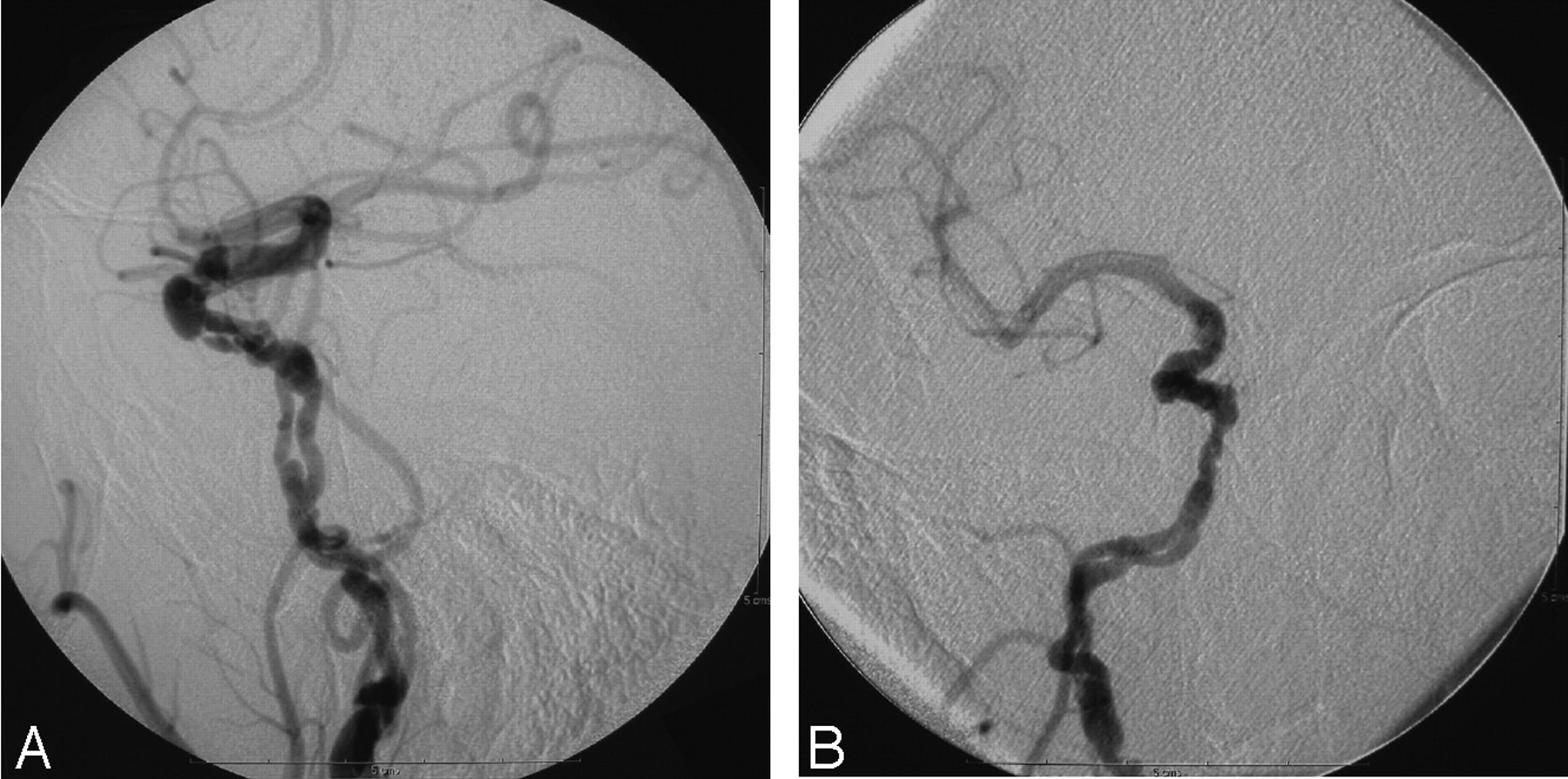
A 69-year-old man was referred for evaluation of two recent transient ischemic attacks. The first attack, which occurred while he was taking aspirin for coronary artery disease, consisted of left hemiparesis. He was placed on Plavix (clopidogrel), but 2 months later experienced a transient episode of diplopia. MR angiography findings obtained at an outside institution were interpreted as showing carotid stenosis, so he underwent diagnostic cerebral angiography for further evaluation (Fig 1). This study showed the entire right internal carotid artery (ICA) was markedly abnormal, with focal and short segment strictures and areas of fusiform dilatation, which suggest an underlying dysplastic process with superimposed pseudoaneurysms. At the cervicopetrous junction, the ICA divided into two lumens of nearly symmetrical caliber that ran nearly parallel to each other, approximately 2 mm apart, and then joined distally at approximately the level of the anterior carotid genu. The limbs of this apparent fenestration were also irregular, with a small pseudoaneurysm at the proximal bifurcation. The angiogram also showed that the other ICA had a small ulcerated plaque at the bulb, and both vertebral arteries were occluded at their origins and segmentally reconstituted from muscular branches from the ascending cervical artery. This patient failed a balloon occlusion test and is scheduled to undergo a right extracranial-to-intracranial arterial bypass followed by sacrifice of the diseased vessel.

A 69-year-old man with right ICA pseudofenestration. Digital subtraction angiography (DSA), right common carotid injection, lateral (A) and frontal (B) views. Both lumens of the fenestrated segment are markedly irregular with focal stenoses alternating with areas of fusiform dilatation. The ICA has a narrow caliber and striking dysplastic changes throughout its entirety.
Case 2.
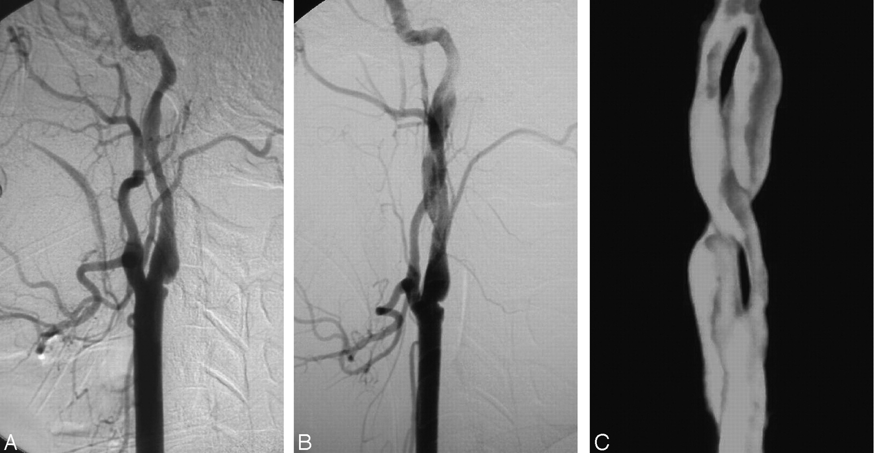
A 33-year-old woman developed “pressure” headaches after a difficult delivery of twins, followed 4 days later by vision changes, difficulties understanding, and numbness over the left face and hand. Angiography (Fig 2A) demonstrated dissections of both ICAs at the craniocervical junction as well as the proximal left vertebral artery, accompanied by a dysplastic appearance of the distal left ICA consistent with fibromuscular dysplasia, and a small, unruptured, wide-necked anterior communicating artery aneurysm. This aneurysm was uneventfully clipped, after which she was anticoagulated with coumadin for 6 months and then switched to aspirin. Her symptoms resolved during this time. Follow-up angiography showed the left ICA dissection had healed with two smooth lumens of nearly equal caliber, spiraling around each other and communicating both proximally and distally without any restriction in flow (Fig 2B, -C). The dissections in other vessels had also improved in appearance. This patient continues to be conservatively treated with aspirin and 10 months later remains asymptomatic with stable anatomy on follow-up studies.

A 33-year-old woman with left ICA dissection, characterized by irregular caliber changes of the single lumen just below the skull base (A, left common carotid injection, lateral DSA). After 6 months of anticoagulation, the dissection has healed with the formation of a relatively smooth, spiraling, double lumen configuration communicating both proximally and distally without any flow limitation (B, lateral DSA; C, 3D rotational angiogram with the external carotid artery cut away to show the ICA).
The location of an apparent fenestration at a site prone to spontaneous dissections, in the setting of an underlying vasculopathy, further supports the theory that a “double-barreled” carotid artery is likely the result of an injury that dissected through a segment of the vessel to communicate both proximally and distally. Our second case illustrates a progression of changes that lead to this phenomenon. It is instructive to note the high frequency of strokes in Gailloud’s series and ours to remind us of the need to notify our clinical colleagues quickly of the potential danger if left untreated. Not all diseased vessels, however, must be necessarily sacrificed. Angiographic features that would suggest a more worrisome course would include marked irregularities in the vessel walls, restricted flow, the presence of a pseudoaneurysm, and extension over time. Without these features, a patient may be conservatively treated with an anticoagulant or antiplatelet regimen as in our second case.
References
- Copyright © American Society of Neuroradiology
In this issue


Related Articles
Cited By...
- No citing articles found.