
Article Figures & Data
Figures
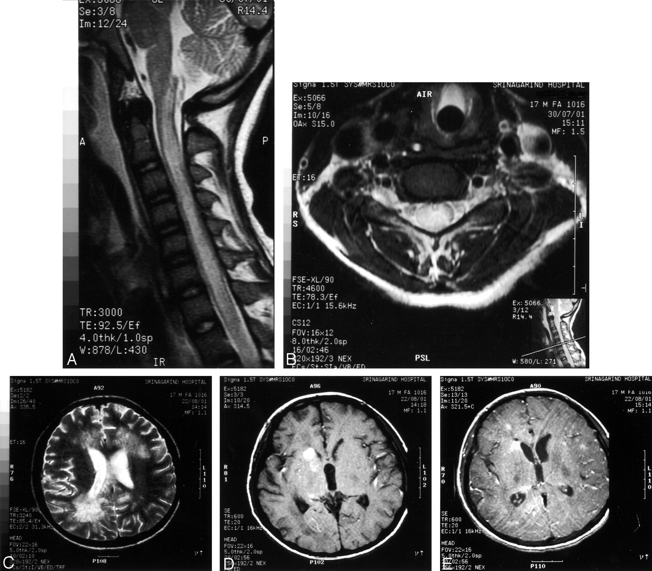
- Fig 1.
Case 1. MR images of spinal cord and brain.
A and B, Sagittal T2-weighted images, showing diffuse cord enlargement with abnormal high signal intensities.
C, Axial T1-weighted image, showing hemorrhagic spot at posterior midpons.
D, Coronal T1-weighted postgadolinium image, showing hemorrhagic tract at posterior midpons level.
- Fig 2.
Case 2. MR images of cervical cord and brain.
A, Sagittal T2-weighted image, showing diffuse cord enlargement with ill-defined area of increased signal intensity.
B, Axial T2-weighted image, showing hyperintense lesion within central gray matter.
C, Axial T2-weighted image, showing fuzzy hyperintense lesion at both periventricular white matter regions.
D, Axial T1-weighted image, showing intracerebral hemorrhage at right caudate nucleus and posterior part of basal ganglia.
E, Axial T1-weighted postgadolinium image, showing scattered tiny nodular enhancement at both frontoparietal regions.


In this issue


Jump to section
Related Articles
Cited By...
- Thoracic Myelopathy Due to Gnathostomiasis Acquired in New Zealand
- Immunoblot Diagnostic Test for Neurognathostomiasis
- In Response
- Gnathostomiasis, Another Emerging Imported Disease
- Update on Eosinophilic Meningoencephalitis and Its Clinical Relevance
- Survival of a suspected case of central nervous system cuterebrosis in a dog: clinical and magnetic resonance imaging findings.