
Article Figures & Data
Figures
- Fig 1.
Correlation analysis of rCBV, K trans, and glioma grade.
A, rCBV and glioma grade were strongly correlated, with Spearman r = 0.817 (P = .0001) and Pearson r = 0.771 (P = .0004). The slope and its gradient are consistent with a strong positive correlation with highly significant P values.
B, A weaker correlation was observed between K trans and glioma grade: Spearman r = 0.234 (P = .046) and Pearson r = 0.277 (P = .017). Although the slope indicates a positive correlation, its gradient is less pronounced, with less significant P values than in A. Furthermore, more overlap is noted in K trans measurements than in the rCBV measurements in A.
C, Relationship between rCBV and Ktrans is characterized by a modest yet statistically significant Spearman rank correlation (r = 0.266; P = .023) and a weak, statistically insignificant Pearson product moment correlation (r = 0.163; P = .168).
D, Receiver operating characteristic curves for rCBV and Ktrans, as estimated from logistic regression. rCBV was a significant predictor of high-grade gliomas with higher sensitivity and specificity than Ktrans.
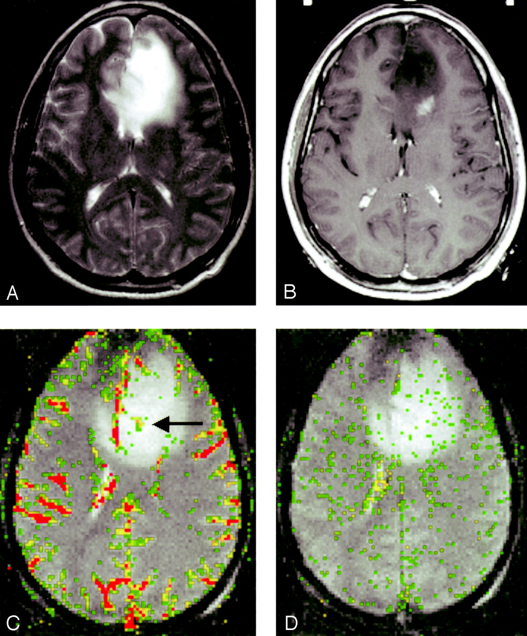
- Fig 2.
Low-grade astrocytoma (grade I/III).
A, T2-weighted image (3158/119) demonstrates bifrontal abnormalities in signal intensity centered primarily in the left frontal lobe.
B, Contrast-enhanced T1-weighted image (600/14/1) demonstrates an ill-defined focus of enhancement.
C, rCBV map demonstrates a few foci of mildly elevated perfusion (arrow), which are in a location different from the region of maximal enhancement in B.
D, SD25 color map suggests low permeability throughout the lesion.
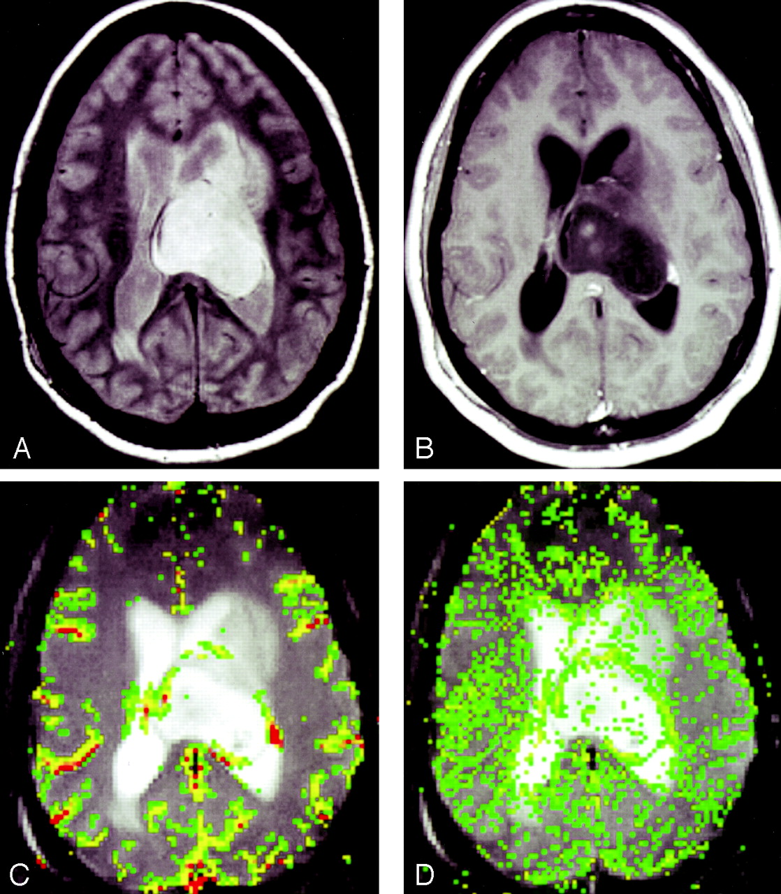
- Fig 3.
Anaplastic astrocytoma (grade II/III).
A, Proton density–weighted image (3158/17) demonstrates heterogeneous, left-sided paraventricular lesion extending into the left lateral ventricle. There is a small amount of vasogenic edema.
B, Contrast-enhanced T1-weighted image (600/14/1) demonstrates a small focus of peripheral enhancement.
C, rCBV map demonstrates elevated perfusion. On this occasion, the finding corresponds to the enhancing focus in B.
D, SD25 color map suggests intermediate permeability in the solid portions of this tumor.
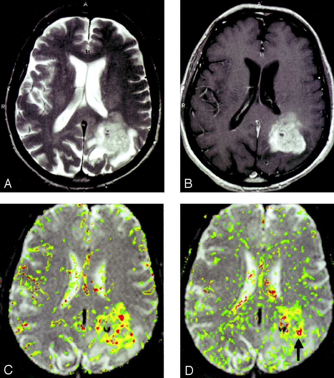
- Fig 4.
GBM (grade III/III).
A, T2-weighted image (3158/119/1) demonstrates a left parietal lesion with mass effect, edema, and signal-intensity heterogeneity. These are features of a high-grade glioma, such as a GBM.
B, Contrast-enhanced T1-weighted (600/14/1) with extensive, heterogeneous contrast enhancement.
C, rCBV map shows markedly elevated perfusion.
D, SD25 color map suggests markedly elevated permeability. Note that the areas of highest rCBV elevation do not directly correspond to the regions of highest SD25 (arrow).
- Fig 5.
Typical normalized signal-intensity curves for the three glioma grades studied. Grade I glioma demonstrates a shallow perfusion signal-intensity curve with SD25 after the bolus peak; this was relatively close to the prebolus baseline, suggesting relatively low permeability. Grade II glioma demonstrates a more substantial initial signal intensity drop, indicating higher rCBV with slower return to baseline. SD25 is considerably larger than that seen in grade I gliomas. Grade III glioma shows a larger area above the curve, indicating high rCBV with a similarly delayed return to baseline; this suggests high permeability.


Tables
Decade (year) Low-Grade Glioma (n = 21) Low-Grade Oligodendroglioma (n = 10) Anaplastic Astrocytoma (n = 16) GBM (n = 26) 0–9 3 1 0 0 10–19 1 0 3 0 20–29 4 4 2 1 30–39 0 3 6 4 40–49 5 2 2 4 50–59 5 0 1 9 60–69 3 0 1 3 70–79 0 0 1 3 80–89 0 0 0 2 Tumor Grade rCBV* Ktrans (seconds) I/III 1.75±0.85 0.00053 ± 0.0013 II/III 3.79 ± 1.48 0.0011 ± 0.0015 III/III 6.05 ± 2.22 0.0020 ± 0.003 Note.—Data are the mean ± standard deviation. Grade I = low-grade glioma, grade II = anaplastic astrocytoma, and grade III = GBM.
* P < .0005.
† P < .4662.
Tumor Grade P Value rCBV Ktrans (second) I versus II* <.0001 0.4662 I versus III* <.0001 0.0912 II versus III* .0005 0.4424 I versus II and III† <.0001 0.027 I and II versus III† <.0001 0.068 Note.—Grade I = low-grade glioma, grade II = anaplastic astrocytoma, and grade III = GBM.
* Tukey honestly significant difference procedure.
† Unequal variance t test.
In this issue


Jump to section
Related Articles
Cited By...
- Volumetric Measurement of Relative CBV Using T1-Perfusion-Weighted MRI with High Temporal Resolution Compared with Traditional T2*-Perfusion-Weighted MRI in Postoperative Patients with High-Grade Gliomas
- Imaging-Based Algorithm for the Local Grading of Glioma
- Prognostic Predictions for Patients with Glioblastoma after Standard Treatment: Application of Contrast Leakage Information from DSC-MRI within Nonenhancing FLAIR High-Signal-Intensity Lesions
- Machine Learning-Based Radiomics for Molecular Subtyping of Gliomas
- 3D Pseudocontinuous Arterial Spin-Labeling MR Imaging in the Preoperative Evaluation of Gliomas
- Comparison of the Effect of Vessel Size Imaging and Cerebral Blood Volume Derived from Perfusion MR Imaging on Glioma Grading
- The Added Prognostic Value of Preoperative Dynamic Contrast-Enhanced MRI Histogram Analysis in Patients with Glioblastoma: Analysis of Overall and Progression-Free Survival
- Glioma Angiogenesis and Perfusion Imaging: Understanding the Relationship between Tumor Blood Volume and Leakiness with Increasing Glioma Grade
- Detection, Characterization, and Inhibition of FGFR-TACC Fusions in IDH Wild-type Glioma
- Prognostic Value of Dynamic Susceptibility Contrast-Enhanced and Diffusion-Weighted MR Imaging in Patients with Glioblastomas
- Pixel-by-Pixel Comparison of Volume Transfer Constant and Estimates of Cerebral Blood Volume from Dynamic Contrast-Enhanced and Dynamic Susceptibility Contrast-Enhanced MR Imaging in High-Grade Gliomas
- Bayesian Estimation of Cerebral Perfusion Using Reduced-Contrast-Dose Dynamic Susceptibility Contrast Perfusion at 3T
- Advanced Magnetic Resonance Imaging of the Physical Processes in Human Glioblastoma
- A Prognostic Model Based on Preoperative MRI Predicts Overall Survival in Patients with Diffuse Gliomas
- Arterial Spin-Labeling Assessment of Normalized Vascular Intratumoral Signal Intensity as a Predictor of Histologic Grade of Astrocytic Neoplasms
- Arterial Spin-Labeled Perfusion of Pediatric Brain Tumors
- Exploratory Evaluation of MR Permeability with 18F-FDG PET Mapping in Pediatric Brain Tumors: A Report from the Pediatric Brain Tumor Consortium
- Multimodal MR Imaging (Diffusion, Perfusion, and Spectroscopy): Is It Possible to Distinguish Oligodendroglial Tumor Grade and 1p/19q Codeletion in the Pretherapeutic Diagnosis?
- The Effect of Pulse Sequence Parameters and Contrast Agent Dose on Percentage Signal Recovery in DSC-MRI: Implications for Clinical Applications
- Interstitial Flow in a 3D Microenvironment Increases Glioma Invasion by a CXCR4-Dependent Mechanism
- Correlation of Perfusion Parameters with Genes Related to Angiogenesis Regulation in Glioblastoma: A Feasibility Study
- The Role of Preload and Leakage Correction in Gadolinium-Based Cerebral Blood Volume Estimation Determined by Comparison with MION as a Criterion Standard
- The Added Value of Apparent Diffusion Coefficient to Cerebral Blood Volume in the Preoperative Grading of Diffuse Gliomas
- Does MR Perfusion Imaging Impact Management Decisions for Patients with Brain Tumors? A Prospective Study
- Quantitative Blood Flow Measurements in Gliomas Using Arterial Spin-Labeling at 3T: Intermodality Agreement and Inter- and Intraobserver Reproducibility Study
- Specific biomarkers of receptors, pathways of inhibition and targeted therapies: clinical applications
- Imaging biomarkers of angiogenesis and the microvascular environment in cerebral tumours
- Clinical applications of imaging biomarkers. Part 1. The neuroradiologist's perspective
- Perfusion CT Imaging of Brain Tumors: An Overview
- Multimodality Assessment of Brain Tumors and Tumor Recurrence
- Permeability Estimates in Histopathology-Proved Treatment-Induced Necrosis Using Perfusion CT: Can These Add to Other Perfusion Parameters in Differentiating from Recurrent/Progressive Tumors?
- Quantitative Diffusion-Weighted and Dynamic Susceptibility-Weighted Contrast-Enhanced Perfusion MR Imaging Analysis of T2 Hypointense Lesion Components in Pediatric Diffuse Intrinsic Pontine Glioma
- In Vivo Correlation of Tumor Blood Volume and Permeability with Histologic and Molecular Angiogenic Markers in Gliomas
- Increased Blood-Brain Barrier Permeability on Perfusion CT Might Predict Malignant Middle Cerebral Artery Infarction
- Combination of high-resolution susceptibility-weighted imaging and the apparent diffusion coefficient: added value to brain tumour imaging and clinical feasibility of non-contrast MRI at 3 T
- Enhancing Fraction in Glioma and Its Relationship to the Tumoral Vascular Microenvironment: A Dynamic Contrast-Enhanced MR Imaging Study
- Semiquantitative Assessment of Intratumoral Susceptibility Signals Using Non-Contrast-Enhanced High-Field High-Resolution Susceptibility-Weighted Imaging in Patients with Gliomas: Comparison with MR Perfusion Imaging
- Cerebral Blood Volume Measurements by Perfusion-Weighted MR Imaging in Gliomas: Ready for Prime Time in Predicting Short-Term Outcome and Recurrent Disease?
- Predicting Human Tumor Drug Concentrations from a Preclinical Pharmacokinetic Model of Temozolomide Brain Disposition
- The Extent and Severity of Vascular Leakage as Evidence of Tumor Aggressiveness in High-Grade Gliomas
- Usefulness of diffusion/perfusion-weighted MRI in patients with non-enhancing supratentorial brain gliomas: a valuable tool to predict tumour grading?
- Physiologic and Metabolic Magnetic Resonance Imaging in Gliomas