
Article Figures & Data
Figures
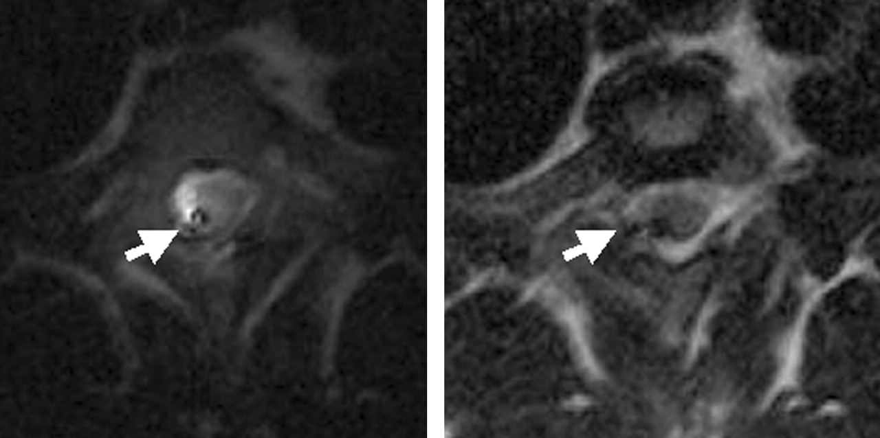
- Fig 1.
Axial FSE T2-weighted images of the canine spinal cord obtained at the T4 level (acquisition, 1; TR/TE/flip angle, 1150/110/90°; TSE factor, 35; bandwidth, 375.6 Hz; matrix, 256 × 256 with 90% Fourier in phase [90% image]; section thickness, 3 mm; spatial resolution, 0.62 mm × 0.62 mm and FOV, 16 cm with a reduced field of view [RFOV] of 80% obtained with the endospinal coil [left] and a surface coil [right]). Note the presence of higher cord signal intensity in the endospinal coil images. Arrow denotes artifact from the endospinal coil.
- Fig 2.
Axial FSE T2-weighted images of a cadaver spine obtained at the T4 level (acquisitions, 1; TR/TE/flip angle, 4055.8/100/90°; TSE factor, 14; bandwidth, 144.7 Hz; matrix, 256 × 256 with 70% image; section thickness, 5 mm; spatial resolution, 0.39 mm × 0.39 mm; and FOV, 10 cm with RFOV of 80%) obtained with endospinal coil [left] and surface coil [right]). Arrow denotes artifact from the endospinal coil. Although the image obtained with endospinal coil has smaller usable FOV, there is greater calculated SNR of the spinal cord.
- Fig 3.
Sagittal SSFP imaging of the cadaver spine (acquisitions, 3; TR/TE/flip angle, 3.7/1.85/55°; bandwidth, 997.8 Hz; matrix, 192 × 256 with 50% image; projection slab, 15 mm; spatial resolution, 0.98 mm × 0.98 mm; and FOV, 25 cm with RFOV of 75% performed with endospinal coil [left] and surface coil [right]). Arrow denotes the endospinal coil positioned within the spinal canal. Note the greater presence of signal intensity available within the spinal canal in the endospinal coil images. Increased CSF signal intensity in endospinal coil image improves cord delineation.
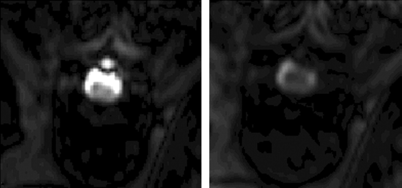
- Fig 4.
Axial SSFP images of the cadaver spine obtained at the T4 level (acquisitions, 3; TR/TE/flip angle, 3.7/1.85/55°; bandwidth, 997.8 Hz; matrix, 192 × 256 with 50% image; projection slab, 15 mm; spatial resolution, 0.98 mm x 0.98 mm; and FOV, 25 cm with RFOV of 75%). Note the significantly greater amount of canal and cord signal intensity present with the endospinal coil (left) versus the surface coil (right).
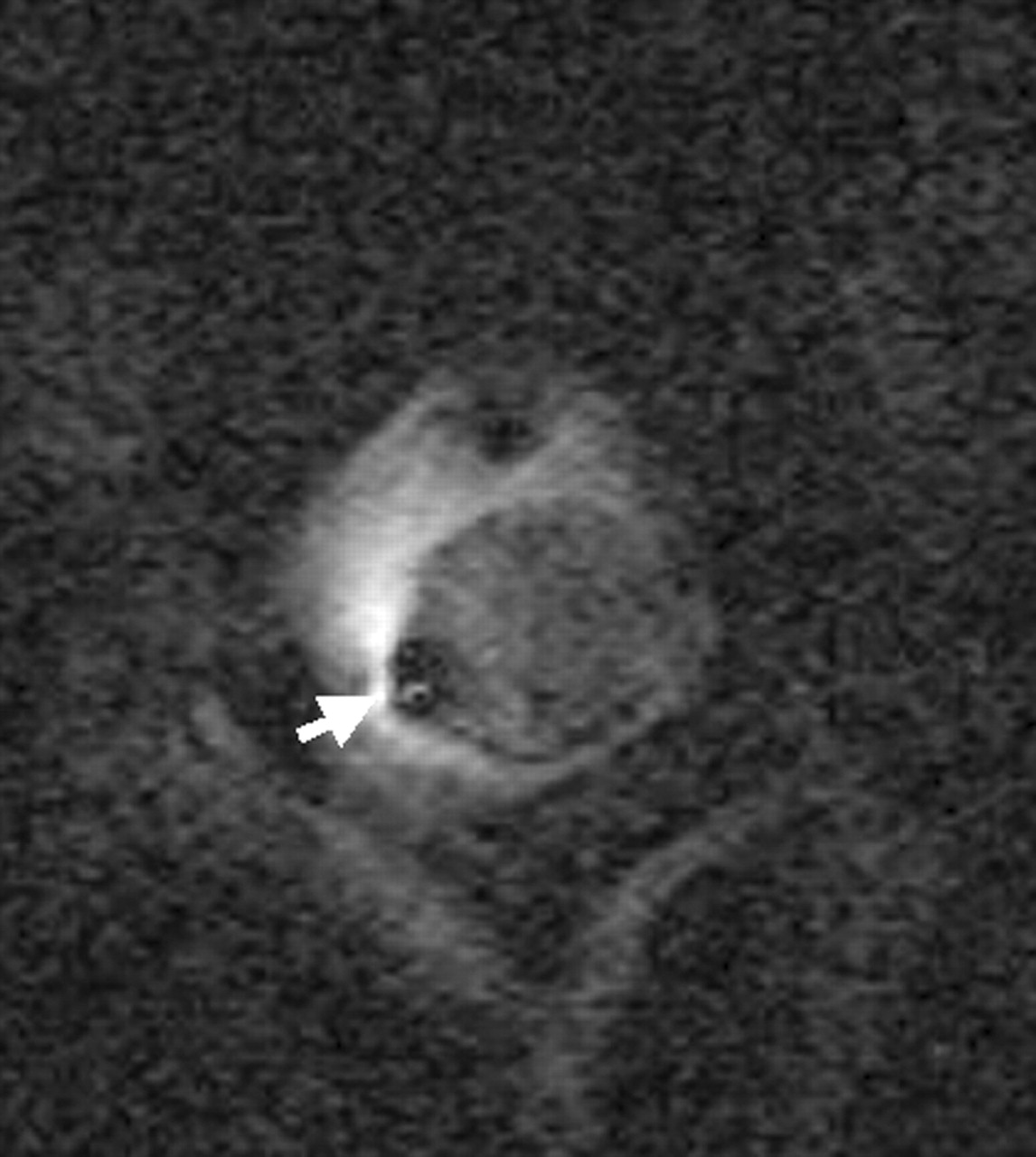
- Fig 5.
Axial high-resolution FSE T2-weighted image of the canine cord obtained at the T4 level (acquisitions, 1; TR/TE/flip angle, 4000/100/90°; TSE factor, 12; bandwidth, 128.9 Hz; matrix, 256 × 256 with 70% image; section thickness, 3 mm; spatial resolution, 0.23 mm × 0.23 mm; and FOV, 6 cm with RFOV of 70%) obtained with endospinal coil. Arrow denotes artifact from endospinal coil in dorsal subarachnoid space.
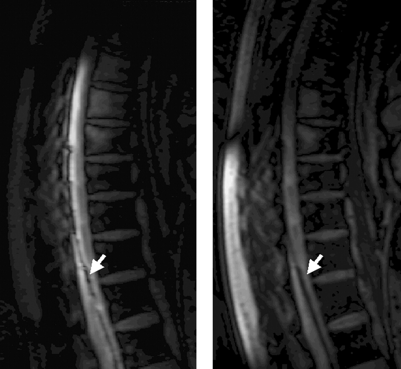
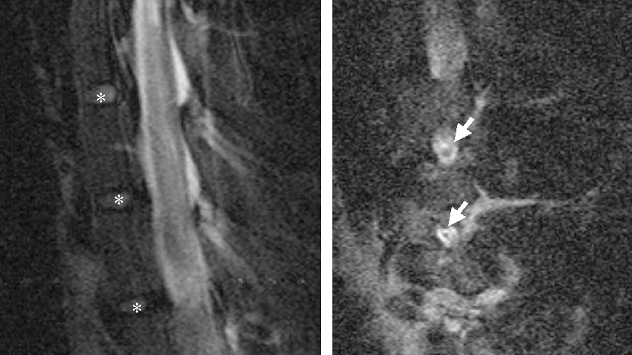
- Fig 6.
Sagittal (right) and parasagittal (left) FSE T2-weighted images of the canine spine (acquisitions, 1; TR/TE/flip angle, 3000/100/90°; TSE factor, 12; bandwidth, 121 Hz; matrix, 256 × 256 with 80% image; section thickness, 3 mm; spatial resolution, 0.39 mm × 0.31 mm; and FOV, 8 cm with RFOV of 70%) obtained with endospinal coil. Asterisks denote disk spaces and arrows denote neural foramina.
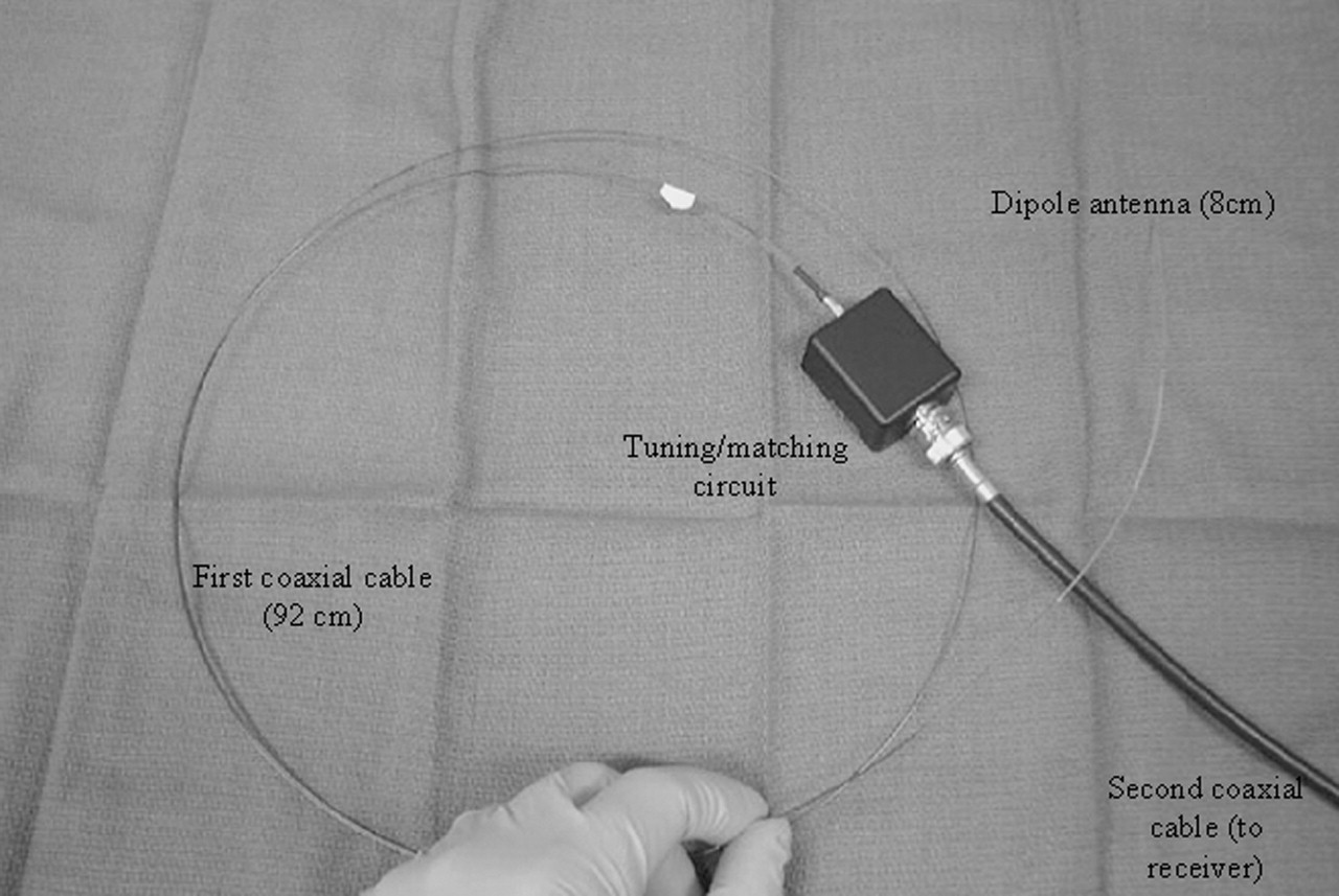
- Fig 7.
The endospinal coil used in this experiment is a 0.032-inch loopless antenna/guidewire consisting of an 8-cm-long dipole antenna connected to a 92-cm first coaxial cable. Tuning and matching circuits are external. Second coaxial cable connects the apparatus to MR receiver by means of an adapter.


Tables
Type NEX TR (ms) TE (ms) Flip Angle TSE Factor Bandwidth (Hz/pixel) Matrix Segmental Image Section Thickness (mm) FOV RFOV SR (mm) TSE 1 4000 100 90 12 128.9 256 ×256 70% 3 6 70% 0.23 ×0.23 TSE 1 4055.8 100 90 14 144.7 256 ×256 70% 5 10 80% 0.39 ×0.39 TSE 1 1150 110 90 35 375.6 256 ×256 90% 3 16 80% 0.62 ×0.62 SSFP 3 3.7 1.85 55 NA 997.8 192 ×256 50% 15 25 75% 0.98 ×0.98 Note.—TSE = turbo spin echo, SSFP = Steady State Free Precession, FOV = field of view, RFOV = reduced field of view, SR = spatial resolution.
TSE 0.62 mm × 0.62 mm SR TSE 0.39 mm × 0.39 mm SR SSFP Surface coil 22.46 13.5 69.23 Endospinal coil 65.89 35.67 132.63 % SNR gain 193.37 164.22 91.58
In this issue


Jump to section
Related Articles
Cited By...
- No citing articles found.