
Article Figures & Data
Figures
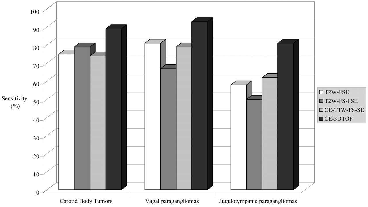
- Fig 1.
Bar graph shows sensitivity (mean for both observers) for detection of carotid body tumors, vagal paragangliomas, and jugulotympanic paragangliomas for each MR imaging technique. T2W-FSE, T2-weighted fast spin-echo imaging; T2W-FS-FSE, T2-weighted fat-suppressed fast spin-echo imaging; CE-T1W-FS-SE, contrast-enhanced T1-weighted fat-suppressed spin-echo imaging; CE-3DTOF, contrast-enhanced 3D time-of-flight imaging.
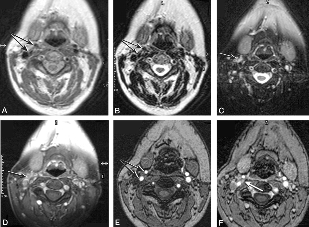
- Fig 2.
Images of a 51-year-old woman with a small right-sided carotid body tumor (confirmed angiographically) and a large left-sided vagal paraganglioma (not shown).
A and B, Axial view dual T2-weighted fast spin-echo image (3750/28/120 [TR/TE1/TE2]) shows only slight splaying of the carotid bifurcation (double arrow).
C, On the axial view T2-weighted fat-suppressed fast spin-echo image (5500/100 [TR/TE]), neither the carotid bifurcation nor a carotid body tumor is visible. High signal intensity in the carotid region is reflecting slow venous flow (arrow).
D, On the axial view contrast-enhanced T1-weighted fat-suppressed spin-echo image (625/17 [TR/TE]), enhancement of veins surrounding the normal left-sided carotid bifurcation (arrow) constrains detection of the small carotid body tumor (arrow).
E, Splaying of the carotid bifurcation (double arrow) can be noticed on the axial view unenhanced 3D time-of-flight MR angiogram (25/6.9/20 [TR/TE/flip angle]).
F, Enhancement of a small carotid body tumor (arrow) is depicted on the axial view contrast-enhanced 3D time-of-flight MR angiogram (25/6.9/20).
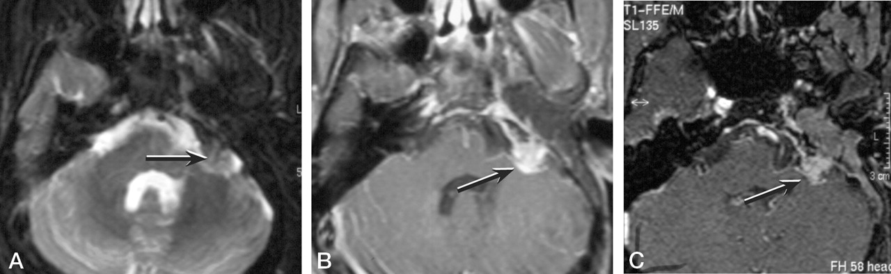
- Fig 3.
Images of a 35-year-old man after surgical resection of a left-sided jugulotympanic paraganglioma with a small residual lesion in the left cerebellopontine angle.
A, Residual paraganglioma in the left cerebellopontine angle is not clearly depicted with the T2-weighted fat-suppressed fast spin-echo sequence (5500/100 [TR/TE]).
B, Contrast-enhanced T1-weighted fat-suppressed spin-echo image (625/17 [TR/TE) clearly shows a small residual paraganglioma (arrow).
C, Contrast-enhanced 3D time-of-flight MR angiogram (25/6.9/20 [TR/TE/flip angle]) clearly shows a small residual paraganglioma (arrow).
- Fig 4.
Unenhanced 3D time-of-flight MR angiogram (25/6.9/20 [TR/TE/flip angle]) of a 39-year-old woman with a left-sided jugulotympanic paraganglioma clearly shows the highly vascular nature of the lesion (arrow).


Tables
TR TE NSA Flip Angle (degrees) Section Thickness (mm) FOV RFOV Matrix Imaging Time T1W-SE 600 20 2 90 5 (gap, 1) 250 70 205 × 256 5:49 T2W-FSE 3750 28/120 3 90 5 (gap, 1) 240 91 177 × 256 4:56 T2W-FS-FSE 5500 100 2 90 5 (gap, 1) 250 91 177 × 256 3:45 CE-T1W-FS-SE 625 17 2 90 5 (gap, 1) 250 70 179 × 256 5:19 3D TOF MRA 25 6.9 1 20 1.5 210 70 196 × 256 5:52 Note.—NSA indicates number of signal averages; FOV, field of view; RFOV, rectangular field of view; T1W-SE, T1-weighted spin-echo; T2W-FSE, dual T2-weighted fast spin-echo; T2W-FS-FSE, T2-weighted fat-suppressed fast spin-echo; CE-T1W-FS-SE, contrast-enhanced T1-weighted fat-suppressed spin-echo; 3D TOF MRA, unenhanced and contrast-enhanced 3D time-of-flight MR angiography.
- TABLE 2:
Data regarding sensitivity, specificity, and negative predictive value [mean (observer 1 − observer 2)]
Sensitivity Fitted Probability* Odds* Specificity NPV T2W-FSE 0.74 (0.64 − 0.83) .735 2.783 0.99 (1.00 − 0.97) 0.86 T2W-FS-FSE 0.70 (0.64 − 0.76) .699 2.333 1.00 (1.00 − 1.00) 0.85 CE-T1 W-FS-SE 0.73 (0.67 − 0.79) .738 2.683 1.00 (1.00 − 1.00) 0.86 3D TOF MRA 0.89 (0.87 − 0.90) .917 7.749 0.99 (0.99 − 0.99) 0.93 * Fitted probability and odds determined with logistic regression model.
Note.—NPV indicates negative predictive value; T2W-FSE, dual T2-weighted fast spin-echo; T2W-FS-FSE, T2-weighted fat-suppressed fast spin-echo; CE-T1W-FS-SE, contrast-enhanced T1-weighted fat-suppressed spin-echo; 3D TOF MRA, unenhanced and contrast-enhanced 3D time-of-flight MR angiography.
- TABLE 3:
Number of detected tumors (mean for observers 1 and 2) in relation to tumor size for each MR imaging technique as compared with criterion standard DSA
0–10 mm 11–20 mm 21–30 mm 31–50 mm ≥51 mm T2W-FSE 1 (0) 11 (3) 10 (2) 22 (17) 8 (8) T2W-FS-FSE 1 (0) 9 (2) 11 (3) 21 (17) 8 (7) CE-T1 W-FS-SE 1 (0) 11 (2) 10 (0) 22 (16) 8 (8) CE-3D TOF 3 (1) 17 (11) 12 (10) 22 (20) 8 (8) DSA 9 19 12 22 8 Note.—T2W-FSE indicates dual T2-weighted fast spin-echo; T2W-FS-FSE, T2-weighted fat-suppressed fast spin-echo; CE-T1 W-FS-SE, contrast-enhanced T1-weighted fat-suppressed spin-echo; 3D TOF MRA, unenhanced and contrast-enhanced 3D time-of-flight MR angiography; DSA, digital subtraction angiography. Data in parenthesis represent presence of intra-tumoral flow for each subgroup and technique.


