Advertisement

Main menu

  • Home
  • Content
    • Current Issue
    • Accepted Manuscripts
    • Article Preview
    • Past Issue Archive
    • AJNR Case Collection
    • Case of the Week Archive
    • Classic Case Archive
    • Case of the Month Archive
  • Special Collections
    • Spinal CSF Leak Articles (Jan 2020-June 2024)
    • 2024 AJNR Journal Awards
    • Most Impactful AJNR Articles
  • Multimedia
    • AJNR Podcast
    • AJNR Scantastics
    • Video Articles
  • For Authors
    • Submit a Manuscript
    • Author Policies
    • Fast publishing of Accepted Manuscripts
    • Graphical Abstract Preparation
    • Manuscript Submission Guidelines
    • Imaging Protocol Submission
    • Submit a Case for the Case Collection
  • About Us
    • About AJNR
    • Editorial Board
  • More
    • Become a Reviewer/Academy of Reviewers
    • Subscribers
    • Permissions
    • Alerts
    • Feedback
    • Advertisers
    • ASNR Home
  • Other Publications
    • ajnr

User menu

  • Alerts
  • Log in

Search

  • Advanced search
American Journal of Neuroradiology

American Journal of Neuroradiology

ASHNR American Society of Functional Neuroradiology ASHNR American Society of Pediatric Neuroradiology ASSR
  • Alerts
  • Log in

Advanced Search

  • Home
  • Content
    • Current Issue
    • Accepted Manuscripts
    • Article Preview
    • Past Issue Archive
    • AJNR Case Collection
    • Case of the Week Archive
    • Classic Case Archive
    • Case of the Month Archive
  • Special Collections
    • Spinal CSF Leak Articles (Jan 2020-June 2024)
    • 2024 AJNR Journal Awards
    • Most Impactful AJNR Articles
  • Multimedia
    • AJNR Podcast
    • AJNR Scantastics
    • Video Articles
  • For Authors
    • Submit a Manuscript
    • Author Policies
    • Fast publishing of Accepted Manuscripts
    • Graphical Abstract Preparation
    • Manuscript Submission Guidelines
    • Imaging Protocol Submission
    • Submit a Case for the Case Collection
  • About Us
    • About AJNR
    • Editorial Board
  • More
    • Become a Reviewer/Academy of Reviewers
    • Subscribers
    • Permissions
    • Alerts
    • Feedback
    • Advertisers
    • ASNR Home
  • Follow AJNR on Twitter
  • Visit AJNR on Facebook
  • Follow AJNR on Instagram
  • Join AJNR on LinkedIn
  • RSS Feeds

Welcome to the new AJNR, Updated Hall of Fame, and more. Read the full announcements.


AJNR is seeking candidates for the position of Associate Section Editor, AJNR Case Collection. Read the full announcement.


OtherHEAD AND NECK

Spontaneous Sphenoid Sinus Mucocele Revealed by Meningitis and Brain Abscess in a 12-Year-Old Child

Olivier Malard, Marie Gayet-Delacroix, Franck Jegoux, Alexis Faure, Philippe Bordure and Claude Beauvillain de Montreuil
American Journal of Neuroradiology May 2004, 25 (5) 873-875;
Olivier Malard
  • Find this author on Google Scholar
  • Find this author on PubMed
  • Search for this author on this site
Marie Gayet-Delacroix
  • Find this author on Google Scholar
  • Find this author on PubMed
  • Search for this author on this site
Franck Jegoux
  • Find this author on Google Scholar
  • Find this author on PubMed
  • Search for this author on this site
Alexis Faure
  • Find this author on Google Scholar
  • Find this author on PubMed
  • Search for this author on this site
Philippe Bordure
  • Find this author on Google Scholar
  • Find this author on PubMed
  • Search for this author on this site
Claude Beauvillain de Montreuil
  • Find this author on Google Scholar
  • Find this author on PubMed
  • Search for this author on this site
  • Article
  • Figures & Data
  • Info & Metrics
  • Responses
  • References
  • PDF
Loading

Abstract

Summary: Sphenoid sinus mucocele is an uncommon lesion related to inflammatory disease that is diagnosed after surgery or a traumatic event. This report describes an unusual case revealed by bacterial meningitis and cerebral abscess in a 12-year-old child. CT and MR imaging allowed precise extension to the skull base in preoperative management and follow-up investigations. Endoscopic transnasal marsupialisation of the mucocele and antibiotic therapy led to complete remission. There was no evidence of recurrence after 6 months, which suggests that sphenoid mucoceles, regardless of size and complications, can be treated by endoscopic sinus surgery.

Sphenoid sinus mucocele is rare (1, 2) and generally caused by direct trauma (3). Patients with these lesions present with various symptoms and signs that may include nasal problems (4) and cranial nerve palsy (5–7). Few cases have been reported in children (8, 9), and the spontaneous case described here appears to be the first diagnosed in a child in conjunction with an infectious brain complication. Since the introduction of endoscopic techniques and instruments, the efficiency of the transnasal route has been evaluated for sphenoid mucocele removal (10, 11). There is increasing evidence in the literature that endoscopic surgery is safe and has a very low rate of recurrence. In our opinion, the transnasal approach is valid even for the management of complicated sphenoid mucocele.

Case Reports

A 12-year-old boy seen in the pediatric emergency department of the Nantes Teaching Hospital complained of severe diffuse headache and persistent fever. He had no history of cranial traumatism, sinus surgery, or chronic sinusitis. Clinical examination revealed typical acute meningitis but no neurologic palsy or ocular complications. CSF assessment confirmed a bacterial pneumococcus–related infection. Treatment was initiated in the emergency department (injection of amoxicillin 200 mg/kg/day). CT and MR imaging revealed a sphenoid mucocele eroding adjacent bone in the roof of the sinus, associated with a 2-cm-diameter brain abscess diameter situated in the right inferior frontal area (Figs 1 and 2).

Fig 1.
  • Download figure
  • Open in new tab
  • Download powerpoint
Fig 1.

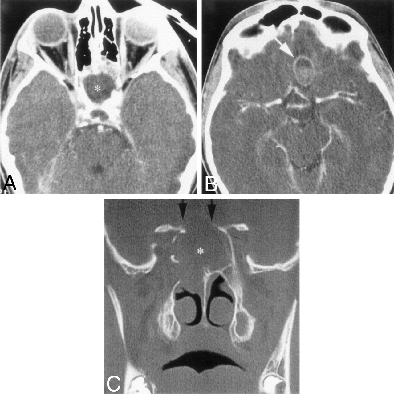
Preoperative CT scans

A, Axial scan showing abnormal opacification of the sphenoid sinus (asterisk [*]).

B, Axial scan showing a right frontal brain abscess (arrow) up to the sphenoid sinus.

C, Coronal scan revealing the sphenoid sinus mucocele (asterisk [*]) with osseous erosion of the sinus roof (arrows).

Fig 2.
  • Download figure
  • Open in new tab
  • Download powerpoint
Fig 2.

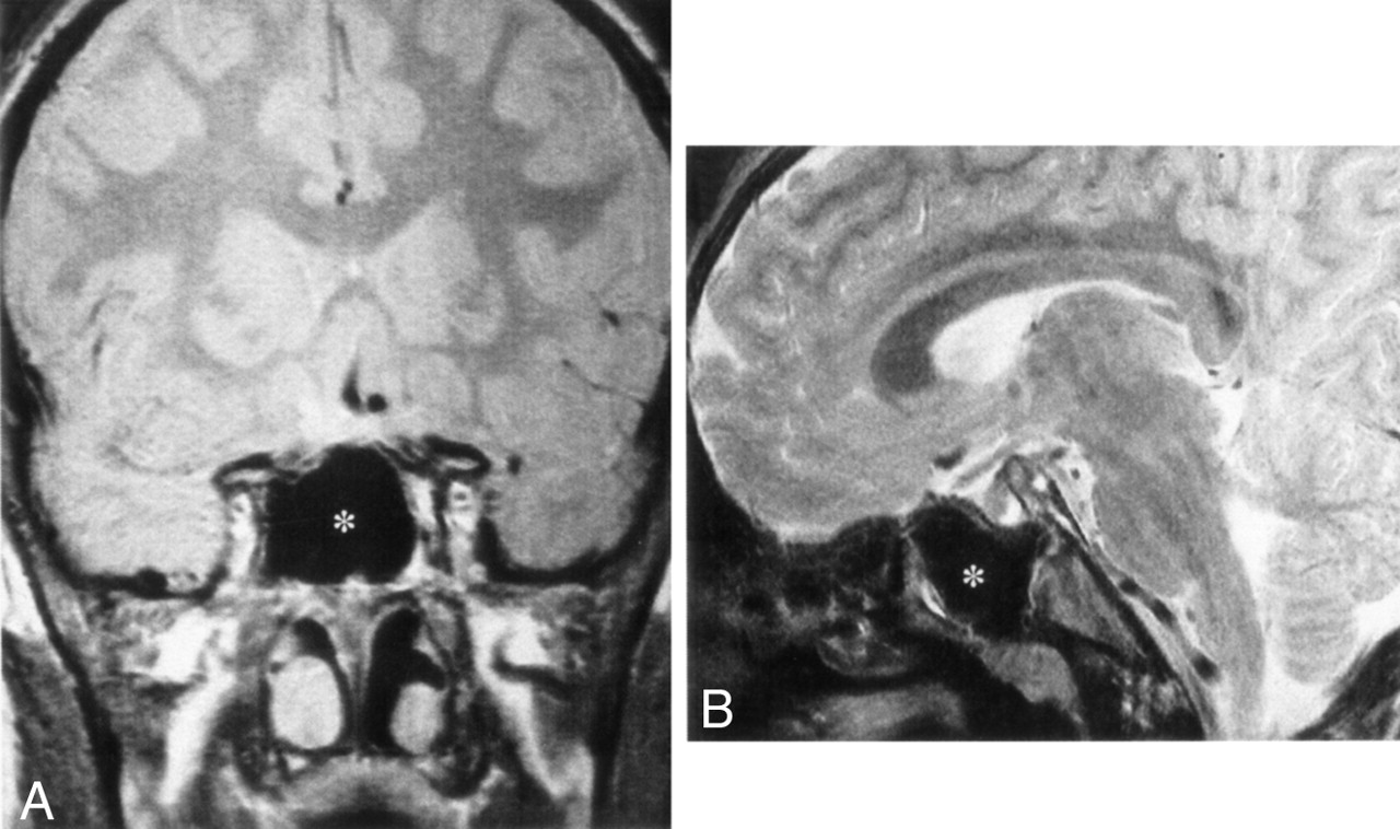
Preoperative T1-weighted MR images.

A, Coronal MR image showing abnormal opacification of the sphenoid sinus (asterisk [*]).

B, Right parasagittal MR image showing the sphenoid sinus mucocele (asterisk [*]) and the adjacent frontal brain abscess (arrow).

Surgical drainage was performed under general anesthesia 48 hours after antibiotic induction. The patient underwent a bilateral endoscopic procedure by the transnasal route, with video monitoring. A huge mucocele was easily located, emerging from the right sphenoid anterior wall above the choana. Bacteriologic analysis of the pus in the mucocele showed numerous leukocytes, but no bacteria. Marsupialisation was gradually achieved after endoscopic instruments were used to enlarge the sphenoid sinus. Complete removal of the mucocele produced a large opening of the rostrum. Bone erosion in the roof of the sinus had allowed the dura mater to enter the sphenoid sinus cavity (Fig 3). There was, however, no interruption of the dura mater and no CSF leakage. At the end of marsupialisation, a Merocel sponge (Medtronic Xomed, Saint Aubin Le Monial, France) was placed in the nasal cavity for 24 hours to prevent bleeding.

Fig 3.
  • Download figure
  • Open in new tab
  • Download powerpoint
Fig 3.

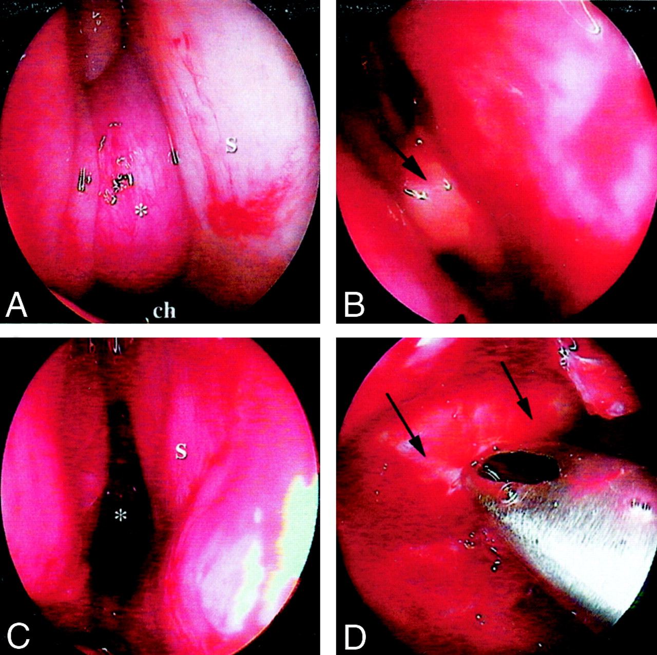
Endoscopic perioperative management.

A, Endoscopic view (0°) by a right transnasal route showing the mucocele (asterisk [*]) up from the choana (ch) and laterally from the nasal septum (s).

B, Endoscopic view (0°) after opening of the mucocele, showing a huge pus discharge (arrow).

C, After marsupialisation of the mucocele, 0° endoscopic view of the right sphenoid sinus (asterisk [*]) lateral and posterior to the nasal septum (s).

D, Endoscopic view (60°) inside the left sphenoid cavity, showing bone erosion of the sinus roof. Dura mater, inspected directly by endoscopy, was not ruptured (arrow).

The patient had no postoperative complications. Because of its depth, the brain abscess was not surgically treated first. The patient’s course was rapidly satisfactory under antibiotics. After 10 days, injections were replaced by oral administration of 6 g/day of amoxicillin, and the patient was allowed to return home. Antibiotics were maintained for 10 weeks, until CT and MR control imaging showed complete extinction of the brain abscess. No signs of recurrence were observed after 6 months of follow-up (Figs 4 and 5).

Fig 4.
  • Download figure
  • Open in new tab
  • Download powerpoint
Fig 4.

Postoperative axial (A) and coronal (B) CT scans 4 months after marsupialisation, showing normal opening and ventilation of the sphenoid (asterisk [*]). There is no evidence of recurrence of the mucocele and no outstanding abscess.

Fig 5.
  • Download figure
  • Open in new tab
  • Download powerpoint
Fig 5.

Preoperative coronal (A) and sagittal (B) T1-weighted MR images showing normal ventilation of the sphenoid sinus (asterisk [*]) and complete abscess extinction.

Discussion

Trauma and chronic inflammation (polypoid sinusitis) are frequent causes of mucocele (3). In addition, many of the mucoceles are simply related to inflammatory disease, without accidental or postsurgical evidence; to our knowledge, mechanisms of expansion have not been elucidated in literature. The development of a sphenoid mucocele in a child has been reported in only a small number of articles (6, 9, 12).

To our knowledge, this clinical case is the first report of sphenoid mucocele in a child revealed by meningitis and a brain abscess. Because sphenoid sinus mucoceles represent only about 1% of all paranasal mucoceles (11), it is difficult to determine their standard clinical features. Pain (vertex or occipital headache) seems to be the most common symptom (1), and eye-threatening complications often reveal the lesion.

Accurate imaging is required to define the precise extension and boundaries of the mucocele. Both CT and MR imaging are recommended before a surgical approach (13). Mucoceles can show various imaging features, depending on the contents (especially water). When the water rate is high, the space occupied by the lesion is hypoattenuated at CT. The sinus wall may be very thin because of constant and prolonged pressure and be conducive to extensive destructive growth on surrounding components. Sinus cavities are hypointense on T1-weighted MR images and hyperintense on T2-weighted MR images but without enhancement. Because of a large increase in the protein rate, a hyperintense signal can be observed on T1-weighted MR images. Thus, in some cases, images can be very difficult to analyze.

An external surgical approach was used in the past for mucocele management, but an exclusively endoscopic transnasal route has become the standard and generally uncontested treatment (4, 11). Some authors, however, have suggested using an endoscopic technique combined with a navigating system (14). In our opinion, the transnasal approach is valid even for the management of complicated sphenoid mucoceles regardless of their size and preoperative complications. This type of treatment is clearly radical, postoperative care is reduced, and sequelae are quite limited.

References

  1. ↵
    Stankiewicz JA. Sphenoid sinus mucocele. Arch Otolaryngol Head Neck Surg 1989;115:735–740
    CrossRefPubMed
  2. ↵
    Sugiura N, Ochi K, Komatsuzaki Y, Koizuka I. Powered endoscopic marsupialization for recurrent sphenoid sinus mucocele: a case report. Auris Nasus Larynx 2003;30(Suppl):S107–S110
  3. ↵
    Iqbal J, Kanaan I, Ahmed M, al Homsi M. Neurosurgical aspects of sphenoid sinus mucocele. Br J Neurosurg 1998;12:527–530
    CrossRefPubMed
  4. ↵
    Hadar T, Shvero J, Nageris B, Yaniv E. The endoscopic approach to isolated sphenoid sinus disease. Harefuah 2002;141:670–672, 764
    PubMed
  5. ↵
    Shikowitz MJ, Goldstein MN, Stegnjajic A. Sphenoid sinus mucocele masquerading as a skull base malignancy. Laryngoscope 1986;96:1405–1410
    PubMed
  6. ↵
    Casteels I, De Loof E, Brock P, et al. Sudden blindness in a child: presenting symptom of a sphenoid sinus mucocele. Br J Ophthalmol 1992;76:502–504
    Abstract/FREE Full Text
  7. ↵
    Bauer B, Evers S, Lindorfer HW, et al. Headache caused by a sphenoid mucocele but presenting as an ergotamine-induced headache. Headache 1997;37:460–462
    CrossRefPubMed
  8. ↵
    Nicolino MP, LeBrigand B, Langevin L, et al. Radiological case of the month: mucocele of the sphenoid sinus. Arch Pediatr Adolesc Med 1997;151:93–94
    CrossRefPubMed
  9. ↵
    Froehlich P, Remond J, Morgon A. Mucocele of the sphenoid sinus in a child. Ann Otol Rhinol Laryngol 1995;104:738–740
    PubMed
  10. ↵
    Har-el G. Telescopic extracranial approach to frontal mucoceles with intracranial extension. J Otolaryngol 1995;24:98–101
    PubMed
  11. ↵
    Lund V. Endoscopic management of paranasal sinus mucoceles. J Laryngol Otol 1998;112:36–40
    PubMed
  12. ↵
    Uren BA, Berkowitz RG. Isolated inflammatory sphenoid sinus disease in children. Ann Otol Rhinol Laryngol 2003;112:370–372
    Abstract/FREE Full Text
  13. ↵
    Sievers KW, Greess H, Baum U, et al. Paranasal sinuses and nasopharynx CT and MRI. Eur J Radiol 2000;33:185–202
    CrossRefPubMed
  14. ↵
    Nagatani T, Saito K, Yoshida J. Treatment of a sphenoid sinus mucocele using an endoscope combined with a navigating system: a case report. J Clin Neurosci 2001;8:456–460
    CrossRefPubMed
  • Received May 21, 2003.
  • Accepted after revision August 26, 2003.
  • Copyright © American Society of Neuroradiology
View Abstract
PreviousNext
Back to top

In this issue

American Journal of Neuroradiology: 25 (5)
American Journal of Neuroradiology
Vol. 25, Issue 5
1 May 2004
  • Table of Contents
  • Index by author
Advertisement
Print
Download PDF
Email Article

Thank you for your interest in spreading the word on American Journal of Neuroradiology.

NOTE: We only request your email address so that the person you are recommending the page to knows that you wanted them to see it, and that it is not junk mail. We do not capture any email address.

Enter multiple addresses on separate lines or separate them with commas.
Spontaneous Sphenoid Sinus Mucocele Revealed by Meningitis and Brain Abscess in a 12-Year-Old Child
(Your Name) has sent you a message from American Journal of Neuroradiology
(Your Name) thought you would like to see the American Journal of Neuroradiology web site.
CAPTCHA
This question is for testing whether or not you are a human visitor and to prevent automated spam submissions.
Cite this article
Olivier Malard, Marie Gayet-Delacroix, Franck Jegoux, Alexis Faure, Philippe Bordure, Claude Beauvillain de Montreuil
Spontaneous Sphenoid Sinus Mucocele Revealed by Meningitis and Brain Abscess in a 12-Year-Old Child
American Journal of Neuroradiology May 2004, 25 (5) 873-875;

Citation Manager Formats

  • BibTeX
  • Bookends
  • EasyBib
  • EndNote (tagged)
  • EndNote 8 (xml)
  • Medlars
  • Mendeley
  • Papers
  • RefWorks Tagged
  • Ref Manager
  • RIS
  • Zotero
0 Responses
Respond to this article
Share
Bookmark this article
Spontaneous Sphenoid Sinus Mucocele Revealed by Meningitis and Brain Abscess in a 12-Year-Old Child
Olivier Malard, Marie Gayet-Delacroix, Franck Jegoux, Alexis Faure, Philippe Bordure, Claude Beauvillain de Montreuil
American Journal of Neuroradiology May 2004, 25 (5) 873-875;
del.icio.us logo Twitter logo Facebook logo Mendeley logo
  • Tweet Widget
  • Facebook Like
  • Google Plus One

Jump to section

  • Article
    • Abstract
    • Case Reports
    • Discussion
    • References
  • Figures & Data
  • Info & Metrics
  • Responses
  • References
  • PDF

Related Articles

  • No related articles found.
  • PubMed
  • Google Scholar

Cited By...

  • No citing articles found.
  • Crossref
  • Google Scholar

This article has not yet been cited by articles in journals that are participating in Crossref Cited-by Linking.

More in this TOC Section

  • Correlation of Apparent Diffusion Coefficient at 3T with Prognostic Parameters of Retinoblastoma
  • Parathyroid Lesions: Characterization with Dual-Phase Arterial and Venous Enhanced CT of the Neck
  • Efficacy of Diffusion-Weighted Imaging for the Differentiation between Lymphomas and Carcinomas of the Nasopharynx and Oropharynx: Correlations of Apparent Diffusion Coefficients and Histologic Features
Show more Head and Neck

Similar Articles

Advertisement

Indexed Content

  • Current Issue
  • Accepted Manuscripts
  • Article Preview
  • Past Issues
  • Editorials
  • Editors Choice
  • Fellow Journal Club
  • Letters to the Editor

Cases

  • Case Collection
  • Archive - Case of the Week
  • Archive - Case of the Month
  • Archive - Classic Case

Special Collections

  • Special Collections

Resources

  • News and Updates
  • Turn around Times
  • Submit a Manuscript
  • Author Policies
  • Manuscript Submission Guidelines
  • Evidence-Based Medicine Level Guide
  • Publishing Checklists
  • Graphical Abstract Preparation
  • Imaging Protocol Submission
  • Submit a Case
  • Become a Reviewer/Academy of Reviewers
  • Get Peer Review Credit from Publons

Multimedia

  • AJNR Podcast
  • AJNR SCANtastic
  • Video Articles

About Us

  • About AJNR
  • Editorial Board
  • Not an AJNR Subscriber? Join Now
  • Alerts
  • Feedback
  • Advertise with us
  • Librarian Resources
  • Permissions
  • Terms and Conditions

American Society of Neuroradiology

  • Not an ASNR Member? Join Now

© 2025 by the American Society of Neuroradiology All rights, including for text and data mining, AI training, and similar technologies, are reserved.
Print ISSN: 0195-6108 Online ISSN: 1936-959X

Powered by HighWire