
Article Figures & Data
Figures
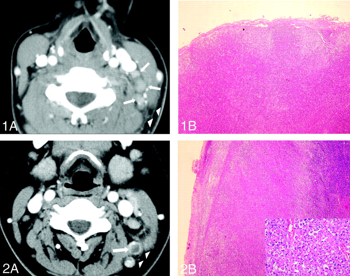
- Fig 1.
Images obtained in a 28-year-old woman presenting with a 2-week history of fever, myalgia, and tender cervical lymphadenopathy.
A, CT scan shows multiple small and medium lymph nodes (arrows) on the left side of the neck (level III). Note the obliteration of perinodal fat at levels III and V and in the adjacent superficial space (arrowheads).
B, Photomicrograph of a surgical specimen from a lymph node shows patchy areas of lymphohistiocytic infiltration that does not distort the otherwise normal lymph node architecture (hematoxylin-eosin stain, original magnification ×40).
- Fig 2.
Images obtained in a 34-year-old woman.
A, CT scan shows small lymph nodes on the left at levels IIB and V. Note the ring-shaped nodal necrosis (arrow) mimicking that of tuberculosis or metastatic lymphadenopathy. Adjacent perinodal infiltration is also seen (arrowheads).
B, Photomicrograph of surgical specimen of lymph node shows a geographic pattern of eosinophilic necrosis and sheets of histiocytes (hematoxylin-eosin stain, original magnification ×40). Inset, High-powered view of a necrotic focus shows abundant karyorrhectic debris (arrows) and crescent-shaped histiocytes (original magnification ×400).


Tables
Feature Value No. of patients 96 Sex M:F 28:68 Ratio 1:2.43 Age, y Mean 12–40 Range 24.4 Leukopenia 30 (31.3%) Fever (temperature >37.5°C) 56 (58.3%) Symptom duration 3 days to 12 weeks Finding Value No. of lymph nodes Total 1196 Range 1–30 No. per patient (n = 96) 12.5 Maximum diameter, cm Range 0.5–3.5 Mean 1.62 Perinodal infiltration 78 (81.3%) Homogeneous contrast enhancement 80 (83.3%) Necrosis Focal 9 (9.4%) Wide or ring-shaped 7 (7.3%) Location Number Neck lymphadenopathy (n = 96) Bilateral 20 (20.8%) Right 34 (35.4%) Left 42 (43.8%) Level of lymph nodes (n = 1194) IA + IB 26 + 34 IIA + IIB 174 + 254 III 222 IV 160 VA + VB 126+ 130 Other* 70 * Level VI, level VII, supraclavicular, parotid, and other superficial nodes.


