
Article Figures & Data
Figures
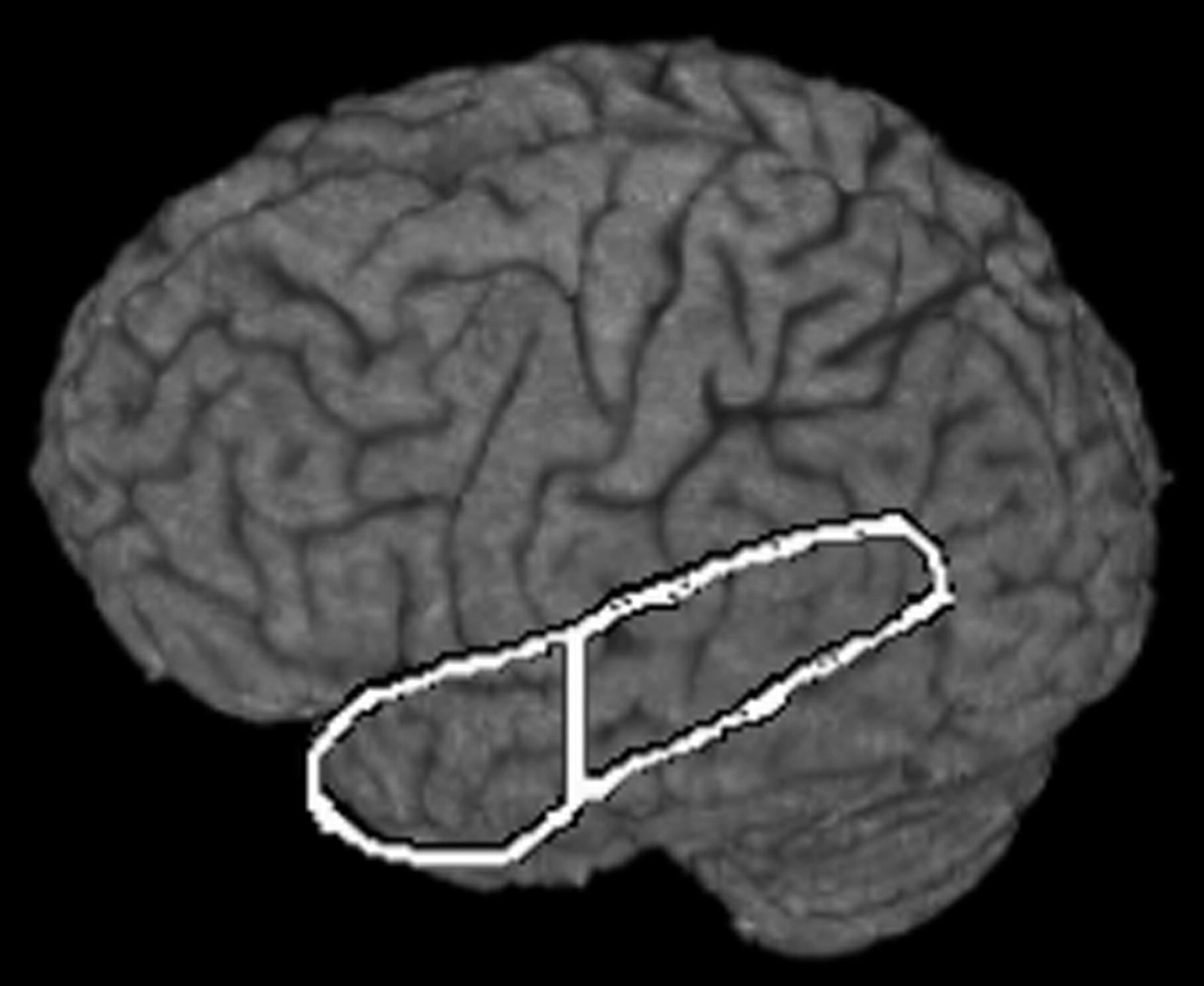
- Fig 1.
Temporal regions of interest.
Vertical line represents Talairach coordinate y = −20, which delineates the anterior region of interest from the posterior region of interest.
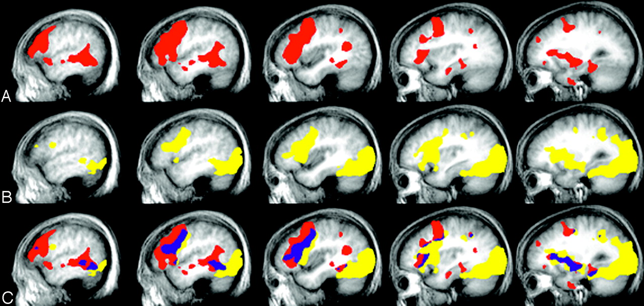
- Fig 2.
Group activation, left hemisphere.
A, Responsive naming; B, confrontation naming; C, overlap: yellow, confrontation naming; red, responsive naming; blue, overlap.
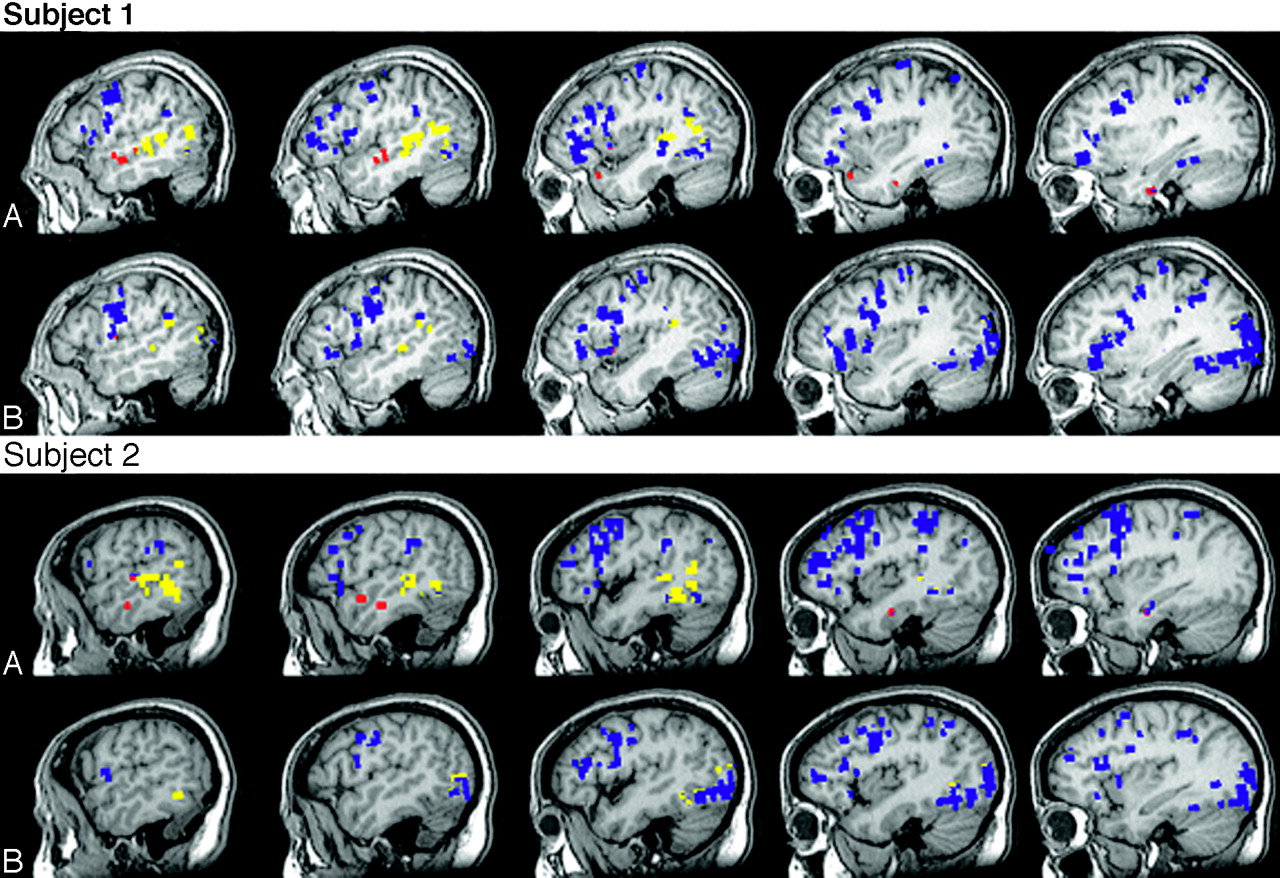
- Fig 3.
Individual activation, left hemisphere.
Activation patterns for the 2 subjects who participated in each experiment for (A) responsive naming and (B) confrontation naming. Blue, activation external to the temporal regions of interest; yellow, activation within the posterior temporal region of interest; and red, activation within the anterior temporal region of interest.


Tables
Cortex Responsive naming Confrontation naming BA x y z z score BA x y z z score Superior temporal 38/22 −50 5 −10 3.5 Middle temporal 21/22 −48 −12 −6 3.9 Middle temporal 21/22/39 −50 −41 −1 5.3 22 −50 −42 −1 4.0 Inferior temporal 20 −27 1 −33 3.0 21 −59 −27 −14 3.1 Responsive naming > Confrontation naming Cortex BA x y z z score Middle superior temporal 22/39 −41 −55 19 4.0 Middle temp/fusiform 20/21 −48 −4 −19 3.9 Superior temporal 22 −60 −42 6 2.8 Inferior temporal 20 −32 −1 −38 2.9 Note.—BA indicates Brodmann area.
There was no significant activation within the temporal regions of interest for confrontation naming compared to responsive naming.
Responsive Naming Confrontation Naming Patient AnteriorTemporal Posterior Temporal Patient Anterior Temporal Posterior Temporal 1* 768 4581 1* 99 1004 2* 1124 2950 2* 0 927 3 518 3479 3 213 1107 4 1117 2280 4 1269 3958 5 628 3939 5 0 870 6 839 2444 6 0 707 7 0 1616 7 0 3328 8 278 492 8 323 1246 9 390 4578 9 11 926 10 111 979 10 108 365 Avg 577.4 2733.8 202.3 1443.8 SD 391.1 1434.2 390.5 1192.0 * Participated in both experiments.


