
Article Figures & Data
Figures
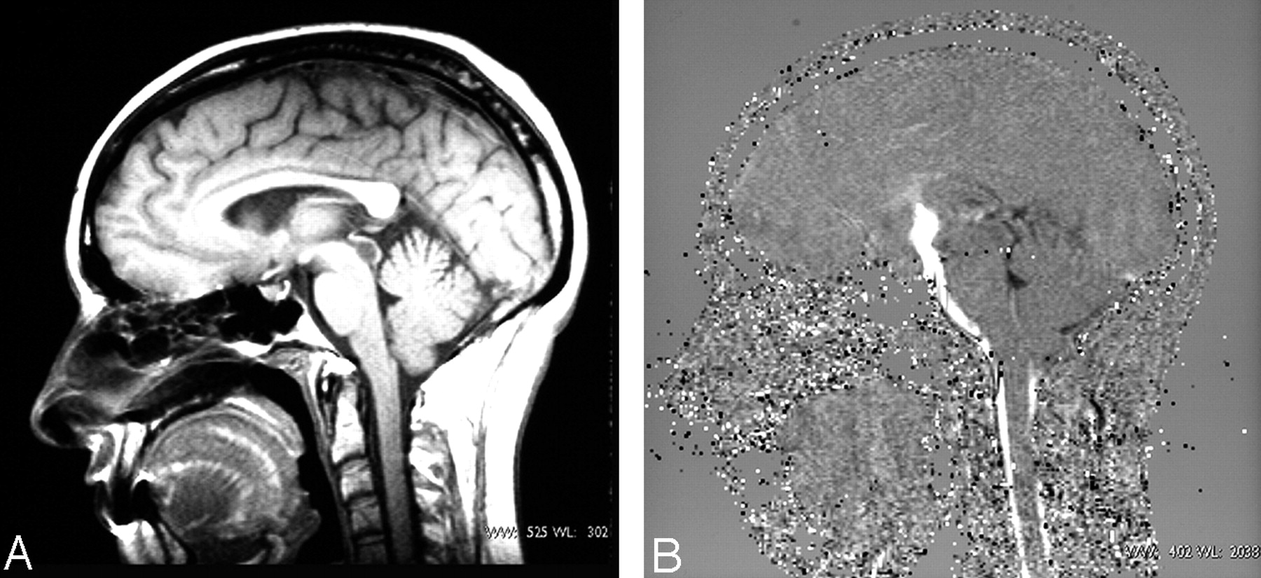
- Fig 1.
A, Sagittal T1-weighted image located in the midsagittal plane shows a cystic lesion inside the aqueduct. B, Sagittal cine phase-contrast image after ventriculostomy shows flow signal intensity between third ventricle and pontine cistern.
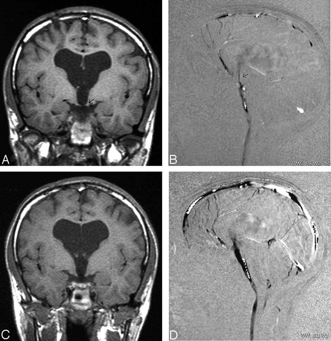
- Fig 2.
An 11-year-old boy with aqueduct stenosis and previous ventriculostomy who had clinical deterioration. A, Coronal T1-weighted image centering in mammillary bodies shows a small defect in the floor of third ventricle (arrow). B, Sagittal cine phase contrast in the ventriculostomy site demonstrates a filiform flow signal intensity (arrow). C and D, Coronal T1-weighted image and sagittal cine phase contrast after new ventriculostomy demonstrate a big defect in the floor of third ventricle and excellent flow passing through the ventriculostomy.
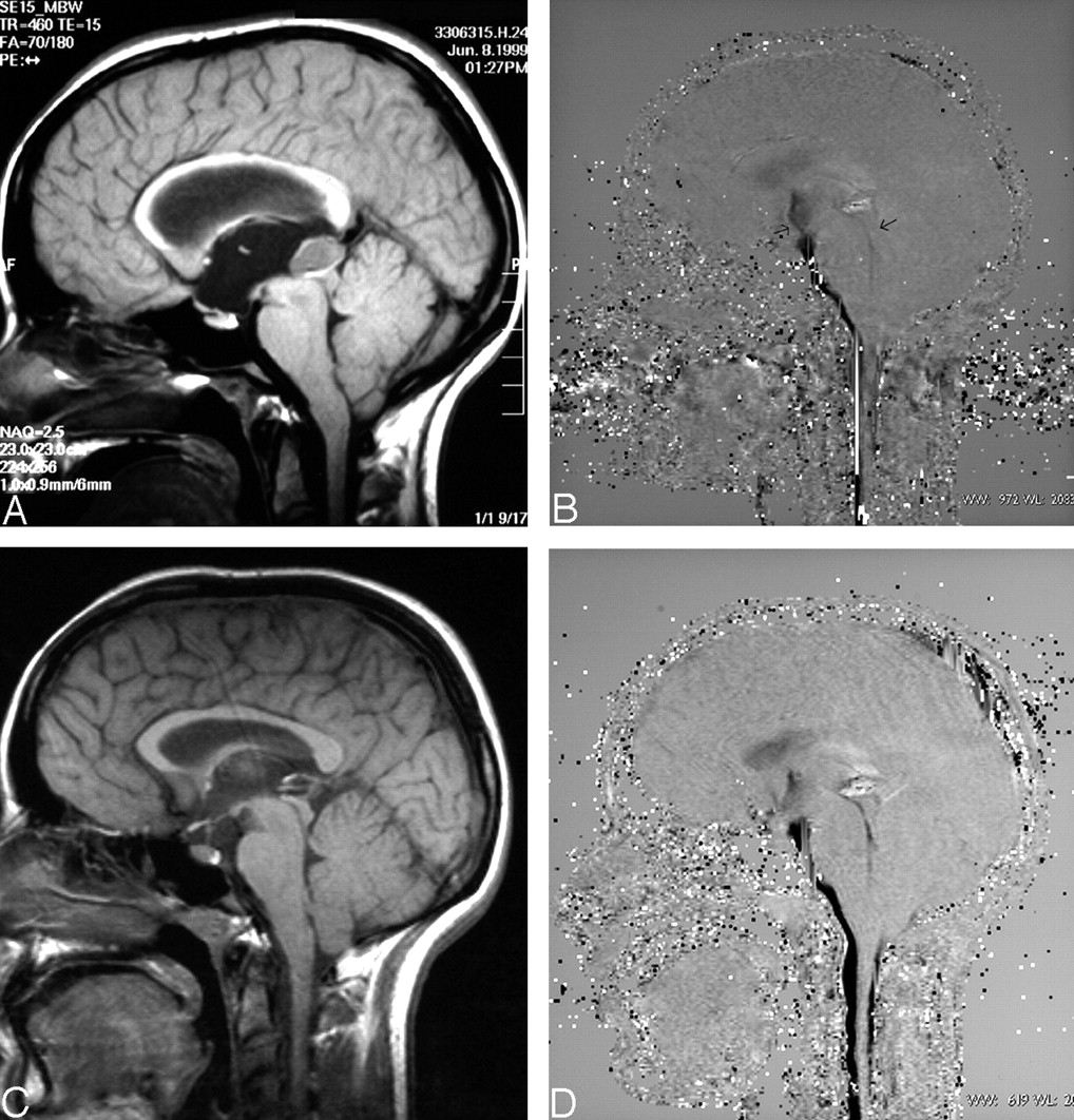
- Fig 3.
Patients with secondary aqueduct stenosis induced by pineal lesion. A, Sagittal T1-weighted image demonstrated a cystic lesion in the pineal region blocking the aqueduct and producing hydrocephalus. B, Sagittal cine phase-contrast in the ventriculostomy site performed shortly after surgery and treatment of pineal lesion demonstrates a filiform flow signal intensity in the ventriculostomy and subtle flow signal intensity in the aqueduct (arrows). C, Sagittal T1-weighted image obtained long after treatment demonstrates reduction of pineal lesion and aqueduct decompression. D, Sagittal cine phase-contrast performed in the same examination shows absence of flow in the ventriculostomy and aqueduct permeability.
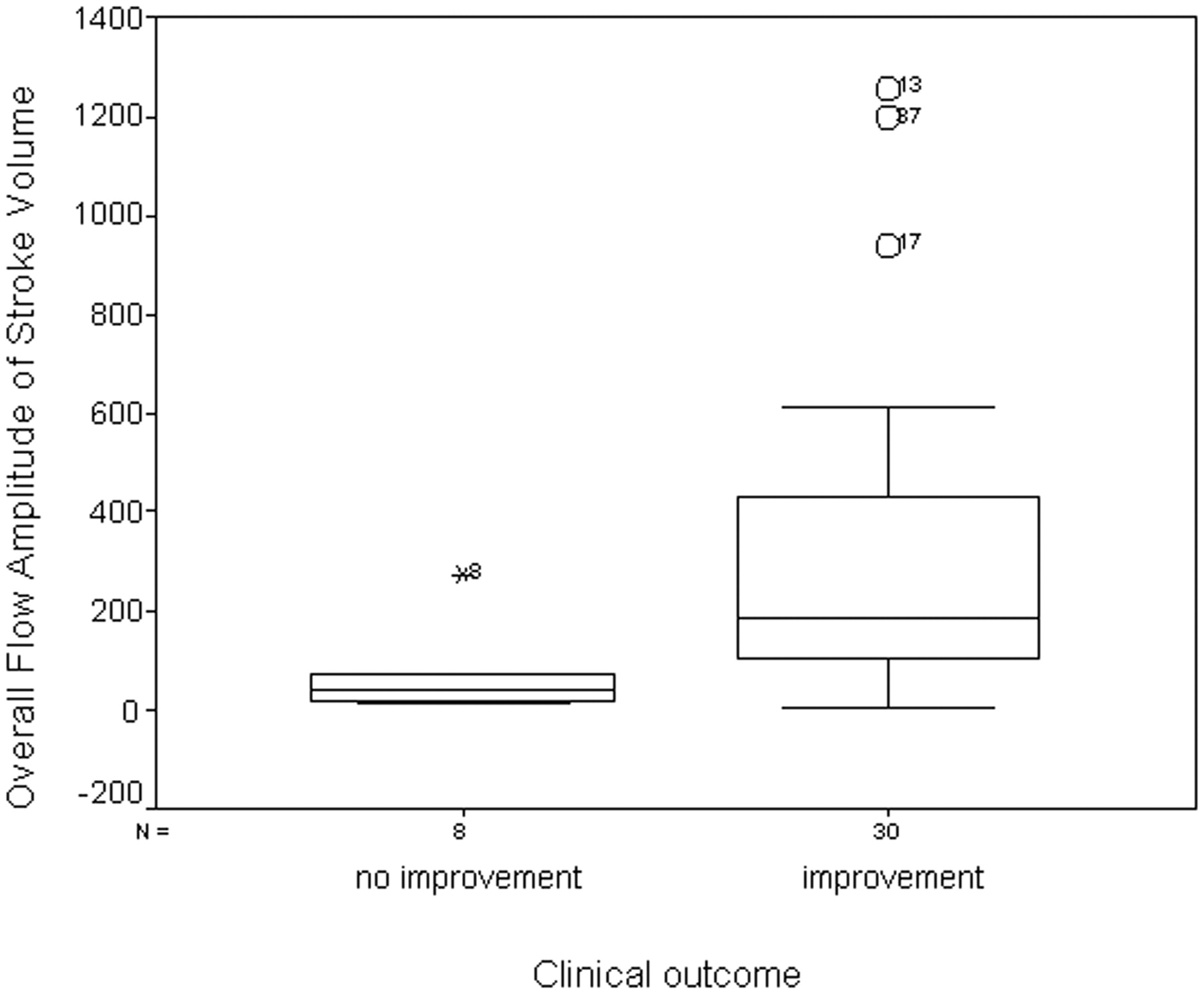
- Fig 4.
Box plot of the OFA of stroke volume related to clinical outcome. Patients with clinical improvement after surgery showed higher stoke volume than patients without clinical improvement.
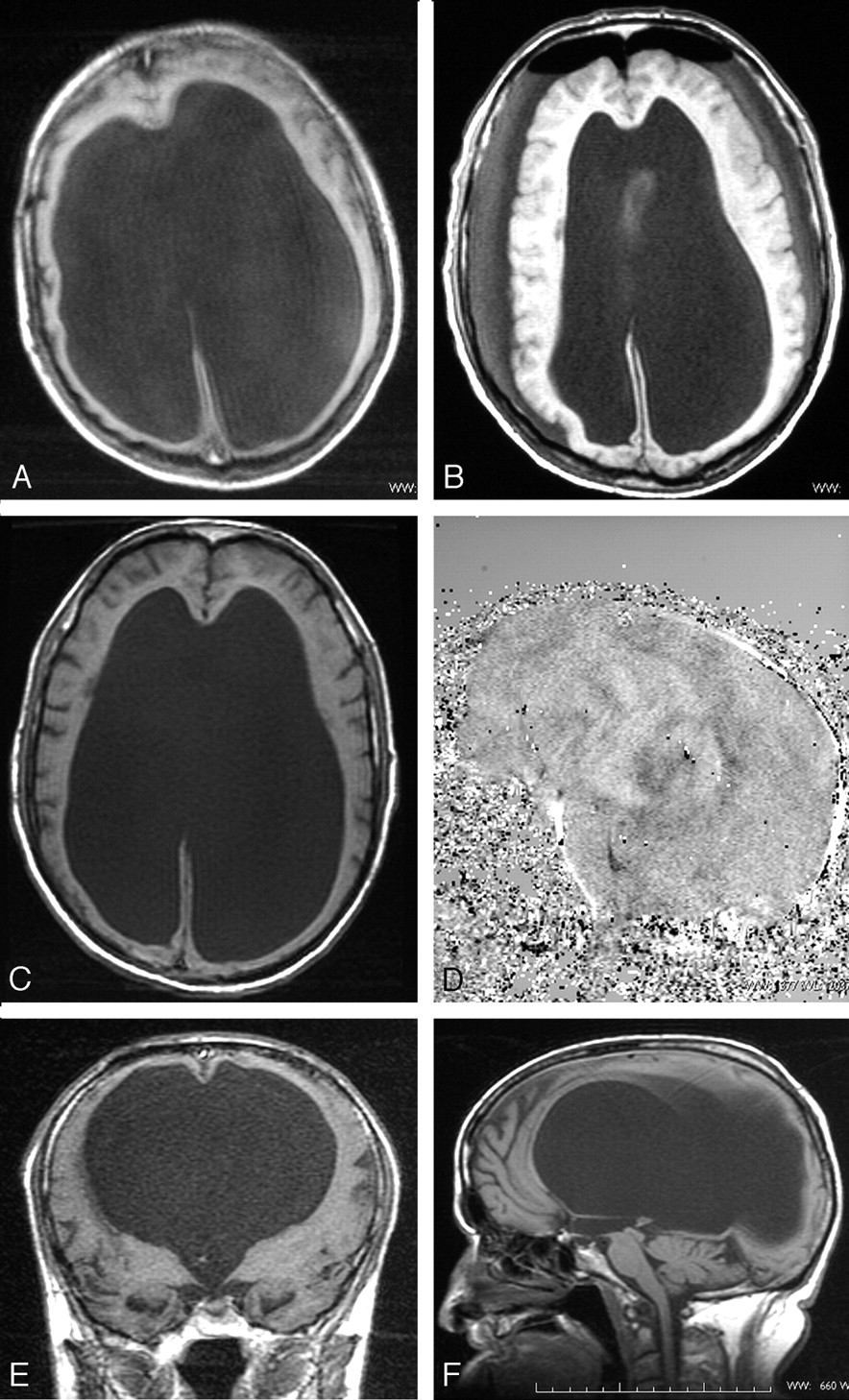
- Fig 5.
Patient with congenital triventricular hydrocephalus. A, Presurgery axial T1-weighted image at the level of ventricular bodies demonstrated an enlarged ventricular size. B, Immediately postsurgery, axial T1-weighted image at the same level shows reduction of ventricular size and 2 subdural collections. Pneumoencephalus was also present. C and D, Axial T1-weighted image, 6 months after surgery, shows that the ventricular bodies regressed to initial size, and sagittal phase-contrast image demonstrates absence of flow in the ventriculostomy. E and F, Coronal and sagittal T1-weighted image, 6 months after surgery, at the level of mammillary bodies demonstrates the persistence of third floor defect.


Tables
Ventricular Size Changes Clinical Outcome NoImprovement(n = 6; 15.8%) PartialImprovement(n = 17; 44.7%) GoodOutcome(n = 13; 34.2%) Clinical Worsening(n = 2; 5.3%) No reduction(n = 23; 60.5%) 5 (13.2%) 8 (21.1%) 8 (21.1%) 2 (5.3%) Reduction(n = 15; 39.5%) 1 (2.6%) 9 (23.7%) 5 (13.2) Clinical Outcome Hydrocephalus PAS(n = 11; 28.9%) SAS(n = 18; 47.4%) AC(n = 6; 15.8%) CH(n = 3; 7.9%) No improvement(n = 6; 15.8%) 1 (2.6%) 4 (10.5%) 1 (2.6%) Partial Improvement(n = 17; 44.7%) 3 (7.9%) 12 (31.6%) 1 (2.6%) 1 (2.6%) Good outcome(n = 13; 34.2%) 7 (18.4%) 5 (13.2%) 1 (2.6%) Clinical worsening(n = 2; 5.3%) 1 (2.6%) 1 (2.6%) Note.—PAS indicates primary aqueduct stenosis; SAS, secondary aqueduct stenosis; AC, Arnold Chiari malformation; CH, communicating hydrocephalus.


