
Article Figures & Data
Figures
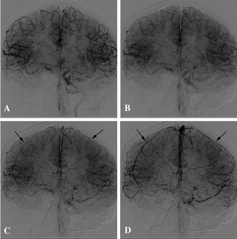
- Fig 1.
Angiogram AP view demonstrates a right carotid occlusion test in a 42-year-old woman with large ICA aneurysm. A and B, Late arterial phase demonstrates excellent cross-filling by Acom. C, Early venous phase—1 second after second image—shows the beginning of venous phase in both sides (black arrows). D, One second later, the venous phase synchronous becomes more evident in both hemispheres (black arrows). This patient represents an example of perfect symmetry of venous phase delay during BTO.
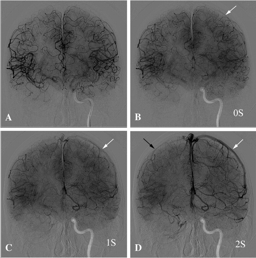
- Fig 2.
Angiographic AP view demonstrates a right carotid occlusion test in a 45-year-old man with a large cervical tumor. A, Late arterial phase demonstrates excellent cross-filling through Acom. B, One second after the previous image, beginning of venous phase on the left side (white arrow) and still some cortical arteries in the right hemisphere (indicated as 0 seconds). C, One second later, there are still cortical arteries on right side and veins on the left side (white arrow). D, One second later (ie, venous phase on 2 seconds) shows the veins filled in both hemispheres (black arrow on the right side and white arrow on the left side). This patient had a 2-second venous drainage delay, and permanent occlusion was performed at the same time.
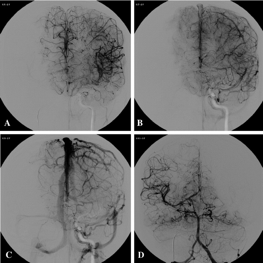
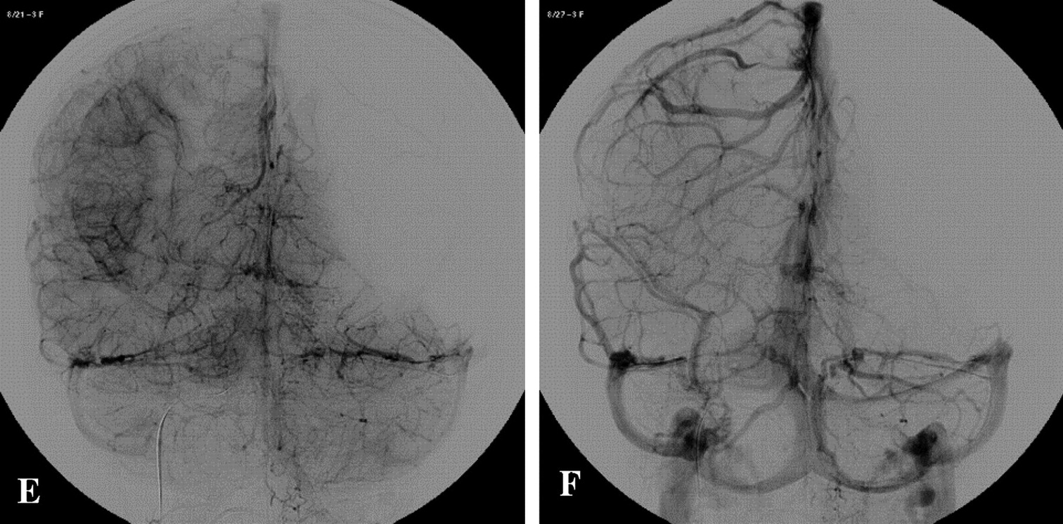
- Fig 3.
A 15-year-old girl test occlusion for a right giant ICA aneurysm. A, Angiogram AP view, injected by left ICA shows late arterial phase. B and C, Early and late venous phases, respectively, demonstrate perfect synchronous venous filling of left hemisphere and territory of right anterior cerebral artery through Acom. D, Arterial phase of the injection through left vertebral artery, AP view, demonstrates excellent cross-filling through Pcom. E and F, Early and late venous phases, respectively, show symmetry of venous filling of vertebrobasilar territory and right MCA territory through the right Pcom. This patient presented a supply of the right internal carotid artery territory via both Acom and Pcom, without venous delay. ICA permanent occlusion was performed at the same time.


Tables
- TABLE 1:
Medical diagnosis in 60 patients undergoing balloon test occlusion of the internal carotid artery followed immediately by therapeutic occlusion, according to gender
Aneurysms Tumors Fistulae Total Female 28 4 1 33 Male 7 19 1 27 Total 35 23 2 60 - TABLE 2:
Venous drainage delay assessed by angiography during balloon test occlusion in 60 patients undergoing permanent internal carotid artery occlusion
Venous Drainage Delay* 0 Seconds 1 Second 2 Seconds 3 Seconds Total Aneurysms 26 6 2 1 35 Tumors 16 4 1 2 23 Direct fistulae 2 0 0 0 2 Total 44 10 3 3 60 * Beginning of venous phase is determined by appearance of first cortical veins at the angiogram. Venous drainage delay corresponds to cortical vein–filling delay of the occluded territory compared to the injected artery (contralateral internal carotid artery or dominant vertebral artery).
In this issue


Jump to section
Related Articles
Cited By...
- Endovascular management of intracranial carotid blowout syndrome in patients with head and neck cancer
- Multicentric Experience in Distal-to-Proximal Revascularization of Tandem Occlusion Stroke Related to Internal Carotid Artery Dissection
- Endovascular Management of Tandem Occlusion Stroke Related to Internal Carotid Artery Dissection Using a Distal to Proximal Approach: Insight from the RECOST Study
- Therapeutic Internal Carotid Artery Occlusion for Large and Giant Aneurysms: A Single Center Cohort of 146 Patients
- Incidence and mechanisms of stroke after permanent carotid artery occlusion following temporary occlusion testing
- Parent Artery Occlusion in Large, Giant, or Fusiform Aneurysms of the Carotid Siphon: Clinical and Imaging Results
- Balloon Occlusion Tests and Therapeutic Vessel Occlusions Revisited: When, When Not, and How
- Reporting standards for balloon test occlusion
- Prognostic Evaluation Based on Cortical Vein Score Difference in Stroke
- Detection of inferolateral trunk syndrome by neuromonitoring during catheter angiography with provocative testing
- Long-Term Clinical and Imaging Follow-Up of Complex Intracranial Aneurysms Treated by Endovascular Parent Vessel Occlusion
- Reply:
- Balloon test occlusion and endosurgical parent artery sacrifice for the evaluation and management of complex intracranial aneurysmal disease
- Endovascular Treatment of Large and Giant Aneurysms