Advertisement

Main menu

  • Home
  • Content
    • Current Issue
    • Accepted Manuscripts
    • Article Preview
    • Past Issue Archive
    • AJNR Case Collection
    • Case of the Week Archive
    • Classic Case Archive
    • Case of the Month Archive
  • Special Collections
    • Spinal CSF Leak Articles (Jan 2020-June 2024)
    • 2024 AJNR Journal Awards
    • Most Impactful AJNR Articles
  • Multimedia
    • AJNR Podcast
    • AJNR Scantastics
    • Video Articles
  • For Authors
    • Submit a Manuscript
    • Author Policies
    • Fast publishing of Accepted Manuscripts
    • Graphical Abstract Preparation
    • Manuscript Submission Guidelines
    • Imaging Protocol Submission
    • Submit a Case for the Case Collection
  • About Us
    • About AJNR
    • Editorial Board
  • More
    • Become a Reviewer/Academy of Reviewers
    • Subscribers
    • Permissions
    • Alerts
    • Feedback
    • Advertisers
    • ASNR Home
  • Other Publications
    • ajnr

User menu

  • Alerts
  • Log in

Search

  • Advanced search
American Journal of Neuroradiology

American Journal of Neuroradiology

ASHNR American Society of Functional Neuroradiology ASHNR American Society of Pediatric Neuroradiology ASSR
  • Alerts
  • Log in

Advanced Search

  • Home
  • Content
    • Current Issue
    • Accepted Manuscripts
    • Article Preview
    • Past Issue Archive
    • AJNR Case Collection
    • Case of the Week Archive
    • Classic Case Archive
    • Case of the Month Archive
  • Special Collections
    • Spinal CSF Leak Articles (Jan 2020-June 2024)
    • 2024 AJNR Journal Awards
    • Most Impactful AJNR Articles
  • Multimedia
    • AJNR Podcast
    • AJNR Scantastics
    • Video Articles
  • For Authors
    • Submit a Manuscript
    • Author Policies
    • Fast publishing of Accepted Manuscripts
    • Graphical Abstract Preparation
    • Manuscript Submission Guidelines
    • Imaging Protocol Submission
    • Submit a Case for the Case Collection
  • About Us
    • About AJNR
    • Editorial Board
  • More
    • Become a Reviewer/Academy of Reviewers
    • Subscribers
    • Permissions
    • Alerts
    • Feedback
    • Advertisers
    • ASNR Home
  • Follow AJNR on Twitter
  • Visit AJNR on Facebook
  • Follow AJNR on Instagram
  • Join AJNR on LinkedIn
  • RSS Feeds

Welcome to the new AJNR, Updated Hall of Fame, and more. Read the full announcements.


AJNR is seeking candidates for the position of Associate Section Editor, AJNR Case Collection. Read the full announcement.


Getting new auth cookie, if you see this message a lot, tell someone!
Research ArticleINTERVENTIONAL

Shapability, Memory, and Luminal Changes in Microcatheters after Steam Shaping: A Comparison of 11 Different Microcatheters

Hiro Kiyosue, Yuzo Hori, Shunro Matsumoto, Mika Okahara, Syuichi Tanoue, Yoshiko Sagara and Hiromu Mori
American Journal of Neuroradiology November 2005, 26 (10) 2610-2616;
Hiro Kiyosue
  • Find this author on Google Scholar
  • Find this author on PubMed
  • Search for this author on this site
Yuzo Hori
  • Find this author on Google Scholar
  • Find this author on PubMed
  • Search for this author on this site
Shunro Matsumoto
  • Find this author on Google Scholar
  • Find this author on PubMed
  • Search for this author on this site
Mika Okahara
  • Find this author on Google Scholar
  • Find this author on PubMed
  • Search for this author on this site
Syuichi Tanoue
  • Find this author on Google Scholar
  • Find this author on PubMed
  • Search for this author on this site
Yoshiko Sagara
  • Find this author on Google Scholar
  • Find this author on PubMed
  • Search for this author on this site
Hiromu Mori
  • Find this author on Google Scholar
  • Find this author on PubMed
  • Search for this author on this site
  • Article
  • Figures & Data
  • Info & Metrics
  • Responses
  • References
  • PDF
Loading

Article Figures & Data

Figures

  • Tables
  • Fig 1.
    • Download figure
    • Open in new tab
    • Download powerpoint
    Fig 1.

    Change of tip angle of a microcatheter.

    A, Initial tip angle. B, Tip angle following the water bath procedure for 10 minutes. C, Tip angle following guidewire procedure. D, Tip angle following the second water bath for 5 minutes.
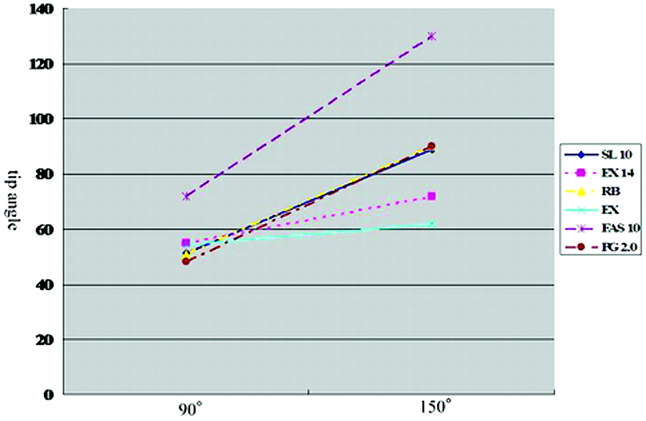
  • Fig 2.
    • Download figure
    • Open in new tab
    • Download powerpoint
    Fig 2.

    Shapability of small-sized catheters.

    Although the mean tip angle is increased according to increasing the intended tip angle in all catheters, Fas10 shows the highest shapability.
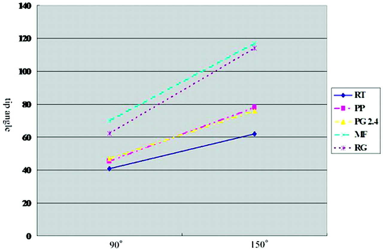
  • Fig 3.
    • Download figure
    • Open in new tab
    • Download powerpoint
    Fig 3.

    Shapability of large-sized catheters. MF and Rg show higher shapability than the others.

  • Fig 4.
    • Download figure
    • Open in new tab
    • Download powerpoint
    Fig 4.

    Durability of the shape of small-sized microcatheters. All microcatheters show reduction of tip angle after the microguidewire procedure (GW). The change rates are >10% in Fas 10 and Pg 2.0. At the final calculation, the Fas10 nonreinforced catheter shows high angle-recovery rates. All reinforced microcatheters except for Pg2.0 show similar change rates, <10%. Pg2.0 shows change rates >10% at the final calculation.
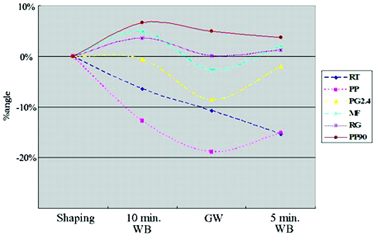
  • Fig 5.
    • Download figure
    • Open in new tab
    • Download powerpoint
    Fig 5.

    Durability of the shape of large-sized microcatheters. RT and PP show reduction of the tip angle before the guidewire procedure. All microcatheters show reduction of the tip angle following the microguidewire procedure (GW). The change rates are >10% in RT and PP. At the final calculation, RT and PP show high reduction rates and final change rates >10%. PP90 and Rg show low reduction rates throughout the examination. The nonreinforced catheter MF shows the highest angle-recover rate.
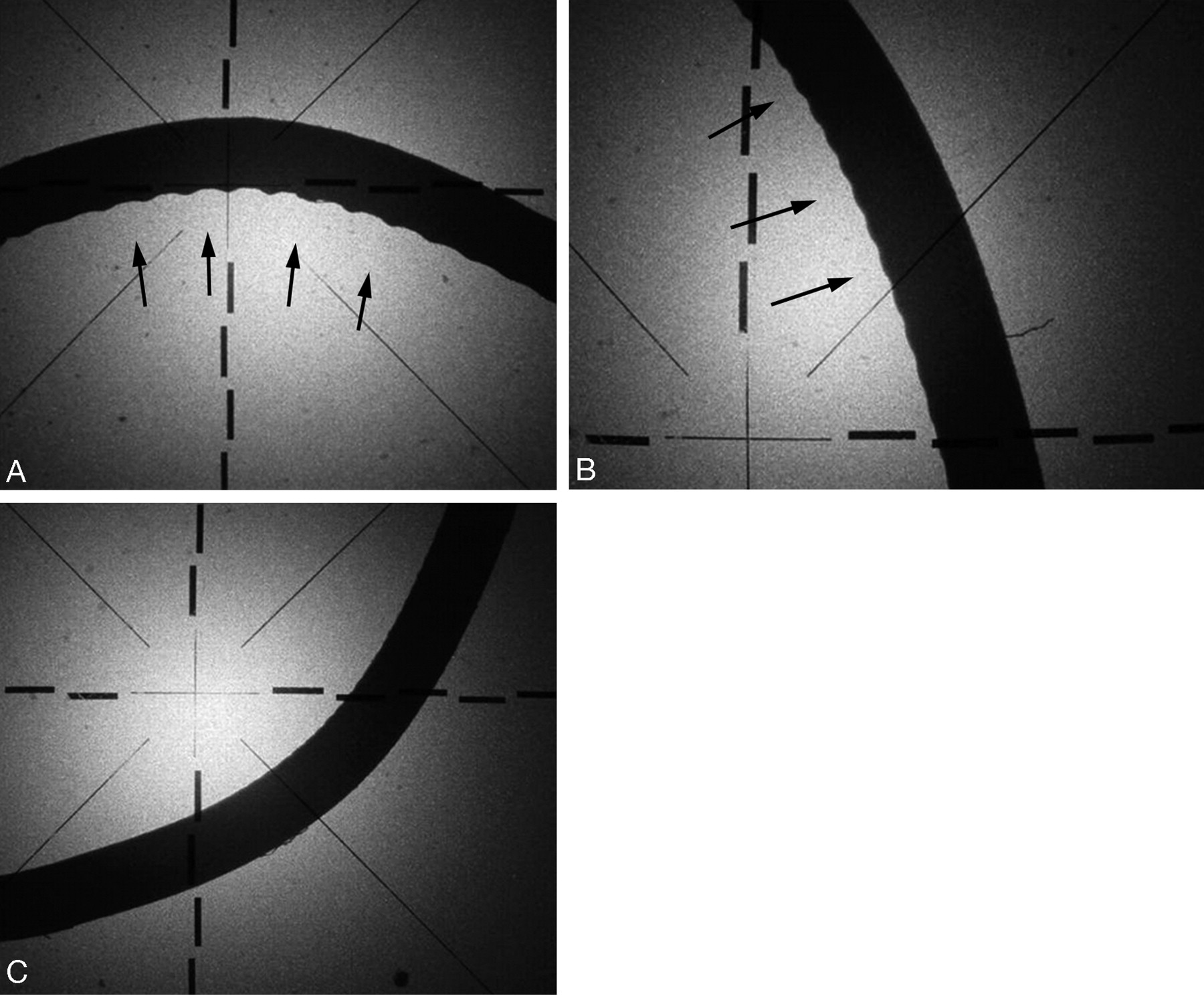
  • Fig 6.
    • Download figure
    • Open in new tab
    • Download powerpoint
    Fig 6.

    Luminal irregularities at the curved portion of the shaped microcatheters.

    A, Moderate irregularity (arrows) on the lesser curvature approximately 0.04 mm in peak-to-valley measurement in RT and Rg.

    B, Mild irregularity (arrows) on the lesser curvature approximately 0.015 mm in peak-to-valley measurement in Ex, Pg2.0, Rb14, and PP.

    C, Smooth surface on the lesser curvature in the other microcatheters.

Tables

  • Figures
    • View popup
    TABLE 1:

    Characteristics of microcatheters

    Proximal OD(French)Distal OD(French)Distal ID(inch)ReinforcementMaterials
    Small size
        Excelsior 10182.62.0.019SS coilPEBAX
        Tracker Excel-142.41.9.017SS coilPEBAX
        Excelsior SL-102.41.7.0165SS coilPEBAX
        Progreat 2.0F2.72.0.020Tungsten coilUE
        Rebar 142.41.9.018SS coilNA
        FasTracker-102.62.0.015–PP + PE
    Large size
        Rapid Transit3.02.3.018SS coilPolyamide
        Prowler Plus3.02.3.018SS coilPolyamide
        Renegade-183.02.5.021Fiberbraid coilPEBAX
        Progreat 2.4F2.92.4.022Tungsten coilUE
        Microferret3.02.4.021–PE
        Prowler Plus MX3.02.3.018SS coilpolyamide
    • Note.—SS coil indicates stainless steel coil; PEBAX, polyether block amides; UE, urethan elastomer; PP, polypropylene; PE, polyethylene; NA, no information available.

    • View popup
    TABLE 2:

    Tip angle of each brand with intended angles of 90° and 150°

    90°150°
    Small size
        Excelsior 101851–57 (54)54–68 (62)
        Tracker Excel-1454–56 (55)70–74 (72)
        Excelsior SL-1051–57 (55)87–91 (89)
        Progreat 2.0F46–51 (48)86–93 (90)
        Rebar 1449–54 (51)88–92 (90)
        FasTracker-1064–77 (72)124–136 (130)
    Large size
        Rapid Transit40–43 (41)58–66 (62)
        Prowler Plus44–47 (45)76–80 (78)
        Renegade-1858–70 (62)111–117 (114)
        Progreat 2.4F47 (47)72–80 (76)
        Microferret64–77 (70)115–121 (117)
    • Notes.—Values are expressed as ranges followed by mean in parentheses.

    • View popup
    TABLE 3:

    Changes of the tip angle of each brand

    Initial Angle (°)10-min WaterbathGuidewire5-min Waterbath
    Small size
        Excelsior 101893–107 (97.2)90–104 (97.2)85–99 (90.6)92–106 (93.2)
        Tracker Excel-1494–105 101.0)97–112 (102.7)91–98 (93.6)92–101 (96.3)
        Excelsior SL-1096–101 (99.0)90–103 (96.3)86–90 (88.3)95–99 (97.0)
        Progreat 2.0F96–105 (100.6)91–108 (99.3)70–98 (84.6)78–97 (87.0)
        Rebar 1496–109 (105.6)96–105 (101.7)98–103 (100.0)94–107 (101.6)
        FasTracker-10110–115 (111.6)107–114 (109.6)93–100 (97.3)105–106 (105.3)
    Large size
        Rapid Transit90–94 (93.0)84–91 (87.0)81–86 (83.0)75–81 (78.6)
        Prowler Plus87–98 (91.3)73–77 (75.6)65–74 (70.6)72–74 (73.0)
        Renegade-1886–102 (94.3)90–120 (105.0)91–117 (100.6)87–114 (99.0)
        Progreat 2.4F93–95 (94.0)90–102 (94.6)82–91 (87.6)92–96 (94.3)
        Microferret99–104 (102.8)99–113 (109.2)91–103 (99.7)93–110 (104.7)
        Prowler Plus MX80–82 (80.7)81–90 (86.0)79–88 (84.6)82–84 (83.3)
    • Note.—Values are expressed as ranges followed by mean in parentheses.

    • View popup
    TABLE 4:

    Changes of the luminal diameter

    % Axial Diameter of Control% Axial Diameter of Shaped Catheters at Angled Portion
    Small size
        Excelsior 1018100.795.5
        Tracker Excel-14100.199.2
        Excelsior SL-1099.399.5
        Progreat 2.0F99.499.1
        Rebar 14100.397.3
        FasTracker-10100.397.3
    Large size
        Rapid Transit99.393.1
        Prowler Plus99.898.8
        Renegade98.990.5
        Progreat 2.4F100.095.4
        Microferret99.488.3
    • Note.—% axial diameter = (axial diameter at 5-mm proximal portion/axial diameter at 2-mm proximal portion) × 100.

PreviousNext
Back to top

In this issue

American Journal of Neuroradiology: 26 (10)
American Journal of Neuroradiology
Vol. 26, Issue 10
1 Nov 2005
  • Table of Contents
  • Index by author
Advertisement
Print
Download PDF
Email Article

Thank you for your interest in spreading the word on American Journal of Neuroradiology.

NOTE: We only request your email address so that the person you are recommending the page to knows that you wanted them to see it, and that it is not junk mail. We do not capture any email address.

Enter multiple addresses on separate lines or separate them with commas.
Shapability, Memory, and Luminal Changes in Microcatheters after Steam Shaping: A Comparison of 11 Different Microcatheters
(Your Name) has sent you a message from American Journal of Neuroradiology
(Your Name) thought you would like to see the American Journal of Neuroradiology web site.
CAPTCHA
This question is for testing whether or not you are a human visitor and to prevent automated spam submissions.
Cite this article
Hiro Kiyosue, Yuzo Hori, Shunro Matsumoto, Mika Okahara, Syuichi Tanoue, Yoshiko Sagara, Hiromu Mori
Shapability, Memory, and Luminal Changes in Microcatheters after Steam Shaping: A Comparison of 11 Different Microcatheters
American Journal of Neuroradiology Nov 2005, 26 (10) 2610-2616;

Citation Manager Formats

  • BibTeX
  • Bookends
  • EasyBib
  • EndNote (tagged)
  • EndNote 8 (xml)
  • Medlars
  • Mendeley
  • Papers
  • RefWorks Tagged
  • Ref Manager
  • RIS
  • Zotero
0 Responses
Respond to this article
Share
Bookmark this article
Shapability, Memory, and Luminal Changes in Microcatheters after Steam Shaping: A Comparison of 11 Different Microcatheters
Hiro Kiyosue, Yuzo Hori, Shunro Matsumoto, Mika Okahara, Syuichi Tanoue, Yoshiko Sagara, Hiromu Mori
American Journal of Neuroradiology Nov 2005, 26 (10) 2610-2616;
del.icio.us logo Twitter logo Facebook logo Mendeley logo
  • Tweet Widget
  • Facebook Like
  • Google Plus One

Jump to section

  • Article
    • Abstract
    • Materials and Methods
    • Results
    • Discussion
    • References
  • Figures & Data
  • Info & Metrics
  • Responses
  • References
  • PDF

Related Articles

  • No related articles found.
  • PubMed
  • Google Scholar

Cited By...

  • No citing articles found.
  • Crossref
  • Google Scholar

This article has not yet been cited by articles in journals that are participating in Crossref Cited-by Linking.

More in this TOC Section

  • SAVE vs. Solumbra Techniques for Thrombectomy
  • CT Perfusion&Reperfusion in Acute Ischemic Stroke
  • Delayed Reperfusion Post-Thrombectomy&Thrombolysis
Show more Interventional

Similar Articles

Advertisement

Indexed Content

  • Current Issue
  • Accepted Manuscripts
  • Article Preview
  • Past Issues
  • Editorials
  • Editors Choice
  • Fellow Journal Club
  • Letters to the Editor

Cases

  • Case Collection
  • Archive - Case of the Week
  • Archive - Case of the Month
  • Archive - Classic Case

Special Collections

  • Special Collections

Resources

  • News and Updates
  • Turn around Times
  • Submit a Manuscript
  • Author Policies
  • Manuscript Submission Guidelines
  • Evidence-Based Medicine Level Guide
  • Publishing Checklists
  • Graphical Abstract Preparation
  • Imaging Protocol Submission
  • Submit a Case
  • Become a Reviewer/Academy of Reviewers
  • Get Peer Review Credit from Publons

Multimedia

  • AJNR Podcast
  • AJNR SCANtastic
  • Video Articles

About Us

  • About AJNR
  • Editorial Board
  • Not an AJNR Subscriber? Join Now
  • Alerts
  • Feedback
  • Advertise with us
  • Librarian Resources
  • Permissions
  • Terms and Conditions

American Society of Neuroradiology

  • Not an ASNR Member? Join Now

© 2025 by the American Society of Neuroradiology All rights, including for text and data mining, AI training, and similar technologies, are reserved.
Print ISSN: 0195-6108 Online ISSN: 1936-959X

Powered by HighWire