
Abstract
Summary: Cross-sectional imaging has demonstrated an increasing role in the evaluation of the orbits and the periorbital structures. The case presented in this article demonstrates the rare finding of a dacryolith by CT. To our knowledge, little has been reported on cross-sectional imaging characteristics of this entity in the recent radiologic literature. We demonstrated that CT can be a useful tool in diagnosing both dacryolithiasis and dacryocystitis. Obtaining an early diagnosis of dacryolithiasis is optimal to avoid potential complications, particularly because treatment is often curative.
Dacryolithiasis is a relatively common disorder and is often underdiagnosed (1). We present an unusual finding of a calcified dacryolith depicted by CT. Dacryoliths are concretions formed in the lacrimal sac from cellular debris and proteins. Dacryoliths may calcify and can cause further obstruction. They are often underlying contributors in patients with intermittent or chronic dacryocystitis. Dacryocystitis is an inflammation of the nasolacrimal sac, which is characterized by epiphora, pain, erythema, dilation of the sac, and swelling of the lacrimal puncta. We report the CT imaging and clinical features of a case of dacryolithiasis with secondary dacryocystitis.
Case Report
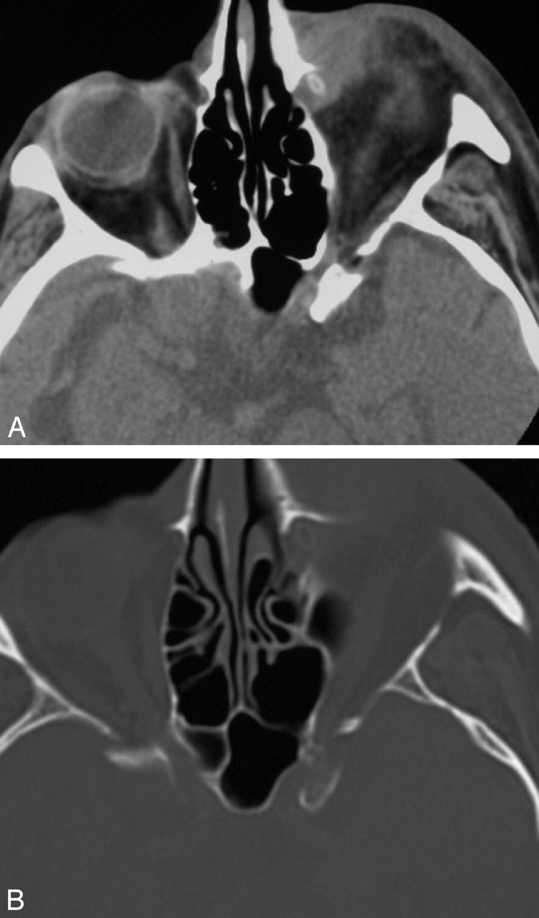
An elderly man presented to the emergency department complaining of swelling and pain of his left eye. He had experienced tearing and swelling for several days, which worsened in the 24 hours before presentation. Findings of the physical examination revealed that the left medial canthal area was warm, erythematous, and edematous. Point tenderness around the medial canthal region extended laterally. A noncontrast CT of the orbits (Fig 1A) showed an inflammatory soft-tissue mass in the region of the left medial epicanthus centered on the lacrimal sac. In addition, extensive soft-tissue edema involved the preseptal tissues. A small oval hyperattenuation with a peripheral rim of calcification was present in the center of the soft-tissue mass. The surrounding osseous structures were intact, with no evidence of bony destruction (Fig 1B).

A, Axial noncontrast CT image obtained at the level of the orbits demonstrates a soft-tissue mass in the region of the left lacrimal sac. There is associated fat stranding of the preseptal soft tissues. A focal hyperattenuation with peripheral calcification within the mass is consistent with a dacryolith. B, Bone window settings of the same patient demonstrate no bony destructive changes.
The patient was admitted to the hospital and received intravenous antibiotics for orbital cellulitis. The lesion was aspirated with a needle, and samples were sent to microbiology. After a short stay, he was discharged to home to finish a course of oral antibiotics. Aspiration cultures grew Streptococcus agalactiae and Staphylococcus aureus. As an outpatient, he eventually underwent a left dacryocystorhinostomy, and the dacryolith was removed. Actinomyces odontolyticus isolates were obtained from the dacryolith. The patient’s symptoms resolved after the surgery.
Discussion
Dacryolithiasis occurs in patients with chronic underlying dacryocystitis. Denuded epithelial cells clump together with exudated proteins and debris, forming a cast in the lacrimal sac. With time, the material eventually mineralizes, most typically with calcium (2). Dacryoliths are typically found in the setting of chronic infections with superimposed fungal colonization. They are found in up to 30% of patients with chronic dacryocystitis (3), are difficult to identify on conventional radiographs, and appear as round or oval filling defects on dacryocystography. On a CT scan, dacryoliths are characterized by focal areas of high attenuation within a soft-tissue attenuation mass in the region of the lacrimal sac (4). They may have a peripheral rim of calcification, giving a “rice kernel” appearance.
Dacryocystitis is characterized by epiphora, erythema, and edema in the region of the medial epicanthus and lacrimal puncta as the result of an infection of the nasolacrimal sac. Often there is mucopurulent discharge from the puncta and associated conjunctivitis. Obstruction or stricture of the nasolacrimal drainage is generally an underlying factor.
A brief review of the normal anatomy of the nasolacrimal system is included for clarity. The lacrimal drainage system consists of upper and lower canaliculi, a lacrimal sac, and a lacrimal duct. The upper and lower canaliculi originate as a small opening in the medial lid margins, which are termed “puncta.” The canaliculi course medially and drain excess tears from the surface of the eyes. They combine to form a common channel called the sinus of Maier, which drains into the posterior wall of the lacrimal sac. The nasolacrimal duct drains the sac via the nasolacrimal canal into the nasal cavity below the inferior turbinate (4).
The incidence of dacryocystitis occurs in a bimodal distribution, with greater frequency in neonates and in people more than 40 years of age (5). Most cases in neonates represent congenital abnormalities. The sexes are affected equally. Most cases are related to incomplete recanalization of the distal nasolacrimal duct in the region where it enters the nasal cavity. A thin membrane is often present covering the opening. The membrane will sometimes rupture on its own, or it may require instrumentation (6).
In the group of patients of more than 40 years of age, the infections relate to acquired abnormalities. Commonly, women are affected more than men, and whites are affected more than blacks. Similar to that in neonates, obstruction at the opening of the nasolacrimal duct into the inferior meatus is often the underlying factor. Contributing conditions may include rhinitis, sinusitis, enlarged turbinates or adenoids, foreign bodies, septal deviation, tumors, mucoceles, nasal septal abscess, iatrogenic causes, and trauma. The obstruction and subsequent stagnation of the tear flow predisposes to infection (2, 3, 5).
The microbiology of dacryocystitis mimics normal conjunctival flora in most instances. The most common aerobic organisms include Staphylococcus epidermidis, S aureus, Streptococcus, Pseudomonas, and Pneumococcal species. The most common anaerobic organisms isolated from the lacrimal sacs in adults with dacryocystitis include Peptostreptococcus, Propionibacterium, Prevotella, and Fusobacterium species. Gram-negative bacteria are associated with copious discharge. The most common gram-negative bacteria isolated are Pseudomonas aeruginosa and Escherichia coli (5). In chronic dacryocystitis, there can be superinfection with fungal species including Actinomyces, Aspergillus, and Candida species (5).
Treatment of acute dacryocystitis typically involves treatment with antibiotics in the acute phase, which may or may not be followed by an external dacryocystorhinostomy or other interventional procedure (3). Patients typically do not require hospitalization unless there are complicating factors such as orbital cellulitis. Other complications include abscess and fistula formation. Chronic dacryocystitis typically requires surgery or an interventional procedure. Occasionally, chronic dacryocystitis relating to allergies may improve with topical steroids (6). Surgical success rates in the treatment of dacryocystitis are approximately 90% (5). Balloon dacryoplasty, with or without stent placement, may be useful in patients with circumscribed focal stenosis or occlusions of the nasolacrimal duct (3).
Not all that swells in the region of the medial epicanthi represents infection. Multiple entities can cause masslike swelling. An enlarged sac that is noninfected is termed a “dacryocystocele.” Epithelial tumors are the most common tumors of the lacrimal sac. The malignant causes include squamous cell carcinoma, adenoid cystic carcinoma, oncocytic carcinoma, and epidermoid carcinoma. The benign lesions include papilloma, dermoid cysts, mucoepidermoid cysts, and adenoma. Lymphoproliferative disorders, primarily lymphoma, are the second most common types of tumors. Rarely mesenchymal tumors occur and include hemangioma, hemangiopericytoma, melanoma, fibroma, fibrous histiocytoma, neurilemmoma, and plexiform neuroma. Granulomatous diseases such as sarcoid and Wegener’s granulomatosis can affect the lacrimal sac. Secondary involvement from cutaneous squamous and basal cell carcinomas can occur. Metastatic disease of the lacrimal sac is rare (7).
- Received January 2, 2005.
- Accepted after revision February 14, 2005.
- Copyright © American Society of Neuroradiology
In this issue


Jump to section
Related Articles
Cited By...
- No citing articles found.