Advertisement

Main menu

  • Home
  • Content
    • Current Issue
    • Accepted Manuscripts
    • Article Preview
    • Past Issue Archive
    • AJNR Case Collection
    • Case of the Week Archive
    • Classic Case Archive
    • Case of the Month Archive
  • Special Collections
    • Spinal CSF Leak Articles (Jan 2020-June 2024)
    • 2024 AJNR Journal Awards
    • Most Impactful AJNR Articles
  • Multimedia
    • AJNR Podcast
    • AJNR Scantastics
    • Video Articles
  • For Authors
    • Submit a Manuscript
    • Author Policies
    • Fast publishing of Accepted Manuscripts
    • Graphical Abstract Preparation
    • Manuscript Submission Guidelines
    • Imaging Protocol Submission
    • Submit a Case for the Case Collection
  • About Us
    • About AJNR
    • Editorial Board
  • More
    • Become a Reviewer/Academy of Reviewers
    • Subscribers
    • Permissions
    • Alerts
    • Feedback
    • Advertisers
    • ASNR Home
  • Other Publications
    • ajnr

User menu

  • Alerts
  • Log in

Search

  • Advanced search
American Journal of Neuroradiology

American Journal of Neuroradiology

ASHNR American Society of Functional Neuroradiology ASHNR American Society of Pediatric Neuroradiology ASSR
  • Alerts
  • Log in

Advanced Search

  • Home
  • Content
    • Current Issue
    • Accepted Manuscripts
    • Article Preview
    • Past Issue Archive
    • AJNR Case Collection
    • Case of the Week Archive
    • Classic Case Archive
    • Case of the Month Archive
  • Special Collections
    • Spinal CSF Leak Articles (Jan 2020-June 2024)
    • 2024 AJNR Journal Awards
    • Most Impactful AJNR Articles
  • Multimedia
    • AJNR Podcast
    • AJNR Scantastics
    • Video Articles
  • For Authors
    • Submit a Manuscript
    • Author Policies
    • Fast publishing of Accepted Manuscripts
    • Graphical Abstract Preparation
    • Manuscript Submission Guidelines
    • Imaging Protocol Submission
    • Submit a Case for the Case Collection
  • About Us
    • About AJNR
    • Editorial Board
  • More
    • Become a Reviewer/Academy of Reviewers
    • Subscribers
    • Permissions
    • Alerts
    • Feedback
    • Advertisers
    • ASNR Home
  • Follow AJNR on Twitter
  • Visit AJNR on Facebook
  • Follow AJNR on Instagram
  • Join AJNR on LinkedIn
  • RSS Feeds

Welcome to the new AJNR, Updated Hall of Fame, and more. Read the full announcements.


AJNR is seeking candidates for the position of Associate Section Editor, AJNR Case Collection. Read the full announcement.


Research ArticleBRAIN

Dilatation of the Virchow-Robin Space Is a Sensitive Indicator of Cerebral Microvascular Disease: Study in Elderly Patients with Dementia

Tufail F. Patankar, Dipayan Mitra, Anoop Varma, Julie Snowden, David Neary and Alan Jackson
American Journal of Neuroradiology June 2005, 26 (6) 1512-1520;
Tufail F. Patankar
  • Find this author on Google Scholar
  • Find this author on PubMed
  • Search for this author on this site
Dipayan Mitra
  • Find this author on Google Scholar
  • Find this author on PubMed
  • Search for this author on this site
Anoop Varma
  • Find this author on Google Scholar
  • Find this author on PubMed
  • Search for this author on this site
Julie Snowden
  • Find this author on Google Scholar
  • Find this author on PubMed
  • Search for this author on this site
David Neary
  • Find this author on Google Scholar
  • Find this author on PubMed
  • Search for this author on this site
Alan Jackson
  • Find this author on Google Scholar
  • Find this author on PubMed
  • Search for this author on this site
  • Article
  • Figures & Data
  • Info & Metrics
  • Responses
  • References
  • PDF
Loading

Article Figures & Data

Figures

  • Tables
  • Fig 1.
    • Download figure
    • Open in new tab
    • Download powerpoint
    Fig 1.

    Matching axial MR images show severe (grade 5) dilatation of the VRS (matrix = 256 × 256, FOV = 230 × 230 mm): A, T2-weighted variable echo (TR/TE1/TE2 = 5500/20/90); B, T1-weighted high-spatial-resolution T1-weighted 3D gradient echo (TR/TE = 24/18, section thickness = 0.89 mm, flip angle = 30°); and C, inversion recovery (TR/TE/TE = 6850/18/300). Calculated CNRs for VRS versus WM are 64.1 for inversion recovery, 24.8 for fast field-echo, 19.1 for the variable-echo second-echo imaging.

  • Fig 2.
    • Download figure
    • Open in new tab
    • Download powerpoint
    Fig 2.

    Axial inversion recovery MR images (TR/TE/TI = 6850/18/300, matrix = 256 × 256, FOV = 230 × 230 mm) show grade 3 VRS dilatation (arrows).
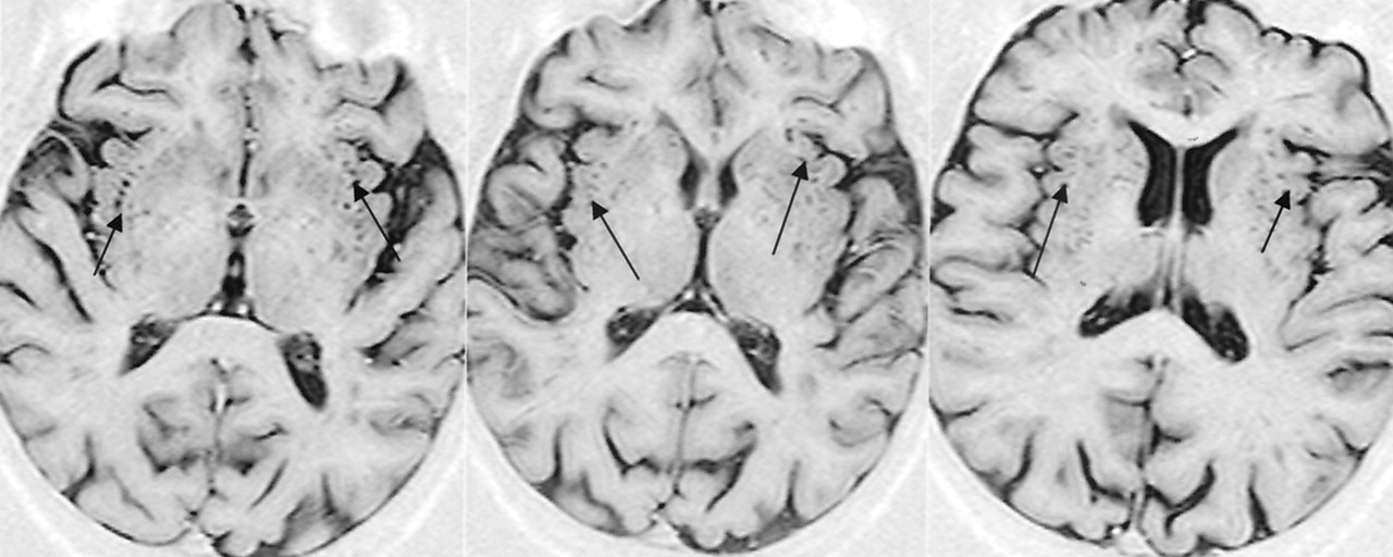
  • Fig 3.
    • Download figure
    • Open in new tab
    • Download powerpoint
    Fig 3.

    Axial inversion recovery MR images (TR/TE/TI = 6850/18/300, matrix = 256 × 256, FOV = 230 × 230 mm) show extensive VRS dilatation (arrows) throughout the BG (grade 5).

  • Fig 4.
    • Download figure
    • Open in new tab
    • Download powerpoint
    Fig 4.

    Axial Inversion recovery MR images (TR/TE/TI = 6850/18/300, matrix = 256 × 256, FOV = 230 × 230 mm) show the VRS (arrows) as linear structure passing through several sections (A–C) in the imaging volume.

Tables

  • Figures
    • View popup
    TABLE 1:

    Adjusted Cohen κ scores for each component of the VRS scoring system

    ComponentIntraobserver 1Intraobserver 2Interobserver
    BG
        Scheme 10.970.890.91
        Scheme 21.00.960.98
    Centrum semiovale0.780.820.84
    Subinsular0.890.910.90
    Mesencephalon0.940.840.82
    • Note.—Intraobserver and interobserver variability assessed by using the Wilcoxon matched-pairs test.

    • View popup
    TABLE 2:

    WM-lesion and VRS scores

    ResultGroupControlADFTD vs IVD
    Control (n = 35)AD (n = 35)FTD (n = 24)IVD (n = 16)P ValueVs ADVs FTDVs IVDVs FTDVs IVD
    Scheltens score
        Total9.03 (7.31)9.28 (5.24)10.75 (8.02)14.3 (10.1)<.05NSNS<.01NS<.01NS
        Deep WM total5.14 (4.09)5.26 (3.87)5.65 (4.21)9.26 (6.98)<.05NSNSNSNS<.01NS
        PVH2.66 (1.94)2.95 (2.16)3.14 (3.02)5.02 (4.26)<.05NSNS<.01NS<.01NS
        BG0.77 (2.30) *0.72 (1.89)0.63 (1.28)1.74 (2.15)<.01NSNS<.01NSNSNS
        Infratentorial0.46 (0.74)0.37 (0.64)0.32 (0.30)0.45 (0.61)NSNSNSNSNSNSNS
    BG
        Scheme 10.86 (0.88)0.82 (0.88)1.06 (0.80)1.92 (0.83)<.005NSNS<.005NS<.001NS
        Scheme 21.46 (1.20)1.29 (1.26)2.0 (1.12)3.92 (0.99)<.001NSNS<.001NS<.001<.01
    Centrum semiovale0.51 (0.85)0.74 (0.60)1.33 (0.70)1.03 (0.39)<.01NS<0.01<.01NSNSNS
    Subinsular0.96 (1.95)0.47 (0.51)0.67 (0.72)1.17 (0.39)NSNSNSNSNSNSNS
    Mesencephalic0.51 (0.51)0.65 (0.49)0.73 (0.46)0.92 (0.28)NSNSNSNSNSNSNS
    • Note.—Differences in ages were compared by using ANOVA (at P < .05) with an a posteriori Tukey test (at P < .01). Other variables were assessed by using the Kruskal-Wallis test (at P < .05) with an a posteriori Mann-Whitney U test (at P < .01) to identify between-group differences. NS = not significant. Data in parentheses are total number of patients in each group. The score is the SD of the measurement.

    • View popup
    TABLE 3:

    Sensitivities and specificities based on BG2 cutoff values as indicators of vascular dementia

    ComparisonBG2 VRS Score
    >2>3>4
    IVD vs AD and FTD
        Sensitivity0.670.540.25
        Specificity0.700.871
    IVD vs AD
        Sensitivity0.670.540.25
        Specificity0.890.931
    IVD vs FTD
        Sensitivity0.670.540.25
        Specificity0.760.931
    • View popup
    TABLE 4:

    Sensitivities and specificities based on centrum-semiovale cutoff values as indicators of vascular dementia

    ComparisonCentrum Semiovale VRS Score
    >0>1
    FTD vs AD and IVD
        Sensitivity0.810.38
        Specificity0.270.25
    FTD vs AD
        Sensitivity0.810.38
        Specificity0.390.50
    FTD vs IVD
        Sensitivity0.810.38
        Specificity0.450.33
PreviousNext
Back to top

In this issue

American Journal of Neuroradiology: 26 (6)
American Journal of Neuroradiology
Vol. 26, Issue 6
1 Jun 2005
  • Table of Contents
  • Index by author
Advertisement
Print
Download PDF
Email Article

Thank you for your interest in spreading the word on American Journal of Neuroradiology.

NOTE: We only request your email address so that the person you are recommending the page to knows that you wanted them to see it, and that it is not junk mail. We do not capture any email address.

Enter multiple addresses on separate lines or separate them with commas.
Dilatation of the Virchow-Robin Space Is a Sensitive Indicator of Cerebral Microvascular Disease: Study in Elderly Patients with Dementia
(Your Name) has sent you a message from American Journal of Neuroradiology
(Your Name) thought you would like to see the American Journal of Neuroradiology web site.
CAPTCHA
This question is for testing whether or not you are a human visitor and to prevent automated spam submissions.
Cite this article
Tufail F. Patankar, Dipayan Mitra, Anoop Varma, Julie Snowden, David Neary, Alan Jackson
Dilatation of the Virchow-Robin Space Is a Sensitive Indicator of Cerebral Microvascular Disease: Study in Elderly Patients with Dementia
American Journal of Neuroradiology Jun 2005, 26 (6) 1512-1520;

Citation Manager Formats

  • BibTeX
  • Bookends
  • EasyBib
  • EndNote (tagged)
  • EndNote 8 (xml)
  • Medlars
  • Mendeley
  • Papers
  • RefWorks Tagged
  • Ref Manager
  • RIS
  • Zotero
0 Responses
Respond to this article
Share
Bookmark this article
Dilatation of the Virchow-Robin Space Is a Sensitive Indicator of Cerebral Microvascular Disease: Study in Elderly Patients with Dementia
Tufail F. Patankar, Dipayan Mitra, Anoop Varma, Julie Snowden, David Neary, Alan Jackson
American Journal of Neuroradiology Jun 2005, 26 (6) 1512-1520;
del.icio.us logo Twitter logo Facebook logo Mendeley logo
  • Tweet Widget
  • Facebook Like
  • Google Plus One

Jump to section

  • Article
    • Abstract
    • Methods
    • Results
    • Discussion
    • Conclusion
    • Footnotes
    • References
  • Figures & Data
  • Info & Metrics
  • Responses
  • References
  • PDF

Related Articles

  • No related articles found.
  • PubMed
  • Google Scholar

Cited By...

  • Enlarged Perivascular Spaces are Associated with White Matter Injury, Brain Atrophy, Cognitive Decline and Markers of Inflammation in an Autosomal Dominant Vascular Neurodegenerative Disease (CADASIL)
  • Divergent enlarged perivascular spaces volumes in early versus late age-of-onset Alzheimers disease
  • On the detectability and accuracy of computational measurements of enlarged perivascular spaces from magnetic resonance images
  • Lesion Volume in Relapsing Multiple Sclerosis is Associated with Perivascular Space Enlargement at the Level of the Basal Ganglia
  • Physiology and Clinical Relevance of Enlarged Perivascular Spaces in the Aging Brain
  • Perivascular Space Semi-Automatic Segmentation (PVSSAS): A Tool for Segmenting, Viewing and Editing Perivascular Spaces
  • Characterization of MR Imaging-Visible Perivascular Spaces in the White Matter of Healthy Adolescents at 3T
  • Ontario Neurodegenerative Disease Research Initiative (ONDRI): Structural MRI methods & outcome measures
  • Altered cognitive function in systemic lupus erythematosus and associations with inflammation and functional and structural brain changes
  • Perivascular spaces contribute to cognition beyond other small vessel disease markers
  • Perivascular Spaces in Old Age: Assessment, Distribution, and Correlation with White Matter Hyperintensities
  • Vascular Cognitive Impairment
  • Dilated Perivascular Spaces in the Basal Ganglia Are a Biomarker of Small-Vessel Disease in a Very Elderly Population with Dementia
  • Hydrocephalus due to extreme dilation of Virchow-Robin spaces
  • Subcortical Cystic Lesions within the Anterior Superior Temporal Gyrus: A Newly Recognized Characteristic Location for Dilated Perivascular Spaces
  • Perivascular Spaces Are Associated with Atherosclerosis: An Insight from the Northern Manhattan Study
  • Enlarged perivascular spaces as a marker of underlying arteriopathy in intracerebral haemorrhage: a multicentre MRI cohort study
  • Rating Method for Dilated Virchow-Robin Spaces on Magnetic Resonance Imaging
  • Topography of dilated perivascular spaces in subjects from a memory clinic cohort
  • Assessment of the Virchow-Robin Spaces in Alzheimer Disease, Mild Cognitive Impairment, and Normal Aging, Using High-Field MR Imaging
  • Frequency and Location of Dilated Virchow-Robin Spaces in Elderly People: A Population-Based 3D MR Imaging Study
  • Cerebrovascular Damage in Late-Life Depression Is Associated With Structural and Functional Abnormalities of Subcutaneous Small Arteries
  • Enlarged Perivascular Spaces on MRI Are a Feature of Cerebral Small Vessel Disease
  • Neuropathological Correlates of Temporal Pole White Matter Hyperintensities in CADASIL
  • Biomarkers of cerebrovascular disease in dementia
  • Blood Pressure Lowering in PROGRESS (Perindopril Protection Against Recurrent Stroke Study) and White Matter Hyperintensities: Should This Progress Matter to Patients?
  • Crossref
  • Google Scholar

This article has not yet been cited by articles in journals that are participating in Crossref Cited-by Linking.

More in this TOC Section

  • Multimodal CT Provides Improved Performance for Lacunar Infarct Detection
  • Statin Therapy Does Not Affect the Radiographic and Clinical Profile of Patients with TIA and Minor Stroke
  • Optimal MRI Sequence for Identifying Occlusion Location in Acute Stroke: Which Value of Time-Resolved Contrast-Enhanced MRA?
Show more Brain

Similar Articles

Advertisement

Indexed Content

  • Current Issue
  • Accepted Manuscripts
  • Article Preview
  • Past Issues
  • Editorials
  • Editors Choice
  • Fellow Journal Club
  • Letters to the Editor

Cases

  • Case Collection
  • Archive - Case of the Week
  • Archive - Case of the Month
  • Archive - Classic Case

Special Collections

  • Special Collections

Resources

  • News and Updates
  • Turn around Times
  • Submit a Manuscript
  • Author Policies
  • Manuscript Submission Guidelines
  • Evidence-Based Medicine Level Guide
  • Publishing Checklists
  • Graphical Abstract Preparation
  • Imaging Protocol Submission
  • Submit a Case
  • Become a Reviewer/Academy of Reviewers
  • Get Peer Review Credit from Publons

Multimedia

  • AJNR Podcast
  • AJNR SCANtastic
  • Video Articles

About Us

  • About AJNR
  • Editorial Board
  • Not an AJNR Subscriber? Join Now
  • Alerts
  • Feedback
  • Advertise with us
  • Librarian Resources
  • Permissions
  • Terms and Conditions

American Society of Neuroradiology

  • Not an ASNR Member? Join Now

© 2025 by the American Society of Neuroradiology All rights, including for text and data mining, AI training, and similar technologies, are reserved.
Print ISSN: 0195-6108 Online ISSN: 1936-959X

Powered by HighWire