
AG Osborn, KL Salzman, G Katzman, J Provenzale, M Castillo, G Hedlund, A Illner, HR Harnsberger, J Cooper, BV Jones, B Hamilton. 1st ed. Salt Lake City: Amirsys; 2004. 992 pages, 4,400 illustrations. $249.
Diagnostic Imaging: Spine
J Ross, M Brant-Zawadski, KR Moore, J Crim, MZ Chen, and G Katzman. 1st ed. Salt Lake City: Amirsys; 2004. 992 pages, 4,400 illustrations. $249.
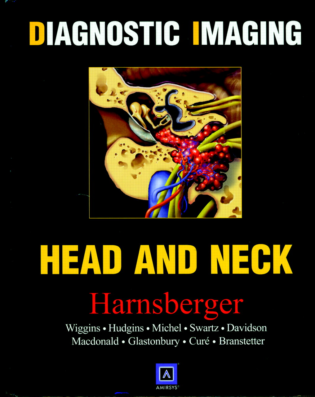
Diagnostic Imaging: Head and Neck
HR Harnsberger, RH Wiggins, PA Hudgins, M Michel, J Swartz, HC Davidson, A Macdonald, C Glastonbury, J Curé, BF Branstetter. 1st ed. Salt Lake City: Amirsys; 2004. 1,000 pages, 4,000 illustrations. $225.
In a marked expansion of the successful Pocket Radiologist series, separate volumes of Diagnostic Imaging: Brain, Diagnostic Imaging: Spine, and Diagnostic Imaging: Head and Neck have been published by Amerisys. They present the reader with a remarkable set of beautifully illustrated and highly educational textbooks. All three follow exactly the same pattern for dissemination of important clinical and imaging information. The premise is that the reader should be presented with easily digestible and complete information in a bulleted format, high-quality MR/CT images, and color plate drawings of anatomy and pathology, and, with that, the information will “stick” far better than the more traditional (prose-driven) texts.
In the world of medical publishing we are witnessing new trends in education with the sale of “books” on compact disks, books with attention-getting humor (as in the text Neuroradiology: The Requisites, by Grossman and Yousem), the proliferation of teaching atlases of unknown cases (there are many of these), and on-line, subject-oriented material. Despite the many new ways of dispensing medical information, books, with their portability and high-quality images, still remain the coin of the realm.
As far as the books listed above are concerned, the color illustrations alone are worth the price of admission, not only because they convey the essentials of the diagnosis, but also because they spectacularly correlate with the CT and MR images. After reading through these books, one is left wondering what is the best way to present information in diagnostic imaging. Is it by the staccato-like details we see in these books or is it by the information presented in the more traditional manner? In the end, this reviewer concludes that both styles work, although the audiences to which each may appeal could differ.
The books in this series are edited by well-recognized authorities in their fields—brain (Anne Osborn), spine (Jeff Ross), head and neck (Ric Harnsberger)—and all three were assisted by a distinguished group of contributors (25 in all), so the reader can be assured of getting accurate information with a wide variety of pathology shown on high-quality images. Remarkably, the three books are not only the same size, they follow the same format. The number of pages in each book is not easily apparent, because there are not page numbers, a major departure from standard texts. When a reader goes to the index to look up a specific entity, he or she is given Roman numerals and Arabic numbers, which in turn correspond to color coding on the pages. Although it takes a couple of minutes to orient oneself to how the sections/parts/chapters are set up and indexed, the colors on the outer leaves of the pages are coordinated with the topics, one concludes that this is a clever way of subdividing the material.
The format is unusual but effective. Each book has major subdivisions, called “parts.” Each part, is subdivided into “sections,” which are further subdivided into “chapters,” and each chapter contains a description of the major diagnoses. This organization makes sense. There is no doubt that between the outside tagging, the color paper on which the anatomy and imaging issues are presented, the anatomic drawings, and the pathologic sections, these are colorful books. This variation in color and the many subdivisions serve to keep one’s attention and can relieve the monotony that often accompanies reading colorless textbooks.
Although there is some minor variation, each section starts out with a set of color pages titled “Anatomy” or “Anatomy and Imaging Issues,” which are then divided into a number of discrete subtitles. There are a generous number of astounding artist’s drawings and these, both in the introductory sections and in the pathology sections, are so good that one cannot help but spend more time looking at the color drawings than the regular images. That is not a drawback at all, but simply an observation related to the magnetic appeal of the drawings. In fact, one is disappointed when a color drawing does not accompany discussion of a given entity. This feeling is simply a tribute to the outstanding quality of the many drawings in the text; it would be unreasonable to expect color drawings of each and every lesion described and shown (to say nothing of the increased cost involved).
As one moves from the introductory pages of each section to specific diagnoses, one encounters the nearly same outline for every lesion—namely, terminology, imaging findings, key facts, differential diagnosis, pathology (often incidentally accompanied by gross/histologic findings), clinical issues, diagnostic checklist, and references. One question that I kept considering as I read through the various portions of all the books was what is the better way to learn about a particular disease? I could have selected any topic for a comparison between a more standard textbook and one from this set. I chose orbital pseudotumors (idiopathic orbital inflammatory disease) in Harnsberger’s volume in comparison to the more classic presentation in Som and Curtin’s Head and Neck Imaging, 4th edition, because pathology is often confusing and there is a rather wide range of diagnostic possibilities. For background information—including how recognition of the disease developed, how the disease presents, what the wide-ranging findings are—and for references cited with specific points, the more traditional text was preferred. On the other hand, for a series of facts and for direct comparison with similar-appearing imaging on the same page (differential diagnosis) Harnsberger’s text succeeds extremely well. One could suggest that diagnoses with a similar appearance to orbital pseudotumor be positioned next to one another so that immediately following the idiopathic orbital inflammatory disease chapter, for example, one would have the chapters on thyroid orbitopathy, lymphoproliferative disorders, and sarcoid, rather than having them separated by entities such as subperiostenal abscess and capillary hemangiomas. This, however, is a minor complaint given the overall high quality of the production and the thought that went into putting each text together.
How thorough are these three books? I looked up some relatively uncommon (but not rare) diseases to see what might be missing, not fully explained, or not well illustrated. Included in this search were chronic inflammatory demyelinating polyneuropathy, lymphocytic hypophysitis, central cord syndrome, posterior reversible encephalopathy syndrome, acute transverse myelitis, Hallervordan-Spatz, hypertrophic olivary degeneration, progressive supranuclear palsy, and endolymphotic sac tumor, among others. As one would expect, not only are they all there, but, most important, each was fully described, accompanied in most cases with color plates, and all were compared with images which may have a similar appearance. There was, for this genre of books, the proper amount of clinical and pathologic information. Do not expect, however, to gather significant information on the state-of-the-art techniques such as MR spectroscopy, diffusion tensor imaging, or perfusion imaging; but, of course, an in-depth analysis of these techniques was not the authors’ intent. Although reading the bullet-like pieces of information can be a little distracting, there is no question that this type of presentation allows the packing in of far more details than would have been possible with ordinary prose.
Needed now are the “final” last steps in which a retrieval of these images and the accompanying vital information are placed in a Web-based format or on a hospital server. When a question arises while reading from a PACS station, one could simply go to a diagnostic imaging icon from Amerisys and readily compare the case at hand with similar images, arrive at a differential diagnosis, and have all the essentials of the abnormality at hand.
It is hard not to be enthusiastic about these texts. They fulfill exquisitely the authors’ intent: to present multitudes of images combined with a barrage of information. Although I personally enjoy reading well-crafted, flowing paragraphs that tie into each other (an old-fashioned view, I am sure) and in which references are directly cited for the stated fact, there is no doubt that the method of teaching and presenting material in these three books have altered the landscape for the way many radiology texts will be written in the future.
Make it a point at the next meeting or continuing medical education course you attend, to pick up and look through one or all three books. You will be sold literally and figuratively on the author’s concept on how to present information and how much vital information you will extract from the book(s). Buy this set of three books, recommend them to all those in neurosciences, make sure your departmental library has every one of the volumes, and refer to them often. This has been a remarkable undertaking by Drs. Harnsberger, Osborn, and Ross and all the editors and contributors to the books.


- Copyright © American Society of Neuroradiology
In this issue


Jump to section
Related Articles
Cited By...
- No citing articles found.