Advertisement

Main menu

  • Home
  • Content
    • Current Issue
    • Accepted Manuscripts
    • Article Preview
    • Past Issue Archive
    • AJNR Case Collection
    • Case of the Week Archive
    • Classic Case Archive
    • Case of the Month Archive
  • Special Collections
    • Spinal CSF Leak Articles (Jan 2020-June 2024)
    • 2024 AJNR Journal Awards
    • Most Impactful AJNR Articles
  • Multimedia
    • AJNR Podcast
    • AJNR Scantastics
    • Video Articles
  • For Authors
    • Submit a Manuscript
    • Author Policies
    • Fast publishing of Accepted Manuscripts
    • Graphical Abstract Preparation
    • Manuscript Submission Guidelines
    • Imaging Protocol Submission
    • Submit a Case for the Case Collection
  • About Us
    • About AJNR
    • Editorial Board
  • More
    • Become a Reviewer/Academy of Reviewers
    • Subscribers
    • Permissions
    • Alerts
    • Feedback
    • Advertisers
    • ASNR Home
  • Other Publications
    • ajnr

User menu

  • Alerts
  • Log in

Search

  • Advanced search
American Journal of Neuroradiology

American Journal of Neuroradiology

ASHNR American Society of Functional Neuroradiology ASHNR American Society of Pediatric Neuroradiology ASSR
  • Alerts
  • Log in

Advanced Search

  • Home
  • Content
    • Current Issue
    • Accepted Manuscripts
    • Article Preview
    • Past Issue Archive
    • AJNR Case Collection
    • Case of the Week Archive
    • Classic Case Archive
    • Case of the Month Archive
  • Special Collections
    • Spinal CSF Leak Articles (Jan 2020-June 2024)
    • 2024 AJNR Journal Awards
    • Most Impactful AJNR Articles
  • Multimedia
    • AJNR Podcast
    • AJNR Scantastics
    • Video Articles
  • For Authors
    • Submit a Manuscript
    • Author Policies
    • Fast publishing of Accepted Manuscripts
    • Graphical Abstract Preparation
    • Manuscript Submission Guidelines
    • Imaging Protocol Submission
    • Submit a Case for the Case Collection
  • About Us
    • About AJNR
    • Editorial Board
  • More
    • Become a Reviewer/Academy of Reviewers
    • Subscribers
    • Permissions
    • Alerts
    • Feedback
    • Advertisers
    • ASNR Home
  • Follow AJNR on Twitter
  • Visit AJNR on Facebook
  • Follow AJNR on Instagram
  • Join AJNR on LinkedIn
  • RSS Feeds

Welcome to the new AJNR, Updated Hall of Fame, and more. Read the full announcements.


AJNR is seeking candidates for the position of Associate Section Editor, AJNR Case Collection. Read the full announcement.


OtherSPINE

Refracture with Cement Extrusion Following Percutaneous Vertebroplasty of a Large Interbody Cleft

A.L. Wagner and E. Baskurt
American Journal of Neuroradiology January 2006, 27 (1) 230-231;
A.L. Wagner
  • Find this author on Google Scholar
  • Find this author on PubMed
  • Search for this author on this site
E. Baskurt
  • Find this author on Google Scholar
  • Find this author on PubMed
  • Search for this author on this site
  • Article
  • Figures & Data
  • Info & Metrics
  • Responses
  • References
  • PDF
Loading

Abstract

SUMMARY: We present a case report of a patient on long-term dialysis who underwent percutaneous vertebroplasty to treat a painful intrabody vertebral cleft and who subsequently experienced a refracture of the posterior portion of the same vertebral body, resulting in anterior displacement of the cement through the anterior cortex. The case raises the question whether, in some patients, the marrow space should be filled with cement in addition to the cleft.

Vertebroplasty has become widespread in the treatment of vertebral compression fractures refractory to medical therapy and has success rates as high as 90% in some studies. Complications from this procedure, while rare, have been described and include bleeding, infection, cement embolism, and various other complications from cement extravasation. Patients with a vertebral cleft, containing either air or fluid, comprise a subset of patients in which vertebroplasty, with filling of the cleft, is typically very effective, and there have been reported cases of height restoration following this type of procedure. This is a report of refracture of the same vertebral body following filling of a large cleft during vertebroplasty in an end-stage renal patient; the case questions whether filling the cleft alone is adequate in some patients.

Case Report

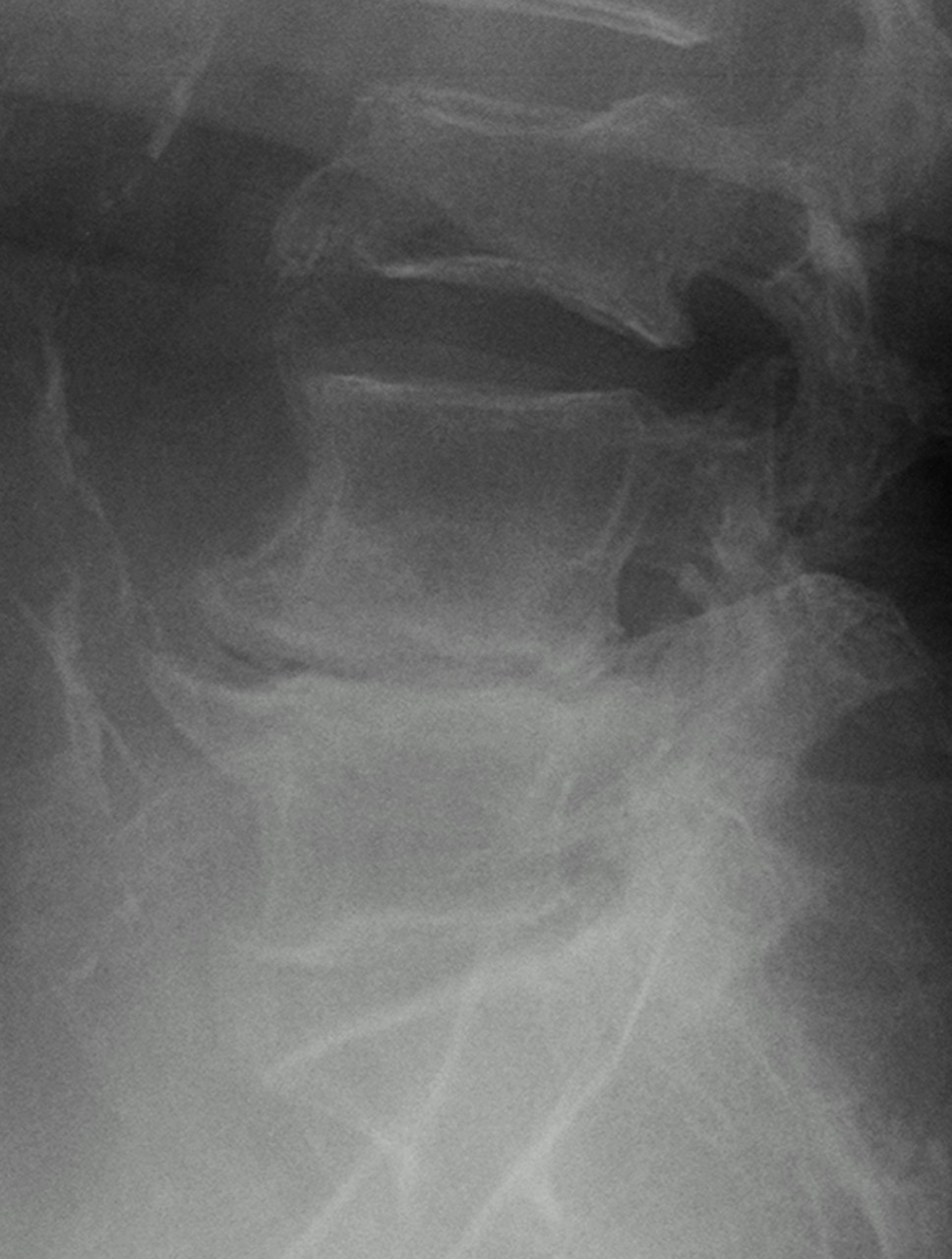
A 75-year-old diabetic woman, who had been on dialysis for >20 years, presented with a 3-month history of severe back pain, unresponsive to bed rest, analgesics, and bracing. Sequential plain films showed severe diffuse osteopenia and a compression fracture at L3 that had progressed from 20% height loss at initial presentation to 60% height loss over a short period of time (Fig 1). Physical examination demonstrated marked tenderness upon palpation of the posterior spinous process of L3, and vertebroplasty was ordered, because of her rapidly progressing fracture. Vertebroplasty was performed by using an 11-gauge needle via a unipedicular approach, and it was immediately noted that the cement was filling a large cavity within the L3 vertebral body.

Fig 1.
  • Download figure
  • Open in new tab
  • Download powerpoint
Fig 1.

Lateral radiograph of the lumbar spine before initial vertebroplasty reveals compression of the L3 vertebral body with little anterior wall bowing.

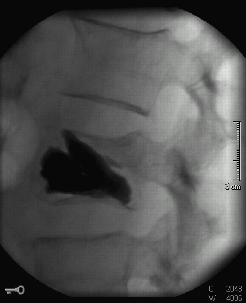
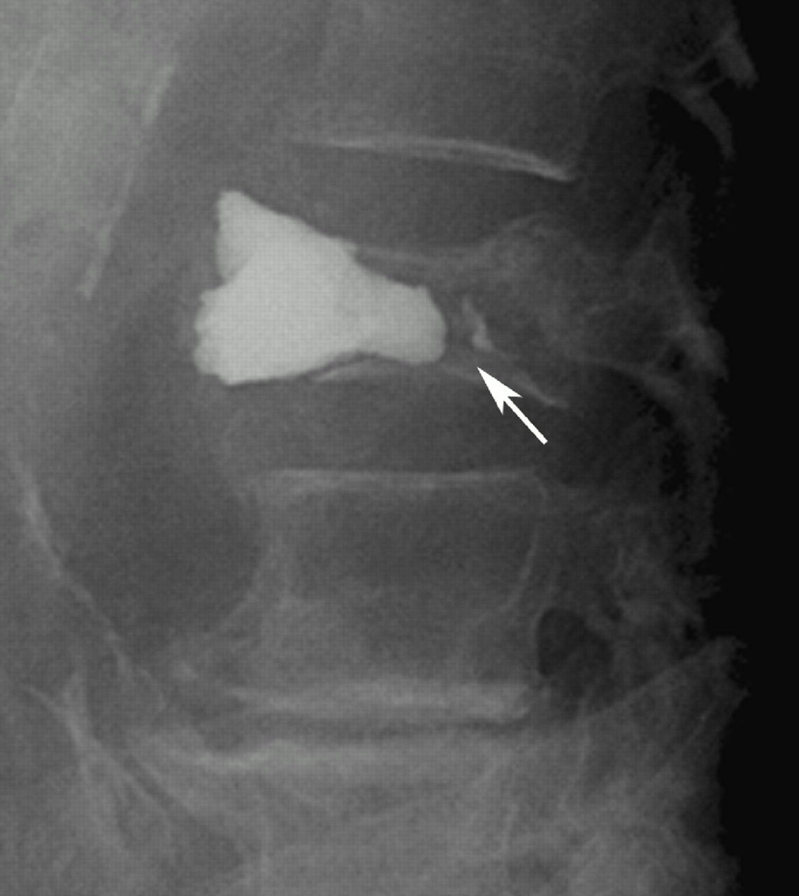
Cement was injected until a pressure end point was reached and was no longer filling the cavity, at which point the procedure was terminated. Although she experienced some mild relief at first, the patient continued to have pain in the same area. MR imaging was performed and demonstrated incomplete filling of the cavity around the existing cement and some marrow edema at the L3 level, with no other explanation for the patient’s pain. A second procedure was performed from the opposite side, with resultant filling of the additional cavity (Fig 2), and repeat MR imaging 2 weeks later demonstrated good filling of the cavity with no significant residual edema. The patient continued to have some back pain until approximately 1 month after the second procedure, when she experienced a sudden, severe onset of unrelenting back pain more severe than any previous episodes. Repeat conventional radiographs at that time demonstrated that the patient had compressed the posterior 25% of the L3 vertebral body, and the cement bolus within the vertebral body had been displaced anteriorly approximately 1 cm into the retroperitoneal space (Fig 3). At this point, the patient refused any more therapy, elected to stop dialysis treatment, and died soon thereafter.

Fig 2.
  • Download figure
  • Open in new tab
  • Download powerpoint
Fig 2.

Lateral view of the L3 vertebral body at the conclusion of vertebroplasty demonstrates cement extending throughout the anterior and middle portions of the vertebral body, filling a large cleft. The presence of the cleft is confirmed by the solid appearance and sharp outer borders of the cement bolus.

Fig 3.
  • Download figure
  • Open in new tab
  • Download powerpoint
Fig 3.

Lateral radiograph of the lumbar spine following a severe acute exacerbation of the patient’s back pain revels that the posterior portion of the vertebral body has collapsed further since the prior MR imaging, with 1 cm of anterior displacement of the intravertebral cement, which now projects well past the anterior spinal line. Note the reduced attenuation in the inferior L3 vertebral body corresponding to where the cement was initially located as well as a small amount of cement remaining in the original location (arrow).

Discussion

Vertebroplasty has become a mainstay in the treatment of osteoporotic fractures, with the first large series in this population appearing in 1997.1 Since then, there have been numerous retrospective and prospective reports in the literature regarding the efficacy and safety of this procedure.2–4 There have been complications, however, as there are in most interventional procedures, including additional fractures of the transverse process or pedicle, bleeding, infection, cement extravasation, and embolization.2, 5

The presence of a vertebral cleft represents a situation that has been well-suited to vertebroplasty, as filling the cleft with cement usually relieves pain6 and has even been reported to increase vertebral body height in some patients.7 The descriptions of the technique in the literature consist of filling of the cleft alone, with no recommendation to also fill marrow space. In our case, filling of the cleft was inadequate to treat the patient, as a refracture at the same level resulted in extrusion of the cement bolus anteriorly approximately 1 month after the procedure. To the best of our knowledge, this complication has been described only once before in the literature,8 and that patient was successfully treated with surgical stabilization. Although it is true that many levels that have undergone vertebroplasties will show some additional collapse on follow-up examinations without associated pain, the risk of symptomatic refracture has not been one commonly associated with vertebroplasty. When filling a vertebral cleft in an osteoporotic patient, it seems reasonable to assume that there is additional risk of refracture of that level, because of the strength of the cement compared with the weakness of the surrounding bone.

In this patient, the presence of long-standing renal failure and dialysis-associated osteonecrosis was the likely etiology of her vertebral cavity. Unfortunately, the marrow does not seem to have been strong enough to support the large volume of cement injected into the vertebral body, with postprocedure posterior vertebral fracture causing anterior cement extrusion. Although this is a rarely seen complication that has only been described twice, it does raise the question whether in certain patients, such as those on dialysis or with secondary drug-induced osteoporosis, and who have vertebral clefts, the marrow space should also be bolstered with cement, to prevent further collapse.

References

  1. ↵
    Jensen ME, Evans AJ, Mathis JM, et al. Percutaneous polymethylmethacrylate vertebroplasty in the treatment of osteoporotic vertebral body compression fractures: technical aspects. AJNR Am J Neuroradiol 1997;18:1897–1904
    Abstract
  2. ↵
    Evans AJ, Jensen ME, Kip KE, et al. Vertebral compression fractures: pain reduction and improvement in functional mobility after percutaneous polymethylmethacrylate vertebroplasty: retrospective report of 245 cases. Radiology 2003;226:366–72
    PubMed
  3. Zoarski GH, Snow P, Olan WJ, et al. Percutaneous vertebroplasty for osteoporotic compression fractures: quantitative prospective evaluation of long term outcomes. J Vasc Interv Radiol 2002;13:139–48
    PubMed
  4. ↵
    Barr JD, Barr MS, Lemley TJ, et al. Percutaneous vertebroplasty for pain relief and spinal stabilization. Spine 2000;15:923–28
  5. ↵
    Laredo JD, Hamze B. Complications of percutaneous vertebroplasty and their prevention. Skeletal Radiol 2004;33:493–505
    PubMed
  6. ↵
    Lane JI, Maus TP, Wald JT, et al. Intravertebral clefts opacified during vertebroplasty: pathogenesis, technical implications, and prognostic significance. AJNR Am J Neuroradiol 2002;23:1642–46
    Abstract/FREE Full Text
  7. ↵
    Teng MM, Wei CJ, Wei LC, et al. Kyphosis and height restoration effects of percutaneous vertebroplasty. AJNR Am J Neuroradiol 2003;24:1893–1900
    Abstract/FREE Full Text
  8. ↵
    Tsai TT, Chen WJ, Lai PL, et al. Polymethylmethacrylate cement dislodgment following percutaneous vertebroplasty: a case report. Spine 2003;28:E457–60
    CrossRefPubMed
  • Received January 25, 2005.
  • Accepted after revision February 15, 2005.
  • Copyright © American Society of Neuroradiology
View Abstract
PreviousNext
Back to top

In this issue

American Journal of Neuroradiology: 27 (1)
American Journal of Neuroradiology
Vol. 27, Issue 1
January, 2006
  • Table of Contents
  • Index by author
Advertisement
Print
Download PDF
Email Article

Thank you for your interest in spreading the word on American Journal of Neuroradiology.

NOTE: We only request your email address so that the person you are recommending the page to knows that you wanted them to see it, and that it is not junk mail. We do not capture any email address.

Enter multiple addresses on separate lines or separate them with commas.
Refracture with Cement Extrusion Following Percutaneous Vertebroplasty of a Large Interbody Cleft
(Your Name) has sent you a message from American Journal of Neuroradiology
(Your Name) thought you would like to see the American Journal of Neuroradiology web site.
CAPTCHA
This question is for testing whether or not you are a human visitor and to prevent automated spam submissions.
Cite this article
A.L. Wagner, E. Baskurt
Refracture with Cement Extrusion Following Percutaneous Vertebroplasty of a Large Interbody Cleft
American Journal of Neuroradiology Jan 2006, 27 (1) 230-231;

Citation Manager Formats

  • BibTeX
  • Bookends
  • EasyBib
  • EndNote (tagged)
  • EndNote 8 (xml)
  • Medlars
  • Mendeley
  • Papers
  • RefWorks Tagged
  • Ref Manager
  • RIS
  • Zotero
0 Responses
Respond to this article
Share
Bookmark this article
Refracture with Cement Extrusion Following Percutaneous Vertebroplasty of a Large Interbody Cleft
A.L. Wagner, E. Baskurt
American Journal of Neuroradiology Jan 2006, 27 (1) 230-231;
del.icio.us logo Twitter logo Facebook logo Mendeley logo
  • Tweet Widget
  • Facebook Like
  • Google Plus One

Jump to section

  • Article
    • Abstract
    • Case Report
    • Discussion
    • References
  • Figures & Data
  • Info & Metrics
  • Responses
  • References
  • PDF

Related Articles

  • No related articles found.
  • PubMed
  • Google Scholar

Cited By...

  • Minimally Invasive Stent Screw-Assisted Internal Fixation Technique Corrects Kyphosis in Osteoporotic Vertebral Fractures with Severe Collapse: A Pilot "Vertebra Plana" Series
  • Response to: the 'armed concrete approach: stent-screw-assisted internal fixation (SAIF) reconstructs and internally fixates the most severe osteoporotic vertebral fractures
  • Subsequent Fracture after Percutaneous Vertebroplasty Can Be Predicted on Preoperative Multidetector Row CT
  • Crossref
  • Google Scholar

This article has not yet been cited by articles in journals that are participating in Crossref Cited-by Linking.

More in this TOC Section

  • Bern Score Validity for SIH
  • MP2RAGE 7T in MS Lesions of the Cervical Spine
  • Deep Learning for STIR Spine MRI Quality
Show more Spine

Similar Articles

Advertisement

Indexed Content

  • Current Issue
  • Accepted Manuscripts
  • Article Preview
  • Past Issues
  • Editorials
  • Editors Choice
  • Fellow Journal Club
  • Letters to the Editor

Cases

  • Case Collection
  • Archive - Case of the Week
  • Archive - Case of the Month
  • Archive - Classic Case

Special Collections

  • Special Collections

Resources

  • News and Updates
  • Turn around Times
  • Submit a Manuscript
  • Author Policies
  • Manuscript Submission Guidelines
  • Evidence-Based Medicine Level Guide
  • Publishing Checklists
  • Graphical Abstract Preparation
  • Imaging Protocol Submission
  • Submit a Case
  • Become a Reviewer/Academy of Reviewers
  • Get Peer Review Credit from Publons

Multimedia

  • AJNR Podcast
  • AJNR SCANtastic
  • Video Articles

About Us

  • About AJNR
  • Editorial Board
  • Not an AJNR Subscriber? Join Now
  • Alerts
  • Feedback
  • Advertise with us
  • Librarian Resources
  • Permissions
  • Terms and Conditions

American Society of Neuroradiology

  • Not an ASNR Member? Join Now

© 2025 by the American Society of Neuroradiology All rights, including for text and data mining, AI training, and similar technologies, are reserved.
Print ISSN: 0195-6108 Online ISSN: 1936-959X

Powered by HighWire