Advertisement

Main menu

  • Home
  • Content
    • Current Issue
    • Accepted Manuscripts
    • Article Preview
    • Past Issue Archive
    • AJNR Case Collection
    • Case of the Week Archive
    • Classic Case Archive
    • Case of the Month Archive
  • Special Collections
    • Spinal CSF Leak Articles (Jan 2020-June 2024)
    • 2024 AJNR Journal Awards
    • Most Impactful AJNR Articles
  • Multimedia
    • AJNR Podcast
    • AJNR Scantastics
    • Video Articles
  • For Authors
    • Submit a Manuscript
    • Author Policies
    • Fast publishing of Accepted Manuscripts
    • Graphical Abstract Preparation
    • Manuscript Submission Guidelines
    • Imaging Protocol Submission
    • Submit a Case for the Case Collection
  • About Us
    • About AJNR
    • Editorial Board
  • More
    • Become a Reviewer/Academy of Reviewers
    • Subscribers
    • Permissions
    • Alerts
    • Feedback
    • Advertisers
    • ASNR Home
  • Other Publications
    • ajnr

User menu

  • Alerts
  • Log in

Search

  • Advanced search
American Journal of Neuroradiology

American Journal of Neuroradiology

ASHNR American Society of Functional Neuroradiology ASHNR American Society of Pediatric Neuroradiology ASSR
  • Alerts
  • Log in

Advanced Search

  • Home
  • Content
    • Current Issue
    • Accepted Manuscripts
    • Article Preview
    • Past Issue Archive
    • AJNR Case Collection
    • Case of the Week Archive
    • Classic Case Archive
    • Case of the Month Archive
  • Special Collections
    • Spinal CSF Leak Articles (Jan 2020-June 2024)
    • 2024 AJNR Journal Awards
    • Most Impactful AJNR Articles
  • Multimedia
    • AJNR Podcast
    • AJNR Scantastics
    • Video Articles
  • For Authors
    • Submit a Manuscript
    • Author Policies
    • Fast publishing of Accepted Manuscripts
    • Graphical Abstract Preparation
    • Manuscript Submission Guidelines
    • Imaging Protocol Submission
    • Submit a Case for the Case Collection
  • About Us
    • About AJNR
    • Editorial Board
  • More
    • Become a Reviewer/Academy of Reviewers
    • Subscribers
    • Permissions
    • Alerts
    • Feedback
    • Advertisers
    • ASNR Home
  • Follow AJNR on Twitter
  • Visit AJNR on Facebook
  • Follow AJNR on Instagram
  • Join AJNR on LinkedIn
  • RSS Feeds

Welcome to the new AJNR, Updated Hall of Fame, and more. Read the full announcements.


AJNR is seeking candidates for the position of Associate Section Editor, AJNR Case Collection. Read the full announcement.


Research ArticleBRAIN

Hippocampal Sulcus Width and Cavities: Comparison Between Patients with Alzheimer Disease and Nondemented Elderly Subjects

A.J. Bastos-Leite, J.H. van Waesberghe, A.L. Oen, W.M. van der Flier, P. Scheltens and F. Barkhof
American Journal of Neuroradiology November 2006, 27 (10) 2141-2145;
A.J. Bastos-Leite
  • Find this author on Google Scholar
  • Find this author on PubMed
  • Search for this author on this site
J.H. van Waesberghe
  • Find this author on Google Scholar
  • Find this author on PubMed
  • Search for this author on this site
A.L. Oen
  • Find this author on Google Scholar
  • Find this author on PubMed
  • Search for this author on this site
W.M. van der Flier
  • Find this author on Google Scholar
  • Find this author on PubMed
  • Search for this author on this site
P. Scheltens
  • Find this author on Google Scholar
  • Find this author on PubMed
  • Search for this author on this site
F. Barkhof
  • Find this author on Google Scholar
  • Find this author on PubMed
  • Search for this author on this site
  • Article
  • Figures & Data
  • Info & Metrics
  • Responses
  • References
  • PDF
Loading

Article Figures & Data

Figures

  • Tables
  • Fig 1.
    • Download figure
    • Open in new tab
    • Download powerpoint
    Fig 1.

    Magnified coronal high-resolution T1-weighted images of the hippocampal region.

    A and B, Coronal T1-weighted image of a 68-year-old nondemented control showing discrete enlargement of the choroidal fissures (cf), suggesting medial temporal lobe atrophy (MTA) grade 1, and hippocampal cavities bilaterally (vertical arrows). Note that both hippocampal sulci are not enlarged (horizontal arrows).

    C and D, Coronal T1-weighted image of a 54-year-old patient with Alzheimer disease showing enlargement of the hippocampal sulcus, measured between the fimbria and the subiculum (vertical measurement overlays), and enlargement of the choroidal fissures (cf) (MTA grade 1).

    E and F, Coronal T1-weighted image of a 76-year-old patient with Alzheimer disease showing enlargement of the hippocampal sulcus (vertical measurement overlays), moderate-to-severe MTA (grade 3) and hippocampal cavities bilaterally (vertical arrows).

    G and H, Coronal T1-weighted image of a 93-year-old patient with Alzheimer disease showing severe MTA (grade 4) and a small hippocampal cavity on the right side (vertical arrow). Note that the fimbria appears laterally displaced; this displacement contributes to an increase of the fimbriosubicular distance (oblique measurement overlays).
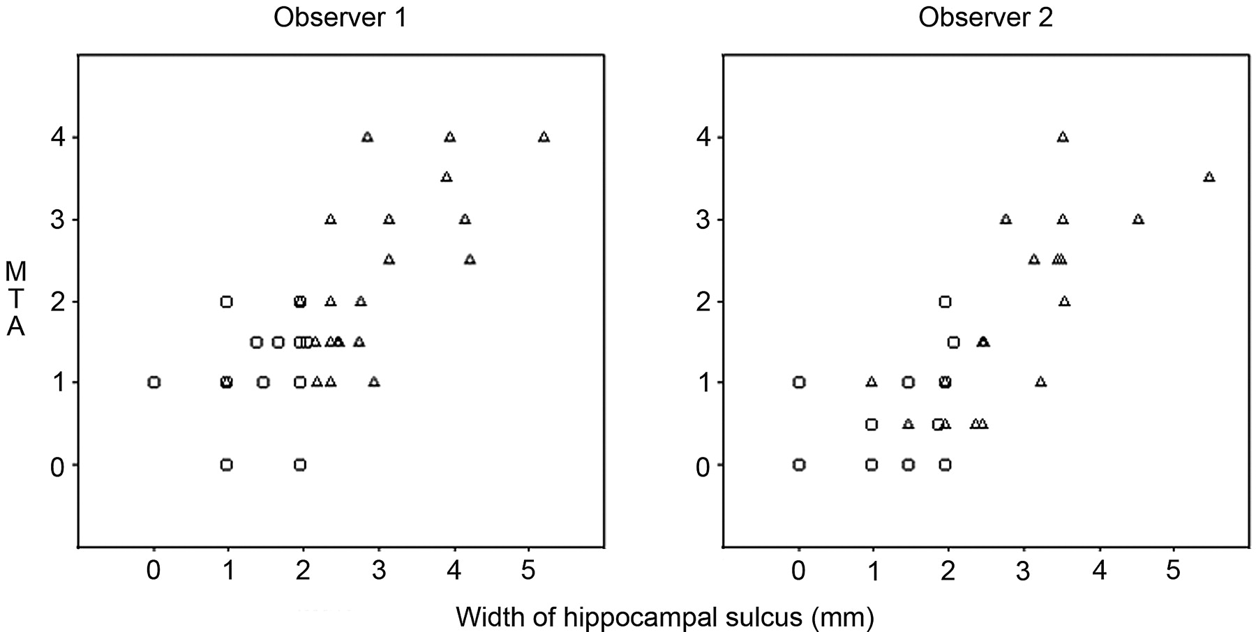
  • Fig 2.
    • Download figure
    • Open in new tab
    • Download powerpoint
    Fig 2.

    Scatterplots displaying the averaged left/right hippocampal sulcus width (fimbriosubicular distance) plotted against the medial temporal atrophy score. Triangles represent patients with Alzheimer disease, and circles correspond to control subjects.

Tables

  • Figures
    • View popup
    Table 1:

    Characteristics of patients with Alzheimer disease (AD) (n = 21) and nondemented elderly control subjects (n = 15) including age, mini-mental state examination (MMSE), hippocampal sulcus (HS) width, number and size of hippocampal cavities (HC), and medial temporal lobe atrophy (MTA) score

    CharacteristicMean (SD)
    AD PatientsControl SubjectsP Value
    Age69.3 (10.9)68.9 (8.0).91
    MMSE score*18.8† (4.5)27.9† (1.9)<.0001
    HS width‡ (mm), observer 12.9 (1.0)1.4 (0.6)<.0001
    HS width‡ (mm), observer 22.8 (1.1)1.4 (0.7)<.0001
    HS width‡ (mm), interobserver average2.8 (0.9)1.4 (0.6)<.0001
    Number of HC, observer 12.6 (3.2)3.6 (2.8).32
    Number of HC, observer 21.9 (2.4)3.6 (2.9).06
    Number of HC, interobserver average2.2 (2.7)3.6 (2.8).15
    Mean size of HC (mm), observer 11.7 (1.0)2.0 (1.1).29
    Mean size of HC (mm), observer 21.3 (1.1)2.0 (0.8).06
    Mean size of HC (mm), interobserver average1.5 (0.9)2.0 (0.9).10
    MTA score‡§, observer 12.2† (1.0)1.1† (0.7)<.01
    MTA score‡§, observer 21.7† (1.1)0.6† (0.7)<.01
    MTA score‡§, interobserver average2.0† (1.1)0.8† (0.6)<.01
    • Note:—

    • * Lower values indicate greater severity.

    • † Please note that means of scores are presented, whereas we used the Mann-Whitney U test to compare differences between groups.

    • ‡ Values are left/right averages.

    • § Higher values indicate greater severity.

    • View popup
    Table 2:

    Interobserver averaged hippocampal sulcus (HS) width (fimbriosubicular distance) according to grade of medial temporal lobe atrophy (MTA) in patients with Alzheimer disease (AD) (n = 21) and nondemented elderly control subjects (n = 15)

    MTA scoreMean HS Width in mm (SD)P Value
    AD PatientsControl Subjects
    0 (n = 3)1.4 (0.5)
    1 (n = 18)2.1 (0.5)1.3 (0.7)P < 0.05
    2 (n = 6)2.5 (0.1)1.7 (0.4)P < 0.05
    3 (n = 6)3.5 (0.4)
    4 (n = 3)4.1 (1.1)
PreviousNext
Back to top

In this issue

American Journal of Neuroradiology: 27 (10)
American Journal of Neuroradiology
Vol. 27, Issue 10
November 2006
  • Table of Contents
  • Index by author
Advertisement
Print
Download PDF
Email Article

Thank you for your interest in spreading the word on American Journal of Neuroradiology.

NOTE: We only request your email address so that the person you are recommending the page to knows that you wanted them to see it, and that it is not junk mail. We do not capture any email address.

Enter multiple addresses on separate lines or separate them with commas.
Hippocampal Sulcus Width and Cavities: Comparison Between Patients with Alzheimer Disease and Nondemented Elderly Subjects
(Your Name) has sent you a message from American Journal of Neuroradiology
(Your Name) thought you would like to see the American Journal of Neuroradiology web site.
CAPTCHA
This question is for testing whether or not you are a human visitor and to prevent automated spam submissions.
Cite this article
A.J. Bastos-Leite, J.H. van Waesberghe, A.L. Oen, W.M. van der Flier, P. Scheltens, F. Barkhof
Hippocampal Sulcus Width and Cavities: Comparison Between Patients with Alzheimer Disease and Nondemented Elderly Subjects
American Journal of Neuroradiology Nov 2006, 27 (10) 2141-2145;

Citation Manager Formats

  • BibTeX
  • Bookends
  • EasyBib
  • EndNote (tagged)
  • EndNote 8 (xml)
  • Medlars
  • Mendeley
  • Papers
  • RefWorks Tagged
  • Ref Manager
  • RIS
  • Zotero
0 Responses
Respond to this article
Share
Bookmark this article
Hippocampal Sulcus Width and Cavities: Comparison Between Patients with Alzheimer Disease and Nondemented Elderly Subjects
A.J. Bastos-Leite, J.H. van Waesberghe, A.L. Oen, W.M. van der Flier, P. Scheltens, F. Barkhof
American Journal of Neuroradiology Nov 2006, 27 (10) 2141-2145;
del.icio.us logo Twitter logo Facebook logo Mendeley logo
  • Tweet Widget
  • Facebook Like
  • Google Plus One

Jump to section

  • Article
    • Abstract
    • Materials and Methods
    • Results
    • Discussion
    • Conclusion
    • References
  • Figures & Data
  • Info & Metrics
  • Responses
  • References
  • PDF

Related Articles

  • No related articles found.
  • PubMed
  • Google Scholar

Cited By...

  • Ontario Neurodegenerative Disease Research Initiative (ONDRI): Structural MRI methods & outcome measures
  • Calcified Neurocysticercosis Associates with Hippocampal Atrophy: A Population-Based Study
  • Frequency and Location of Dilated Virchow-Robin Spaces in Elderly People: A Population-Based 3D MR Imaging Study
  • Crossref
  • Google Scholar

This article has not yet been cited by articles in journals that are participating in Crossref Cited-by Linking.

More in this TOC Section

  • Predictors of Reperfusion in Patients with Acute Ischemic Stroke
  • Qualitative and Quantitative Analysis of MR Imaging Findings in Patients with Middle Cerebral Artery Stroke Implanted with Mesenchymal Stem Cells
  • Multimodal CT Provides Improved Performance for Lacunar Infarct Detection
Show more Brain

Similar Articles

Advertisement

Indexed Content

  • Current Issue
  • Accepted Manuscripts
  • Article Preview
  • Past Issues
  • Editorials
  • Editors Choice
  • Fellow Journal Club
  • Letters to the Editor

Cases

  • Case Collection
  • Archive - Case of the Week
  • Archive - Case of the Month
  • Archive - Classic Case

Special Collections

  • Special Collections

Resources

  • News and Updates
  • Turn around Times
  • Submit a Manuscript
  • Author Policies
  • Manuscript Submission Guidelines
  • Evidence-Based Medicine Level Guide
  • Publishing Checklists
  • Graphical Abstract Preparation
  • Imaging Protocol Submission
  • Submit a Case
  • Become a Reviewer/Academy of Reviewers
  • Get Peer Review Credit from Publons

Multimedia

  • AJNR Podcast
  • AJNR SCANtastic
  • Video Articles

About Us

  • About AJNR
  • Editorial Board
  • Not an AJNR Subscriber? Join Now
  • Alerts
  • Feedback
  • Advertise with us
  • Librarian Resources
  • Permissions
  • Terms and Conditions

American Society of Neuroradiology

  • Not an ASNR Member? Join Now

© 2025 by the American Society of Neuroradiology All rights, including for text and data mining, AI training, and similar technologies, are reserved.
Print ISSN: 0195-6108 Online ISSN: 1936-959X

Powered by HighWire