
Article Figures & Data
Figures
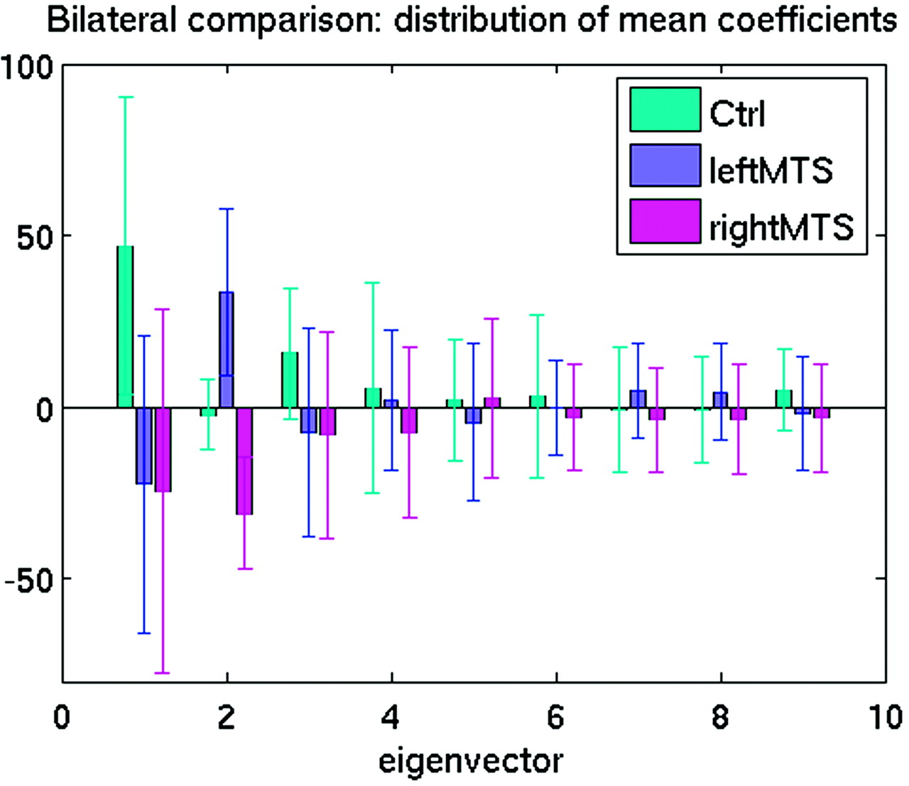
- Fig 1.
Mean coefficients associated with the first 9 eigenvectors for all groups. The displacement of each graphic point on the hippocampal surface from template (or atlas, which was a manually outlined hippocampus of a healthy subject not otherwise included in this study) to target (or subject) is represented by a vector. Crosshairs represent the standard deviation of each vector. The figure shows the first 9 eigenvectors, which explained 75% of the total variance and were used to compute asymmetry measures for each group. Of the first 3 eigenvectors, eigenvectors 1 and 3 showed large differences between the control group and each of the MTS groups but showed very similar values for the MTS groups. Eigenvector 2 showed large differences between the control and MTS groups, as well as between the MTS groups. Because eigenvectors 1 and 3 show only minimal differences between the MTS groups, they minimally contributed to discriminating shape differences between the MTS groups. Eigenvector 2, for the MTS groups, showed similar absolute magnitudes. However, the left MTS groups showed a positive value, whereas the right MTS group showed a negative value. This indicates that the shape changes represented by eigenvector 2, comparing the right and left MTS hippocampi, were highly symmetrical.
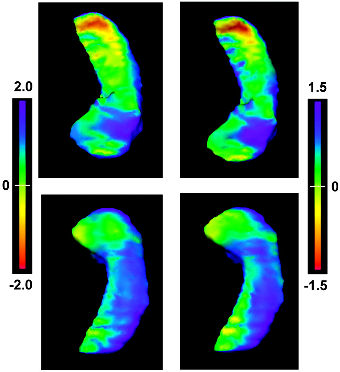
- Fig 2.
The flame-scale hippocampus panels that display deformation patterns of the left MTS hippocampi. The left column shows the deformation pattern using all eigenvector coefficients, whereas the right column shows the deformation pattern after applying only the extreme positive coefficient from eigenvector 2. Top row, view from above. Bottom row, view from below. The deformation patterns are projected on the surfaces of the control hippocampi, with the flame scale representing (in millimeters) the surface differences between the MTS hippocampi and control hippocampi. The patterns of deformation show marked similarity, demonstrating that the positive component of eigenvector 2 largely represents the deformation changes accounting for differences in the left MTS hippocampi.
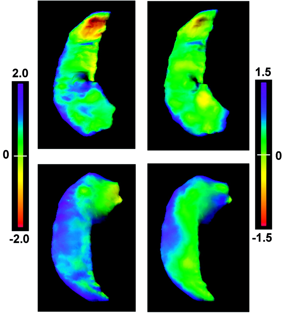
- Fig 3.
The flame scale hippocampus panels, presented in a similar fashion as in Fig 2, showing deformation patterns of the right MTS hippocampi. The left column shows the deformation pattern with application of all eigenvector coefficients, whereas the right column shows the deformation pattern after applying only the extreme negative coefficient from eigenvector 2. Flame scale units are measured in millimeters. As with the positive component of eigenvector 2 for the left MTS hippocampal deformation pattern, the negative component of eigenvector 2 largely represents the deformation changes accounting for the differences in the right MTS hippocampi.


