
Article Figures & Data
Figures
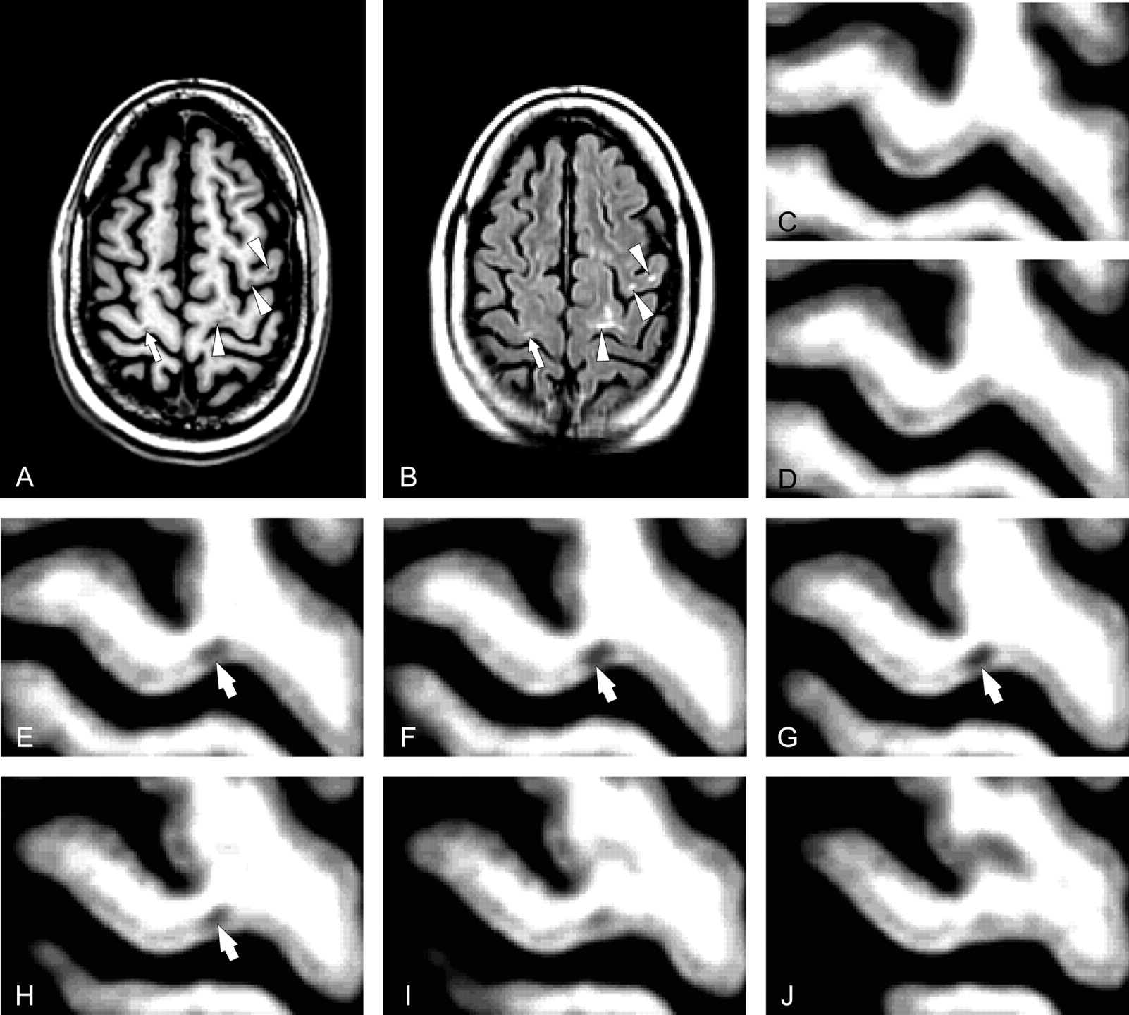
- Fig 1.
Average of 12 coregistered axial SPGR (A) and FLAIR (B) MR images demonstrate cortical MS lesions in patient 3. Several Type B cortical lesions traverse the gray-white boundary (white arrowheads, A, B). A single Type A lesion (white arrows A, B) is confined to the gray matter of the motor strip. Serial magnified images of the precentral gyrus (C-J) conform that this lesion (white arrows D-H) does not extend into the juxtacortical white matter.
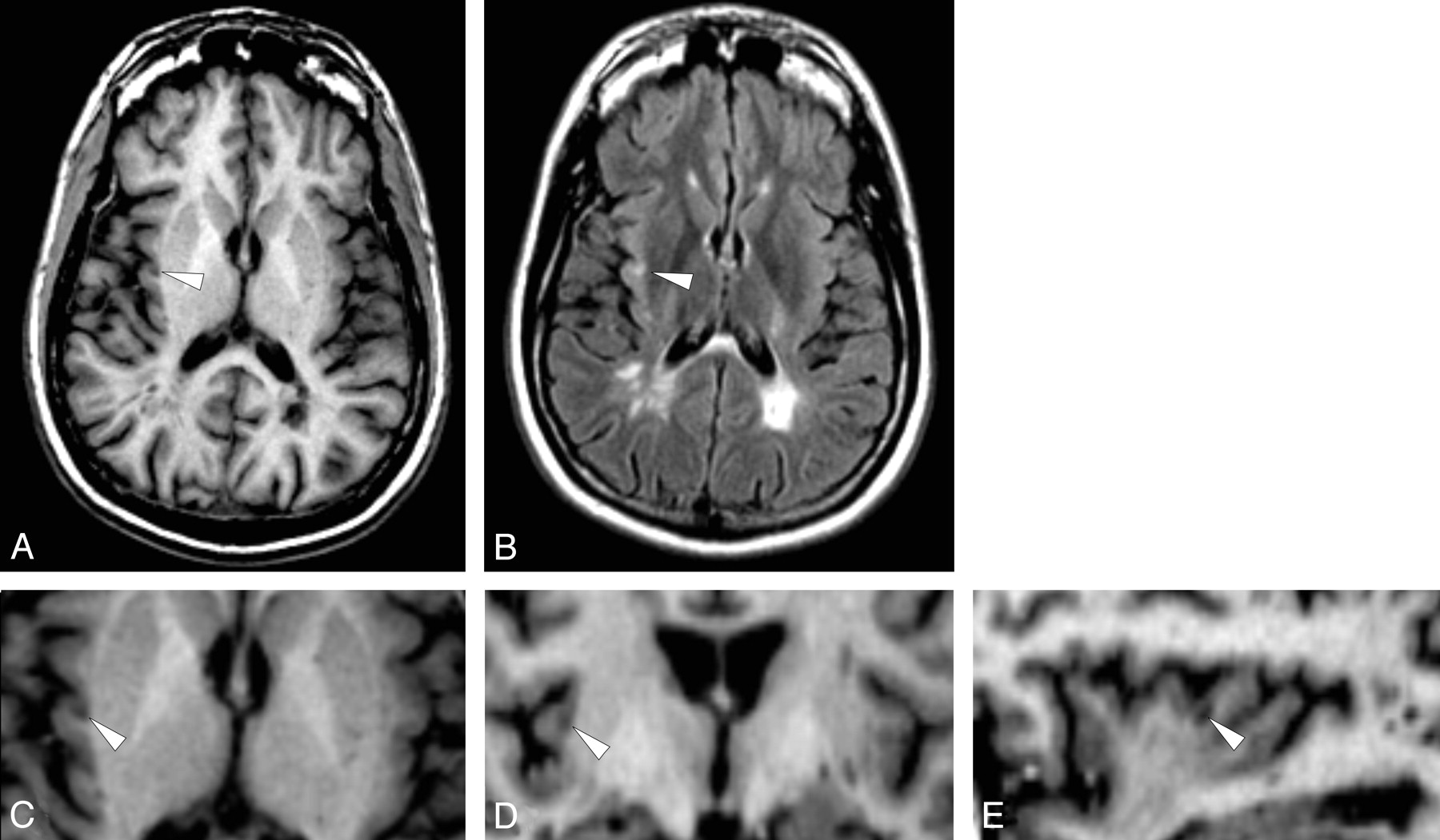
- Fig 2.
Average of 12 coregistered SPGR (A) and FLAIR (B) demonstrate a Type B cortical lesion in the right insula (arrowheads). Higher magnification of the averaged SPGR dataset in the axial (C), coronal (D), and sagittal (E) planes shows the relationship of the lesion to the cortical ribbon. (Patient 2).
- Fig 3.
Coregistration and averaging 6 (B) and 12 (C) T1 SPGR MRI volumes improves conspicuity of cortical lesions (e.g. Type B lesion arrows, A-C) relative to a single SPGR (A). Although the Type B lesion is visible on the single SPGR (A), coregistering and averaging 6 (B) and 12 (C) SPGRs improves lesion conspicuity, and allows clear characterization of lesion location with respect to the gray-white boundary. (Patient 6).


Tables
Patients (n = 22) Healthy Volunteers (n = 12) Age*† 40.3 ± 8.8 (22−57) years 41.5 ± 11.2 (28−58) years Gender† 16 women; 6 men 6 women; 6 men MS type 10 SPMS;12 RRMS Disease duration* 9.8 ± 8.2 (0.2−26.1) years EDSS score* 3.3 ± 1.9 (0−7.0) [baseline] 3.4 ± 2.1 (0−6.5) [end] Number of CELs* 41.2 ± 82.9 (0−345) [total] T2 lesions volume* (cm3) 16.2 ± 16.9 (1.2−68.2) [baseline] 14.38 ± 15.2 (0.7−52.0) [end] T1 BHs number* 10.3 ± 8.9 (0−37) [baseline] 10.5 ± 10.1 (0−39) [end] T1 BHs volume* (cm3) 1.8 ± 2.0 (0−7.5) [baseline] 1.9 ± 2.3 (0−8.3) [end] BPF*‡ 0.80 ± 0.02 (0.76−0.83) [baseline] 0.85 ± 0.01 (0.84−0.88) 0.79 ± 0.02 (0.74−0.83) [end] On-going therapy Cyclophoshamide = 1 (4 untreated patients) Copolimer-1 = 4 Interferon β (IFNβ) = 6 IFNβ + Daclizumab = 7 Note:—MS indicates multiple sclerosis; RRMS, relapsing-remitting multiple sclerosis; SPMS, secondary-progressive multiple sclerosis; EDSS, expanded disability status scale; CEL, contrast-enhancing lesion; BH, black hole; BPF, brain parenchymal fraction.
* mean ± SD (range).
† P values vs healthy volunteers > .05.
‡ P ≤ .0001. See text for comparisons within patients at different time points.
- Table 2:
Demographic, clinical, and MRI characteristics of patients with cortical lesions at the study entry
Pt # Age Gender Years of MS EDSS MS type T1 Les Volume (cm3) T2 Les Volume (cm3) CELs BPF CLs(A) CLs(B) 1 35.2 Male 9 5.5 SP 4.4 12.5 0 0.77 0 2 2 35.7 Male 3.4 0 RR 0.8 19.2 6 0.78 1 6 3 35.8 Male 4.7 1.5 RR 1.4 12.1 0 0.78 3 7 4 36.9 Female 15.4 2.5 RR 1.6 16.9 NA 0.81 1 7 5 50.3 Female 8.9 6 SP 2.4 15.2 4 0.82 0 10 6 48.6 Female 24 2.5 RR 4.1 42.2 2 0.77 2 3 7 42.7 Female 0.3 1.5 RR 0.1 2.6 1 0.82 0 3 8 41.2 Female 25.2 4.5 SP 1.2 18.6 1 0.77 1 5 9 48.3 Female 6.1 2.5 SP* 2.5 68.3 26 0.76 5 1 10 26.5 Female 13 6 SP 7.5 25.6 0 0.76 2 0 11 56.6 Female 18.9 6.5 SP 1.4 6.3 2 0.68 1 7 12 22.3 Male 2 3 RR 4.2 44.70 3 0.81 4 13 13 35.7 Female 11.9 5 SP 0.2 1.2 0 0.82 0 2 Note:—MS indicates multiple sclerosis; RR, relapsing-remitting; SP, secondary-progressive; EDSS, expanded disability status scale; CEL, contrast-enhancing lesion; CL, cortical lesion; BPF, brain parenchymal fraction; NA, not available; Les, lesion.
* Relapsing-progressive MS.
Number (% of total) Cortical Lesions Type A Type B Superior frontal gyrus 17 (19.8%) 2 15 Middle frontal gyrus 25 (29.1%) 6 19 Inferior frontal gyrus 3 (3.5%) 1 2 Precentral gyrus 18 (20.5%) 6 12 Postcentral gyrus 3 (3.5%) 0 3 Precuneus 2 (2.3%) 1 1 Superior parietal lobe 2 (2.3%) 0 2 Supramarginal gyrus 1 (1.2%) 0 1 Inferior parietal lobe 2 (2.3%) 0 2 Insula 2 (2.3%) 0 2 Superior temporal gyrus 5 (6.8%) 2 3 Inferior temporal gyrus 1 (1.2%) 0 1 Parahippocampal gyrus 2 (2.3%) 0 2 Uncus 2 (2.3%) 1 1 Cuneus 1 (1.2%) 1 0 Total 86 20 66
In this issue


Jump to section
Related Articles
Cited By...
- Multimodal Quantitative Magnetic Resonance Imaging of Thalamic Development and Aging across the Human Lifespan: Implications to Neurodegeneration in Multiple Sclerosis
- Identification and Clinical Impact of Multiple Sclerosis Cortical Lesions as Assessed by Routine 3T MR Imaging
- Consensus recommendations for MS cortical lesion scoring using double inversion recovery MRI
- Imaging distribution and frequency of cortical lesions in patients with multiple sclerosis
- MRI criteria for MS in patients with clinically isolated syndromes
- In vivo imaging of cortical pathology in multiple sclerosis using ultra-high field MRI
- Can imaging techniques measure neuroprotection and remyelination in multiple sclerosis?