
Article Figures & Data
Figures
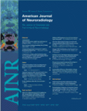
- Fig 1.
Patient 11, a 43-year-old woman with an incidentally discovered fusiform P2 aneurysm.
A, Lateral projection of vertebral aneurysm shows an 8-mm fusiform P2 aneurysm.
B, Simultaneous angiogram of vertebral artery and right internal carotid artery shows complete occlusion of the aneurysm including the parent PCA and good filling of distal PCA branches through leptomeningeal collateral vessels.
C, MR imaging 6 weeks after PCA occlusion shows no infarction in PCA territory.
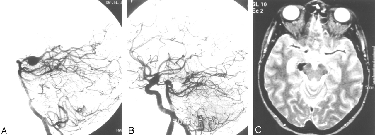
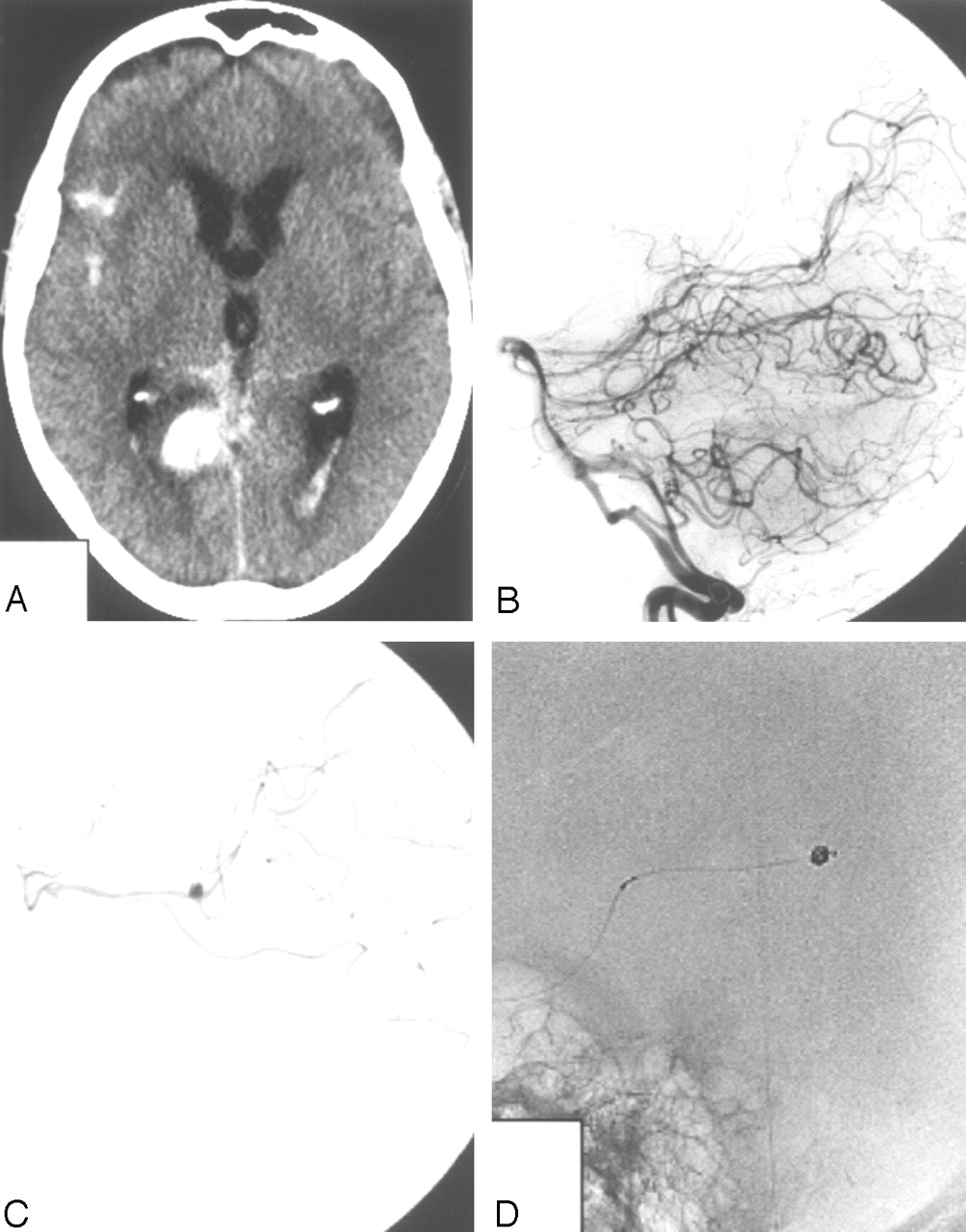
- Fig 2.
Patient 20, a 32-year-old man presenting with HH grade III SAH and hemianopsia.
A, CT scan on the day of admission shows SAH and aneurysm in the left ambient cistern (arrow).
B, Vertebral angiogram shows occluded left PCA beyond the P2, presumably by a dissecting aneurysm. Endovascular therapy was judged not necessary.
C, CT scan after sudden clinical detoriation 4 days after admission shows enlargement of the aneurysm, recurrent SAH with thalamic hematoma and hemorrhagic infarction in the PCA territory.
D, Angiogram after recurrent SAH shows filling of large dissecting aneurysm.
E, Occlusion of the aneurysm with coils including the afferent P2. The patient died 3 days later.
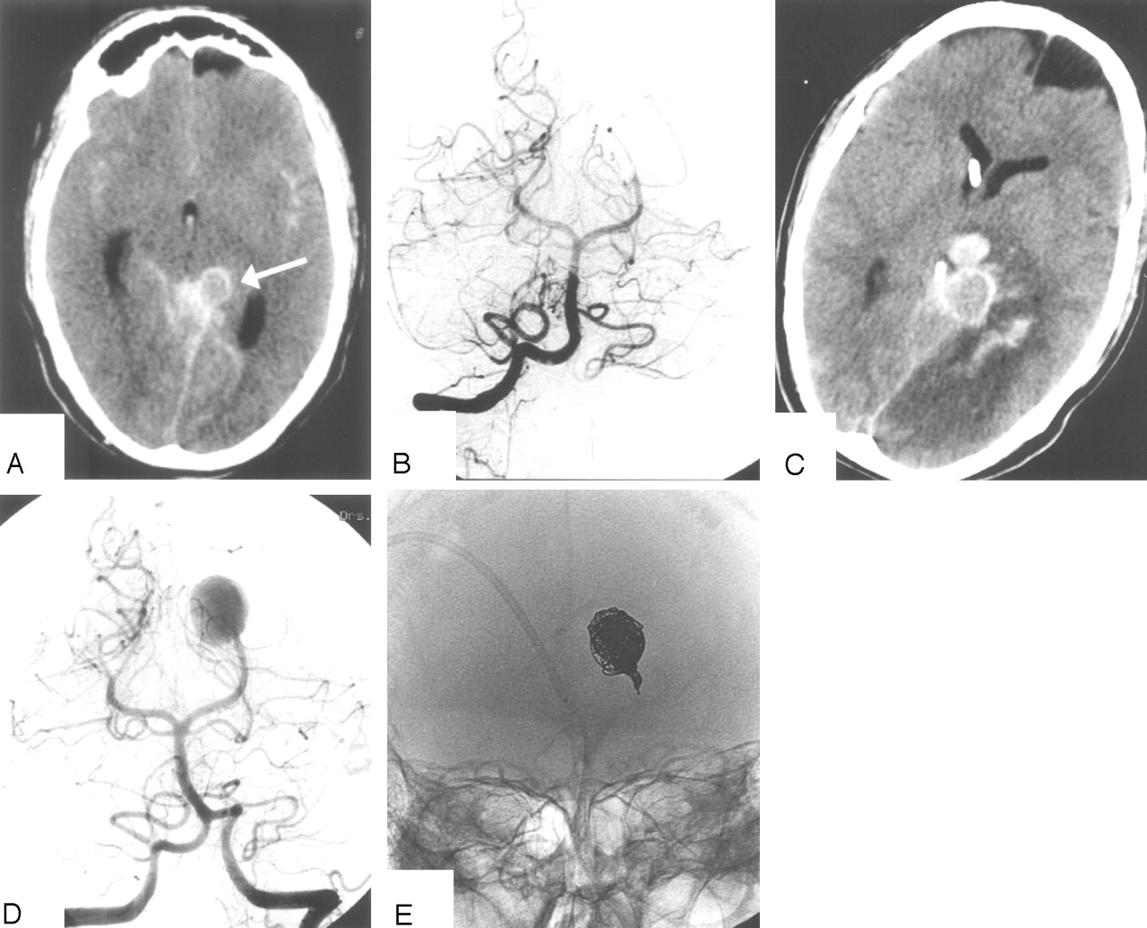
- Fig 3.
Patient 13, a 64-year-old man presenting with HH grade I SAH and right occulomotor palsy.
A and B, MR imaging and angiography show dissecting fusiform P2 aneurysm with intramural thrombus.
C and D, Vertebral (C) and right internal carotid (D) angiogram after occlusion of the aneurysm including the parent PCA show good collateral supply to the occipital lobe through leptomeningeal collaterals.
E, MR imaging 6 weeks after PCA occlusion demonstrates no infarction in right PCA territory.
- Fig 4.
Patient 22, a 64-year-old woman presenting with HH grade I SAH and hemianopsia.
A, CT scan showing subarachnoid and intraventricular blood and a hematoma in the medial occipital lobe.
B, Lateral vertebral angiogram showing a small aneurysm on the P4 (arrow).
C, Superselective angiogram, which better demonstrates the small aneurysm.
D, Occlusion of the aneurysm including the parent artery with coils.
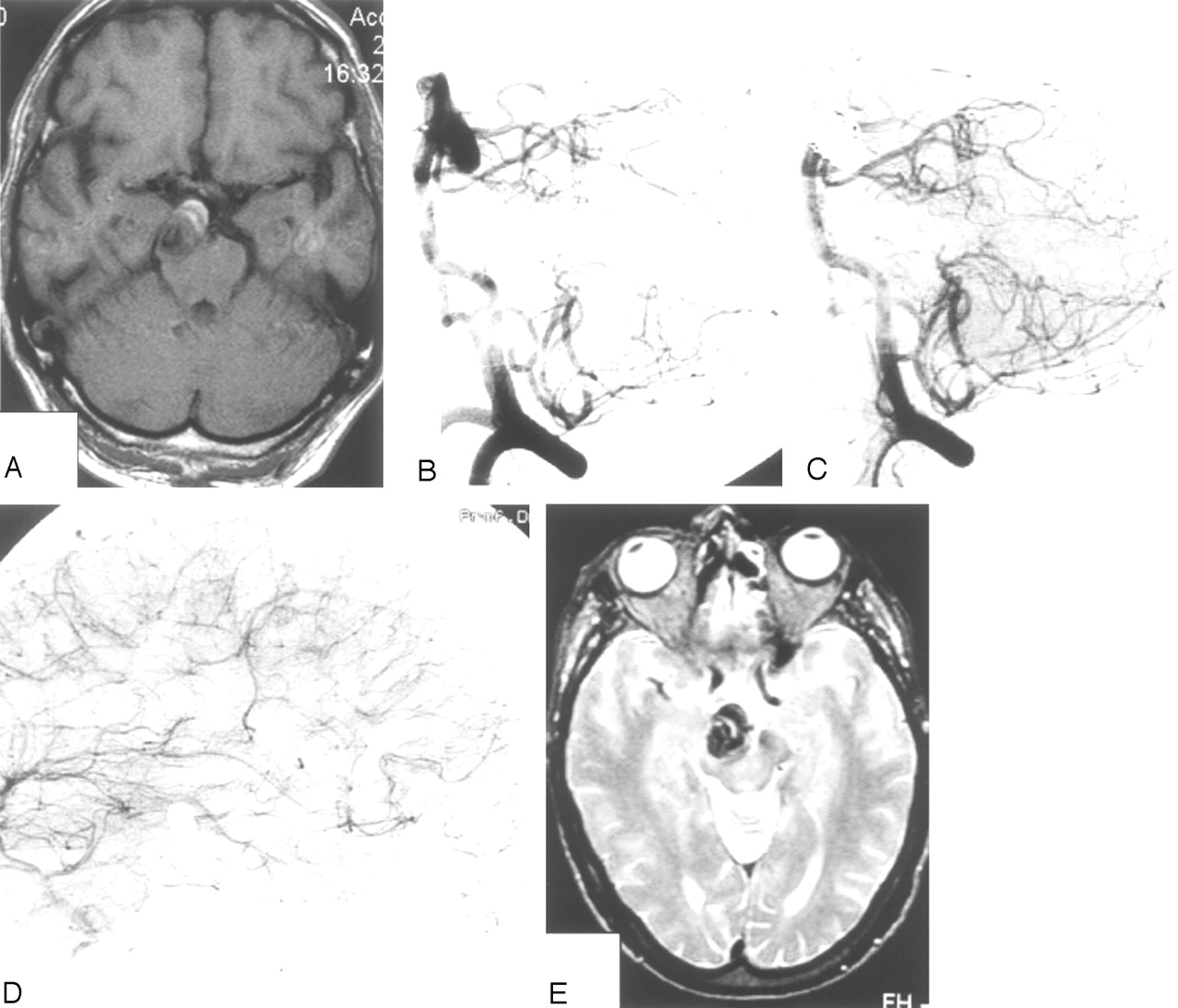
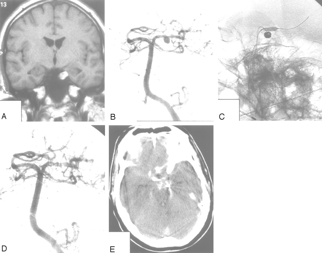
- Fig 5.
Patient 12, a 35-year-old man presenting with acute left occulomotor palsy.
A and B, MR imaging and angiography show a left P1–P2 aneurysm pointing downward with an intramural thrombus.
C and D, Coiling of the aneurysm with the aid of a supporting balloon results in complete occlusion of the lumen.
E, CT scan 2 months later shows SAH from the coiled aneurysm. The patient died the next day.


Tables
- Table 1:
Clinical and aneurysm characteristics of 22 patients with posterior cerebral artery aneurysms
Patient No./Age (y)/Sex Clinical Presentation Site Aneurysm Type Size (mm) Treatment Outcome and Duration of Follow-up Associated Disease 1/F/41 SAH other aneurysm P1–P2 Saccular 6 Coil occlusion GOS 5 12 mo 1 other aneurysm 2/F/72 SAH other aneurysm P1–P2 Saccular 6 Coil occlusion GOS 5 24 mo 1 other aneurysm 3/F/59 SAH other aneurysm P3–P4 Saccular 4 Coil occlusion GOS 5 8 mo 3 other aneurysms 4/F/49 SAH other aneurysm P1–P2 Saccular 3 Coil occlusion GOS 5 10 mo 3 other aneurysms 5/F/47 SAH other aneurysm P1–P2 Saccular 3 Coil occlusion GOS 5 12 mo 5 other aneurysms 6/F/45 SAH other aneurysm P1–P2 Saccular 4 Coil occlusion GOS 5 6 mo 7 other aneurysms 7/F/55 SAH other aneurysm P1–P2 Saccular 4 Coil occlusion GOS 5 8 mo 3 other aneurysms 8/F/51 SAH other aneurysm P1–P2 Saccular 2 Coil occlusion GOS 5 6 mo 2 other aneurysms 9/M/56 SAH other aneurysm P1–P2 Saccular 4 Coil occlusion GOS 5 7 mo 1 other aneurysm 10/M/49 Incidental P1–P2 Saccular 7 Coil occlusion No deficit 6 mo 11/F/43 Incidental P2 Fusiform 8 Coil PVO No deficit 6 mo 12/M/35 CN III palsy P1–P2 Saccular/wall dissection 8 Coil occlusion Died 2 months later of SAH 13/M/64 SAH HH I and CN III palsy P2 Fusiform dissection 20 Coil PVO GOS 5 no deficit 12 mo 14/M/52 SAH HH II and CN III palsy P1 Saccular 6 Coil occlusion (X2) GOS 5 no deficit 18 mo Occluded left internal carotid artery 15/F/44 SAH HH I P1–P2 Saccular 3 Coil occlusion GOS 5 60 mo 1 other aneurysm 16/M/47 SAH HH I P1–P2 Saccular 3 Coil occlusion GOS 5 6 mo 17/F/52 SAH HH II P1–P2 Saccular 35 Coil occlusion (X2) GOS 5 34 mo Ipsilateral occipital AVM 18/F/62 SAH HH I P1–P2 Saccular 6 Coil occlusion GOS 5 20 mo 1 other aneurysm 19/F/41 SAH HHI P1–P2 Saccular 4 Coil occlusion GOS 5 6 mo 20/M/32 SAH HH III, hemianopsia, after rebleeding HH V P2–P3 Dissecting 16 Coil PVO Death 21/M/27 SAH HH IV P4 Mycotic 8 Coil PVO Death AIDS, endocarditis 22/F/64 SAH HH II, hemianopsia P4 Saccular 2 Coil PVO GOS 4 (hemianopsia) 6 mo Note.—SAH indicates subarachnoid hemorrhage; GOS, Glasgow Outcome Score; HH, Hunt and Hess grade; CN III, occulomotor nerve; AVM, arteriovenous malformation; PVO, parent vessel occlusion.
In this issue


Jump to section
Related Articles
Cited By...
- National treatment practices in the management of infectious intracranial aneurysms and infective endocarditis
- Endovascular parent artery occlusion of proximal posterior cerebral artery aneurysms: a report of two cases
- Endoluminal Reconstruction for Nonsaccular Aneurysms of the Proximal Posterior Cerebral Artery with the Pipeline Embolization Device
- Review of 2 Decades of Aneurysm-Recurrence Literature, Part 2: Managing Recurrence after Endovascular Coiling
- Endovascular Treatment of Large and Giant Aneurysms