Advertisement

Main menu

  • Home
  • Content
    • Current Issue
    • Accepted Manuscripts
    • Article Preview
    • Past Issue Archive
    • AJNR Case Collection
    • Case of the Week Archive
    • Classic Case Archive
    • Case of the Month Archive
  • Special Collections
    • Spinal CSF Leak Articles (Jan 2020-June 2024)
    • 2024 AJNR Journal Awards
    • Most Impactful AJNR Articles
  • Multimedia
    • AJNR Podcast
    • AJNR Scantastics
    • Video Articles
  • For Authors
    • Submit a Manuscript
    • Author Policies
    • Fast publishing of Accepted Manuscripts
    • Graphical Abstract Preparation
    • Manuscript Submission Guidelines
    • Imaging Protocol Submission
    • Submit a Case for the Case Collection
  • About Us
    • About AJNR
    • Editorial Board
  • More
    • Become a Reviewer/Academy of Reviewers
    • Subscribers
    • Permissions
    • Alerts
    • Feedback
    • Advertisers
    • ASNR Home
  • Other Publications
    • ajnr

User menu

  • Alerts
  • Log in

Search

  • Advanced search
American Journal of Neuroradiology

American Journal of Neuroradiology

ASHNR American Society of Functional Neuroradiology ASHNR American Society of Pediatric Neuroradiology ASSR
  • Alerts
  • Log in

Advanced Search

  • Home
  • Content
    • Current Issue
    • Accepted Manuscripts
    • Article Preview
    • Past Issue Archive
    • AJNR Case Collection
    • Case of the Week Archive
    • Classic Case Archive
    • Case of the Month Archive
  • Special Collections
    • Spinal CSF Leak Articles (Jan 2020-June 2024)
    • 2024 AJNR Journal Awards
    • Most Impactful AJNR Articles
  • Multimedia
    • AJNR Podcast
    • AJNR Scantastics
    • Video Articles
  • For Authors
    • Submit a Manuscript
    • Author Policies
    • Fast publishing of Accepted Manuscripts
    • Graphical Abstract Preparation
    • Manuscript Submission Guidelines
    • Imaging Protocol Submission
    • Submit a Case for the Case Collection
  • About Us
    • About AJNR
    • Editorial Board
  • More
    • Become a Reviewer/Academy of Reviewers
    • Subscribers
    • Permissions
    • Alerts
    • Feedback
    • Advertisers
    • ASNR Home
  • Follow AJNR on Twitter
  • Visit AJNR on Facebook
  • Follow AJNR on Instagram
  • Join AJNR on LinkedIn
  • RSS Feeds

Welcome to the new AJNR, Updated Hall of Fame, and more. Read the full announcements.


AJNR is seeking candidates for the position of Associate Section Editor, AJNR Case Collection. Read the full announcement.


OtherBRAIN

Intracranial Epidermoid Cyst with Hemorrhage: MR Imaging Findings

C.-Y. Chen, J.-S. Wong, S.-C. Hsieh, J.-S. Chu and W.P. Chan
American Journal of Neuroradiology February 2006, 27 (2) 427-429;
C.-Y. Chen
  • Find this author on Google Scholar
  • Find this author on PubMed
  • Search for this author on this site
J.-S. Wong
  • Find this author on Google Scholar
  • Find this author on PubMed
  • Search for this author on this site
S.-C. Hsieh
  • Find this author on Google Scholar
  • Find this author on PubMed
  • Search for this author on this site
J.-S. Chu
  • Find this author on Google Scholar
  • Find this author on PubMed
  • Search for this author on this site
W.P. Chan
  • Find this author on Google Scholar
  • Find this author on PubMed
  • Search for this author on this site
  • Article
  • Figures & Data
  • Info & Metrics
  • Responses
  • References
  • PDF
Loading

Article Figures & Data

Figures

  •   Fig 1.
    • Download figure
    • Open in new tab
    • Download powerpoint
    Fig 1.

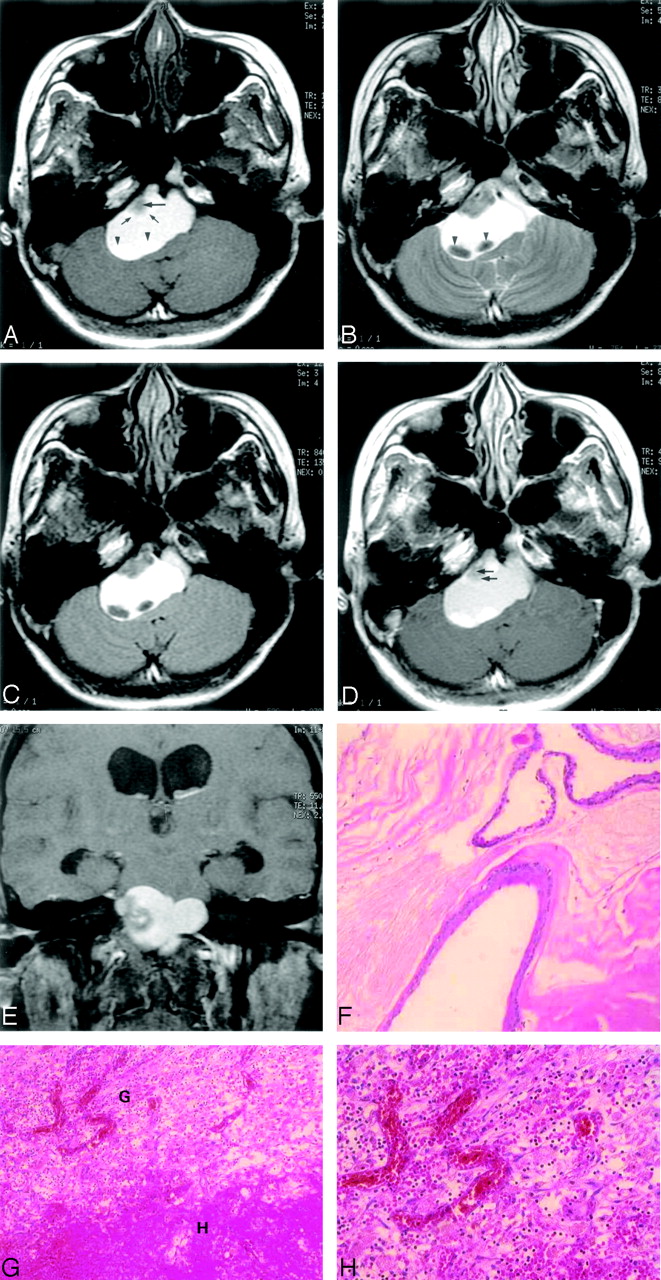
    A 28-year-old woman with posthemorrhagic epidermoid tumor.

    A, Axial T1 noncontrast-weighted fast spin-echo MR image (1823.5 milliseconds/7.4 milliseconds /2/750 milliseconds [TR/TE/excitation/TI]) shows a well-defined hyperintense extra-axial mass in the prepontine area and right cerebellopontine angle. Homogeneous hyperintensity is noted in most parts of the lesion. There is a focal area of heterogeneous intensity in the anterolateral aspect with hypointensity at the middle (arrow) and a crescent hyperintense portion at the periphery (small arrows). There are 2 oval shapes with hyperintensity in the dependent part of the lesion (arrowheads).

    B, Axial T2-weighted fast spin-echo MR image (3500 milliseconds/86 milliseconds/2 [TR/TE/excitations]) shows the hyperintense lesion has a CSF equivalent signal intensity along with focal areas of heterogeneous hypointensity and 2 areas of homogenous hypointense signal intensity (arrowheads).

    C, Axial fluid-attenuated inversion recovery MR image (8402 milliseconds/135 milliseconds/0.5/2100 milliseconds [TR/TE/excitation/TI]) shows no signal intensity drop in the lesion, and a clear border between the mass and CSF space is seen because the water signal intensity has been suppressed.

    D, Axial contrast-enhanced T1-weighted fast spin-echo MR imaging image (450 milliseconds/9 milliseconds/2 [TR/TE/excitation]) of the same level shows faint enhancement (arrows) of the anterior nodule (compare with panel A).

    E, On coronal T1-weighted, fat-suppressed contrast-enhanced MR imaging image, the hyperintensity within the lesion is not suppressed. Hydrocephalus with bilateral temporal horn dilation is noted.

    F, Photomicrograph of the lesion specimen. The cyst is lined by keratinizing squamous epithelium and filled with lamellated keratinous debris. (hematoxylin-eosin [[H&E] stain; original magnification ×100).

    G, Photomicrograph of the lesion specimen. Hemorrhage (H) with surrounding granulation tissue (G) made up about 25% of the specimen volume. (H&E stain; original magnification ×100).

    H, Photomicrograph of the lesion specimen. Mild vascularity, with capillary sized vessels, was seen in the granulation tissue. (H&E stain; original magnification ×200).

PreviousNext
Back to top

In this issue

American Journal of Neuroradiology: 27 (2)
American Journal of Neuroradiology
Vol. 27, Issue 2
February, 2006
  • Table of Contents
  • Index by author
Advertisement
Print
Download PDF
Email Article

Thank you for your interest in spreading the word on American Journal of Neuroradiology.

NOTE: We only request your email address so that the person you are recommending the page to knows that you wanted them to see it, and that it is not junk mail. We do not capture any email address.

Enter multiple addresses on separate lines or separate them with commas.
Intracranial Epidermoid Cyst with Hemorrhage: MR Imaging Findings
(Your Name) has sent you a message from American Journal of Neuroradiology
(Your Name) thought you would like to see the American Journal of Neuroradiology web site.
CAPTCHA
This question is for testing whether or not you are a human visitor and to prevent automated spam submissions.
Cite this article
C.-Y. Chen, J.-S. Wong, S.-C. Hsieh, J.-S. Chu, W.P. Chan
Intracranial Epidermoid Cyst with Hemorrhage: MR Imaging Findings
American Journal of Neuroradiology Feb 2006, 27 (2) 427-429;

Citation Manager Formats

  • BibTeX
  • Bookends
  • EasyBib
  • EndNote (tagged)
  • EndNote 8 (xml)
  • Medlars
  • Mendeley
  • Papers
  • RefWorks Tagged
  • Ref Manager
  • RIS
  • Zotero
0 Responses
Respond to this article
Share
Bookmark this article
Intracranial Epidermoid Cyst with Hemorrhage: MR Imaging Findings
C.-Y. Chen, J.-S. Wong, S.-C. Hsieh, J.-S. Chu, W.P. Chan
American Journal of Neuroradiology Feb 2006, 27 (2) 427-429;
del.icio.us logo Twitter logo Facebook logo Mendeley logo
  • Tweet Widget
  • Facebook Like
  • Google Plus One

Jump to section

  • Article
    • Abstract
    • Case Reports
    • Discussion
    • References
  • Figures & Data
  • Info & Metrics
  • Responses
  • References
  • PDF

Related Articles

  • No related articles found.
  • PubMed
  • Google Scholar

Cited By...

  • No citing articles found.
  • Crossref
  • Google Scholar

This article has not yet been cited by articles in journals that are participating in Crossref Cited-by Linking.

More in this TOC Section

  • Fast Contrast-Enhanced 4D MRA and 4D Flow MRI Using Constrained Reconstruction (HYPRFlow): Potential Applications for Brain Arteriovenous Malformations
  • Quiet PROPELLER MRI Techniques Match the Quality of Conventional PROPELLER Brain Imaging Techniques
  • Predictors of Reperfusion in Patients with Acute Ischemic Stroke
Show more Brain

Similar Articles

Advertisement

Indexed Content

  • Current Issue
  • Accepted Manuscripts
  • Article Preview
  • Past Issues
  • Editorials
  • Editors Choice
  • Fellow Journal Club
  • Letters to the Editor

Cases

  • Case Collection
  • Archive - Case of the Week
  • Archive - Case of the Month
  • Archive - Classic Case

Special Collections

  • Special Collections

Resources

  • News and Updates
  • Turn around Times
  • Submit a Manuscript
  • Author Policies
  • Manuscript Submission Guidelines
  • Evidence-Based Medicine Level Guide
  • Publishing Checklists
  • Graphical Abstract Preparation
  • Imaging Protocol Submission
  • Submit a Case
  • Become a Reviewer/Academy of Reviewers
  • Get Peer Review Credit from Publons

Multimedia

  • AJNR Podcast
  • AJNR SCANtastic
  • Video Articles

About Us

  • About AJNR
  • Editorial Board
  • Not an AJNR Subscriber? Join Now
  • Alerts
  • Feedback
  • Advertise with us
  • Librarian Resources
  • Permissions
  • Terms and Conditions

American Society of Neuroradiology

  • Not an ASNR Member? Join Now

© 2025 by the American Society of Neuroradiology All rights, including for text and data mining, AI training, and similar technologies, are reserved.
Print ISSN: 0195-6108 Online ISSN: 1936-959X

Powered by HighWire