
Article Figures & Data
Figures
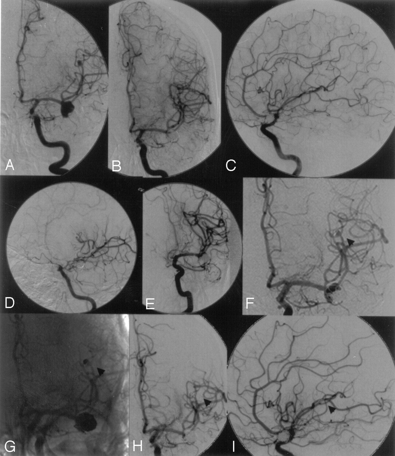
- Fig 1.
A, Left internal carotid artery (ICA) angiogram showing a left middle cerebral artery (MCA) bifurcation aneurysm and an additional distal MCA aneurysm.
B and C, Left ICA angiogram after complete embolization of both MCA aneurysms.
D, Left ICA angiogram 4 days after coil embolization demonstrating severe vasospasms at the distal ICA and proximal MCA.
E, After successful balloon dilation of the ICA and proximal MCA by using a 3-mm balloon occlusion system.
F and G, Six-month follow-up showing partial recanalization of both aneurysms as a result of coil compaction (arrowhead at distal MCA aneurysm).
H and I, Left ICA angiogram after retreatment showing complete occlusion of both MCA aneurysms (arrowhead at distal MCA aneurysm).
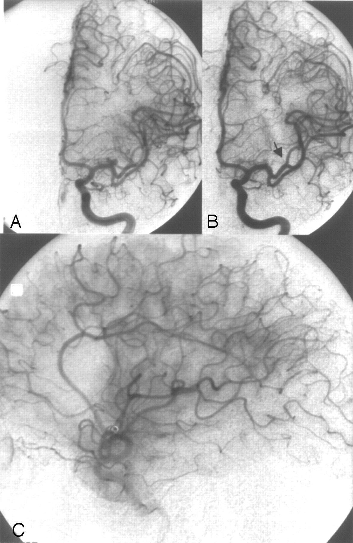
- Fig 2.
Case 1.
A, Left internal carotid artery (ICA) angiogram showing a 3-mm left middle cerebral artery (MCA) bifurcation aneurysm.
B, Left ICA angiogram after complete aneurysm occlusion with 2 coils (GDC-10: 3 × 6, 2 × 4). Note the occlusion of the small neighboring arterial branch M2 (arrowhead).
C, Left ICA angiogram, lateral view, late arterial phase after selective intra-arterial thrombolysis (1 Mio IU urokinase) still showing a small perfusion deficit. The MCA was partially filled in a retrograde manner through cortical vessels, thus avoiding larger infarction. Outcome according GOS at 6 months was GR.
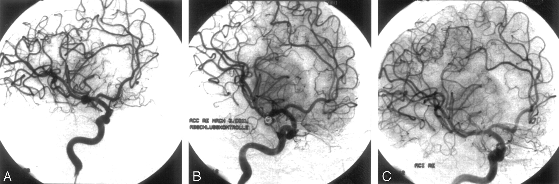
- Fig 3.
Case 2.
A, Right internal carotid artery (ICA) angiogram, oblique view, showing a 4-mm aneurysm at the right middle cerebral artery (MCA) bifurcation.
B, Right ICA angiogram, oblique view, after embolization with 3 platinum coils (GDC-10: 4 × 10, 3 × 8, 2 × 4) still demonstrating a slight opacification of the medial aneurysm rim, classified as subtotal occlusion.
C, Follow-up angiogram 6 months after embolization revealed subsequent thrombosis with total (100%) aneurysm occlusion.
- Fig 4.
Case 3.
A, CT angiography revealing bilateral asymptomatic middle cerebral artery (MCA) bifurcation aneurysms and an additional aneurysm at the left distal superior cerebellar artery.
B and C, In a first procedure, the left MCA and the cerebellar superior artery aneurysms were completely embolized.
D–F, Eight weeks later, the right MCA bifurcation aneurysm was completely occluded with coils.


Tables
- Table 1:
Patient characteristics, clinical data, aneurysm size and location, occlusion rates, and outcome for patients treated by coiling
Patient No./Age (y) SAH HH Bleeding Source Multiple Aneurysms MCA Location Size Occlusion Rate after EVT Occlusion Rate after 6 mo GOS at 30 d GOS at 6 mo 1/31 x 5 LB 3 LB 8 100 100 MD MD 2/73 x 2 ACA (L) 3 L M1 6 95–99 100 GR GR 3/81 x 2 MCA (R) 1 R M1 4 100 * MD GR 4/67 x 3 MCA (L) 1 L M1 4 100 100 SD GR 5/51 x 2 MCA (R) 3 R M1 5 100 100 GR GR 6/46 0 None 2 R M2 3 100 100 GR GR 7/60 0 None 1 R M1 7 95–99 † MD † 8/41 0 None 2 LB 4 95–99 100 GR GR 9/41 x 3 MCA (R) 1 R M1 8 100 95–99 SD MD 10/72 x 3 MCA (L) 1 L M2 6 100 95–99 SD GR 11/64 x 3 MCA (R) 2 L M1 5 100 * MD MD 12/56 x 1 RB 1 RB 4 100 100 GR GR 13/53 x 1 LB 1 LB 4 100 95–99 GR GR 14/69 0 None 1 RB 3 95–99 95–99 GR GR 15/46 0 None 1 L M1 3 100 100 MD GR 16/39 x 1 MCA (R) 3 L M1 15 100 † MD † 17/44 0 None 2 LB 18 95–99 95–99 GR GR 18/46 x 1 MCA (R) 3 R M1 5 100 100 GR GR 19/31 0 None 1 R M1 4 95–99 100 GR GR 20/58 x 3 ICA (L) 2 R M1 7 100 100 SD GR 21/49 0 None 2 RB 3 100 100 GR GR 22/32 x 2 LB 2 LB 10 100 95–99 MD GR L M1 3 100 95–99 23/72 x 2 RB 1 RB 5 95–99 ‡ SD 24/43 0 None 2 LB 5 95–99 * MD D 25/41 0 None 3 RB 4 100 100 GR MD 26/63 0 None 1 RB 5 100 100 GR GR 27/41 0 None 1 LB 4 100 100 GR GR 28/52 0 None 1 RB 3 100 100 GR GR 29/62 0 None 2 L M2 12 100 100 GR GR 30/34 0 None 1 RB 8 100 <95 GR GR 31/43 0 None 3 LB 14 100 100 GR GR RB 16 100 100 Note:—SAH indicates subarachnoid hemorrage; HH, Hunt and Hess classification; MCA, middle cerebral artery; EVT, endovascular therapy; GOS, Glasgow Outcome Scale; L, left; R, right; LB, left MCA bifurcation; RB, right MCA bifurcation; ACA, anterior cerebral artery; ICA, internal carotid artery; R/L M1, right/left MCA, segment M1; R/L M2, right/left MCA, segment 2. For GOS scores, GR indicates good recovery; MD, moderately disabled; SD, severely disabled; D, died.
* Patient did not want additional follow-up.
† Patient was not available.
‡ Patient died before 6-month control angiography.
- Table 2:
Patient characteristics, clinical data, aneurysm size and location, occlusion rates, and outcome for patients with intended, but not performed, coiling (subsequent surgical clipping)
Patient No./Age (y) SAH HH Bleeding Source Multiple Aneurysms MCA Location Size GOS at 30 d GOS at 6 mo 1/51 x 1 MCA (R) 2 R M1 15 SD MD 2/58 0 None 2 L M2 3 GR GR 3/36 x 2 MCA (R) 1 R M1 5 GR GR 4/56 0 None 4 L M1 5 GR GR 5/39 x 2 MCA (L) 1 L M1 5 MD GR Note:—SAH indicates subarachnoid hemorrage; HH, Hunt and Hess classification; MCA, middle cerebral artery; GOS, Glasgow Outcome Scale; L, left; R, right; LB, left MCA bifurcation; RB, right MCA bifurcation; ICA, internal carotid artery; R/L M1, right/left MCA, segment M1; R/L M2, right/left MCA, segment 2. For GOS scores, GR indicates good recovery; MD, moderately disabled; SD, severely disabled.
In this issue


Jump to section
Related Articles
Cited By...
- Long-Term Outcomes of the WEB Device for Treatment of Wide-Neck Bifurcation Aneurysms
- Intra-arterial versus intravenous abciximab therapy for thromboembolic complications of neuroendovascular procedures: case review and meta-analysis
- Stent-Assisted Coiling of Complex Middle Cerebral Artery Aneurysms: Initial and Midterm Results
- Endovascular Treatment of Wide-Neck Middle Cerebral Artery Aneurysms with Stents: A Review of 16 Cases