
Article Figures & Data
Figures
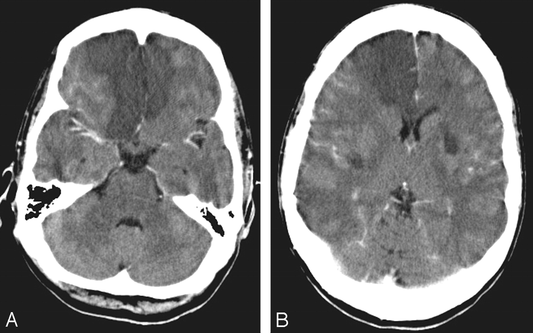
- Fig 1.
Axial cranial CT scans obtained with intravenous contrast enhancement (A, -B) show multiple areas of abnormal low attenuation, which are most dramatic within both frontal lobes as well as within the bilateral basal ganglia.
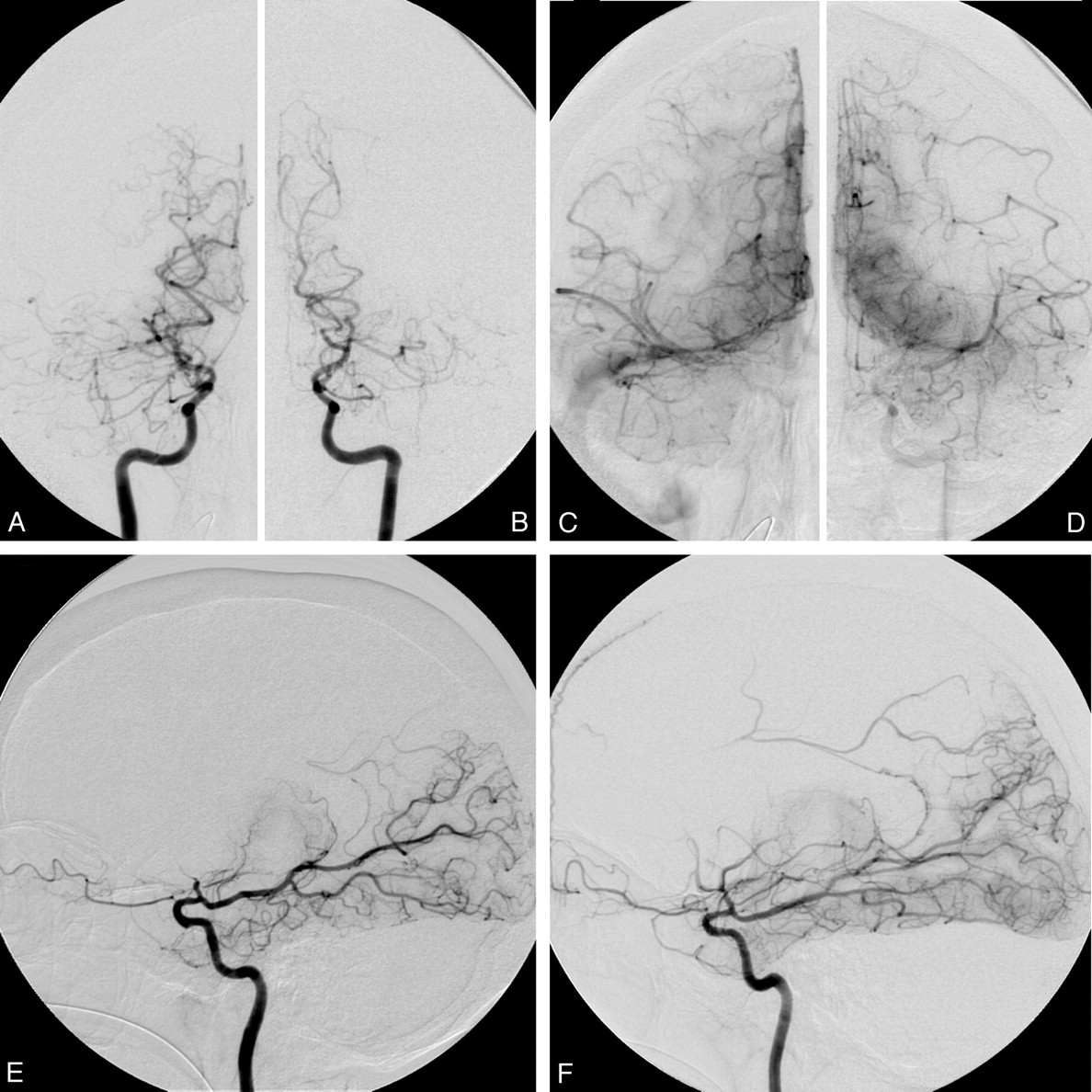
- Fig 2.
The anteroposterior projections of bilateral internal carotid artery (ICA) angiography in early (A, right; -B, left) and late arterial phases (C, right; -D, left) and lateral projections of bilateral ICA angiography (E, right; -F, left) show nearly complete occlusion of the bilateral supraclinoid ICA as well as bilateral proximal segments of the ACAs and MCAs. Only minimal delay antegrade flows of the bilateral MCAs are noted. Collateral flows are seen from the posterior cerebral artery via the posterior choroidal plexus to the pericallosal artery.
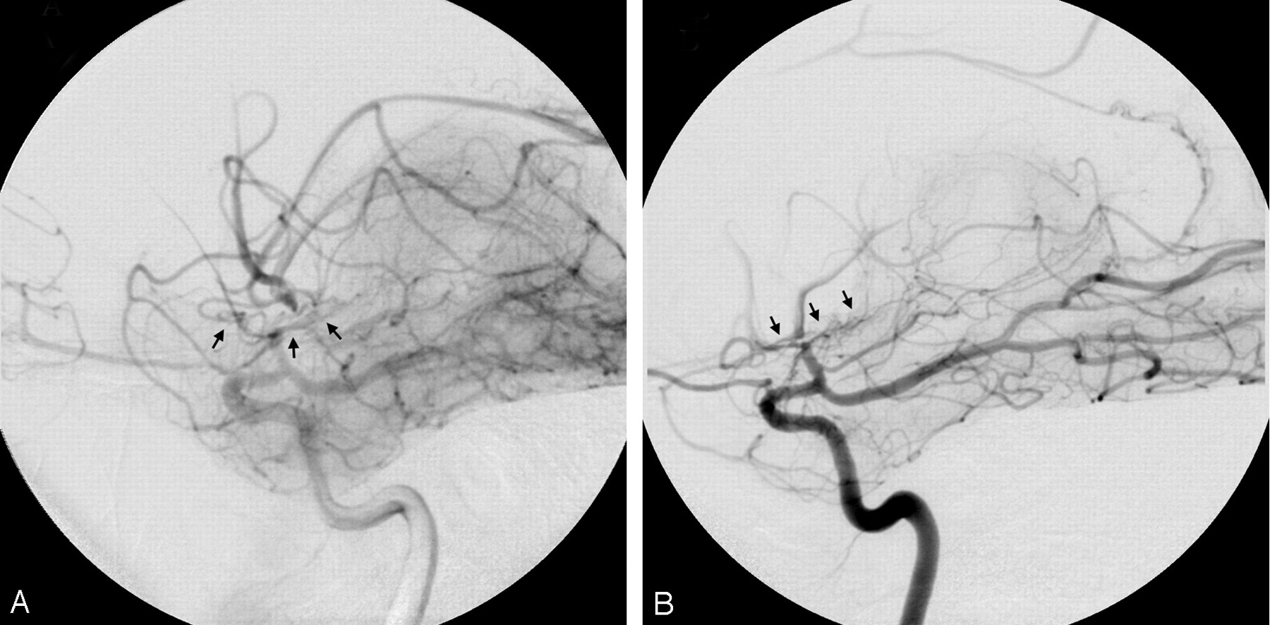
- Fig 3.
Magnified lateral projections of bilateral ICA angiography (A, right; -B, left) show only minimal collateral flows (arrows) from lenticulostriates over the base of brain, which reconstitute into the postocclusive or highly stenotic portions of the MCAs.


Tables
Diseases associated with Graves disease Ulcerative colitis, inflammatory bowel disease, pulmonary hypertension, aplastic anemia, primary sclerosing cholangitis, retroperitoneal fibrosis, membranous gromerulonephritis, immunoglobulin A/minimal change nephropathy, myasthenia gravis, cerebral venous thrombosis, thyroid carcinoma, breast carcinoma, Moyamoya syndrome Diseases associated with Moyamoya syndrome Sickle cell anemia, aplastic anemia, systemic lupus erythematous, antiphospholipid syndrome, ulcerative colitis, tuberculosis, leptospirosis, Down syndrome, Apert syndrome, neurofibromatosis, tuberous sclerosis, cranial irradiation, Graves disease - Table 2:
Summary of reported cases of Moyamoya syndrome in association with Graves disease
Study Age/Sex (y) Race Clinical Presentations Cerebral Angiographic Findings Outcome Kushima et al3 26/F Japanese Thyrotoxicosis, recurrent hemiparesis, speech disturbance Typical netlike vessels at the base of brain, occlusion of right ACA and MCA Recovery Kushima et al3 22/F Japanese Thyrotoxicosis, hemiparesis Narrowing of bilateral ICA, netlike vessels at the base of brain Recovery Liu et al4 28/F Chinese Thyrotoxicosis, hemiparesis Bilateral ACA and MCA occlusion with Moyamoya vessels, tubular stenosis of bilateral cervical ICA Not described Tendler et al5 37/F Hispanic Thyrotoxicosis, hemiparesis Right distal ICA, proximal MCA, and ACA occlusion, collateral from PCA Recovery Tendler et al5 47/F Caucasian Moyamoya disease was diagnosed 10 years before thyrotoxicosis Bilateral MCA occlusion, “puff of smoke” vessels on left basal ganglia Recovery Leno et al6 21/M Hispanic Down syndrome, hemiparesis, thyrotoxicosis Marked stenoses of both supraclinoid ICA, decreased flow over MCA, occluded ACA, prominent lenticulostrates Recovery Kim et al7 37/F Korean Thyrotoxicosis, cardiomegaly, pulmonary edema, seizure Bilateral ICA and left MCA occlusion (by MR angiography) Recovery Nakamura et al 8 23/F Japanese Thyrotoxicosis, cardiomegaly, cerebral infarction Multiple intracranial arterial stenoses, netlike collaterals around circle of Willis Recovery after revasculization Nakamura et al8 54/F Japanese Upper limits of normal range of thyroid function, hemiparesis Multiple intracranial arterial stenosis around circle of Willis Recovery after revasculization Hsu et al (this study) 40/F Caucasian Thyrotoxicosis, rapid progressive bilateral cerebral infarction Bilateral distal ICA and proximal MCA and ACA occlusion Died Note:—ACA indicates anterior cerebral artery; MCA, middle cerebral artery; ICA, internal carotid artery; PCA, posterior cerebral artery.


