
Article Figures & Data
Figures
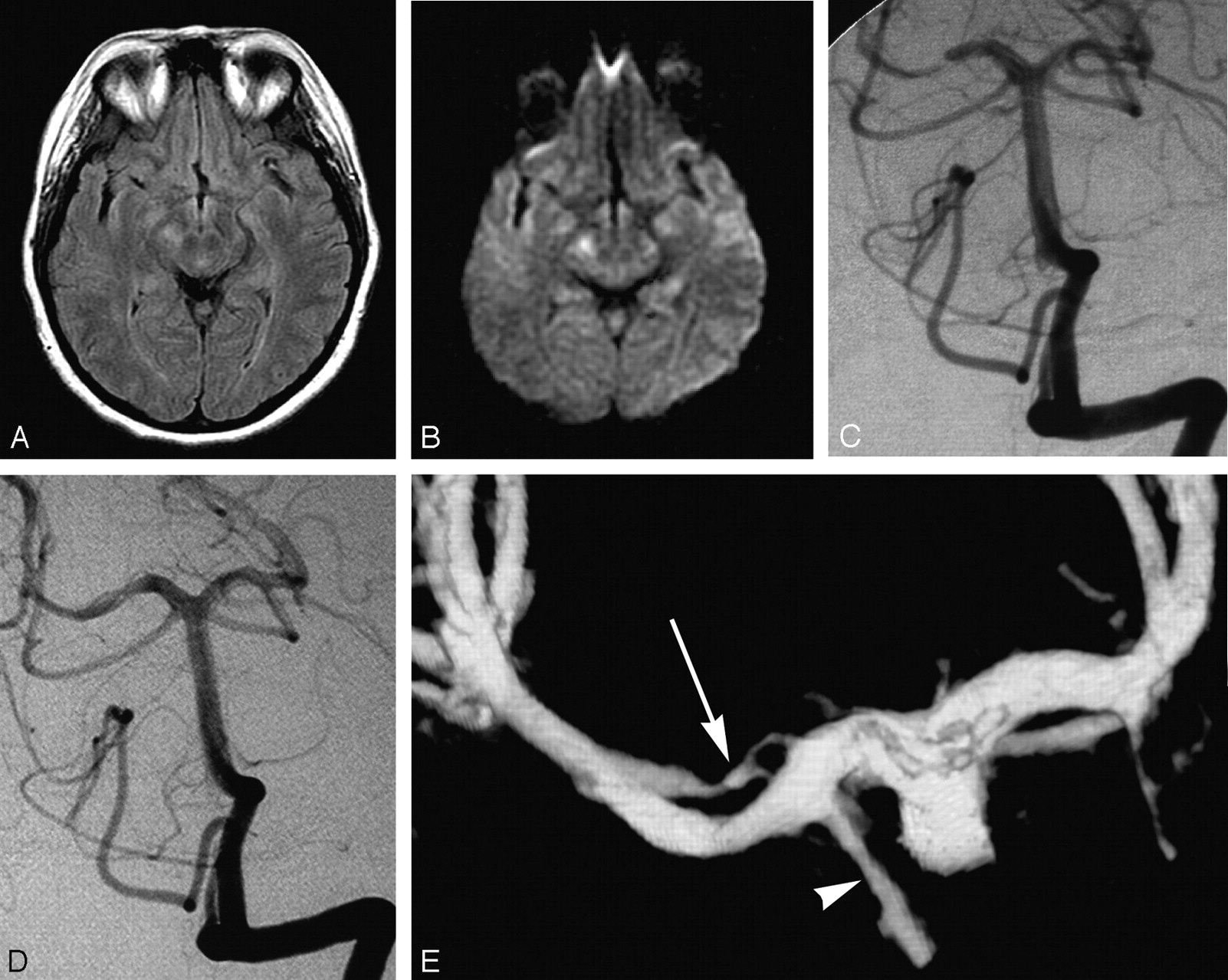
- Fig 1.
47-year-old woman presenting with quadranopsia.
A, Axial FLAIR image demonstrating hyperintensity in the right cerebral peduncle.
B, Axial diffusion-weighted image (DWI) image showing corresponding abnormal increased signal intensity. There was respective low signal intensity on the apparent diffusion coefficient map (not shown).
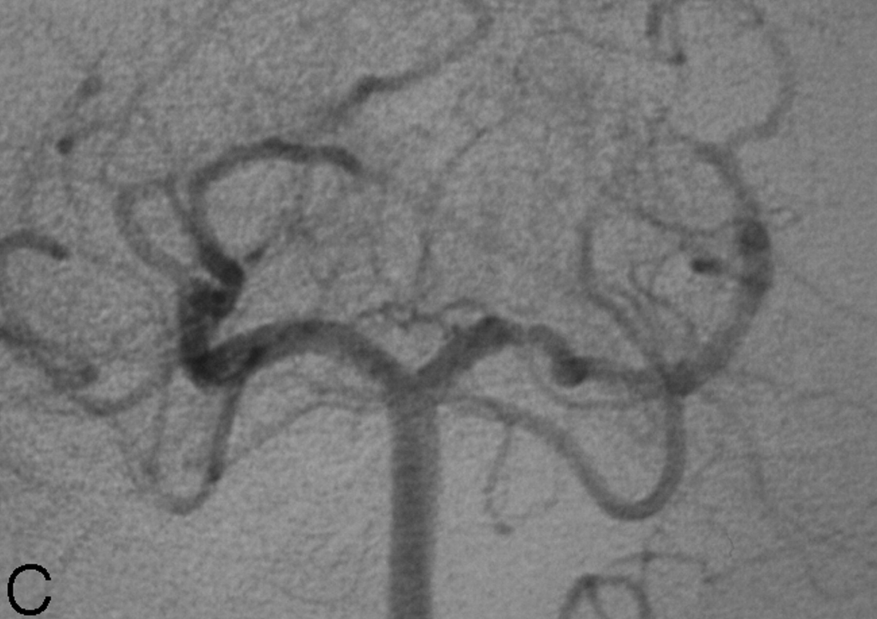
C, DSA, left vertebral artery injection, transfacial view, demonstrates a smoothly tapered focal, near-occlusive narrowing of the proximal right PCA.
D, DSA, left vertebral artery injection, transfacial view. One-year follow-up demonstrating a “double lumen” sign of the right P2 segment.
E, 3D reconstruction of left vertebral angiogram rotational DSA which shows 2 patent lumens (double lumen sign) distal to the posterior communicating artery (arrowhead). Note the irregularly recanalized false lumen (arrow) with 2 separate inflow channels.
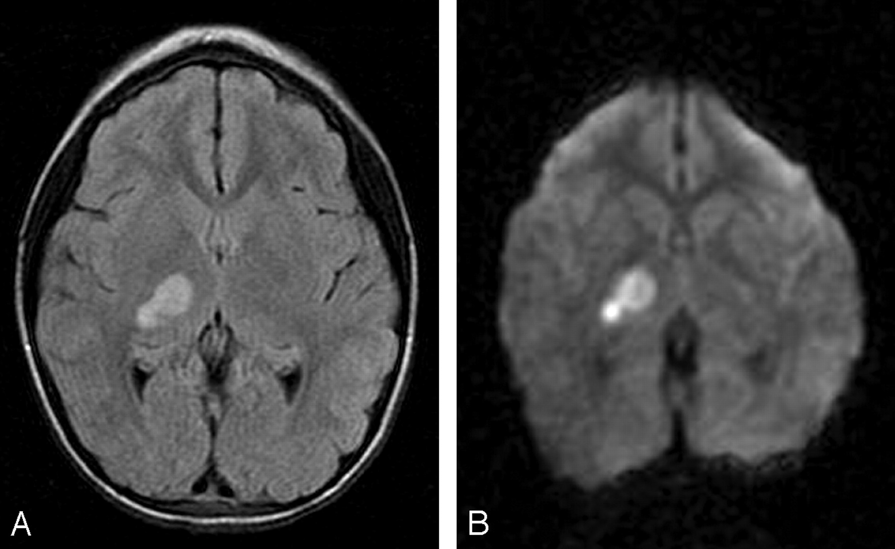
- Fig 2.
7-year-old girl presenting with left hemiparesis.
A, Axial FLAIR image demonstrating hyperintensity in the right thalamus and posterior limb of the internal capsule.
B, Axial DWI image showing corresponding restricted diffusion. There was respective low signal intensity on the apparent diffusion coefficient map (not shown).
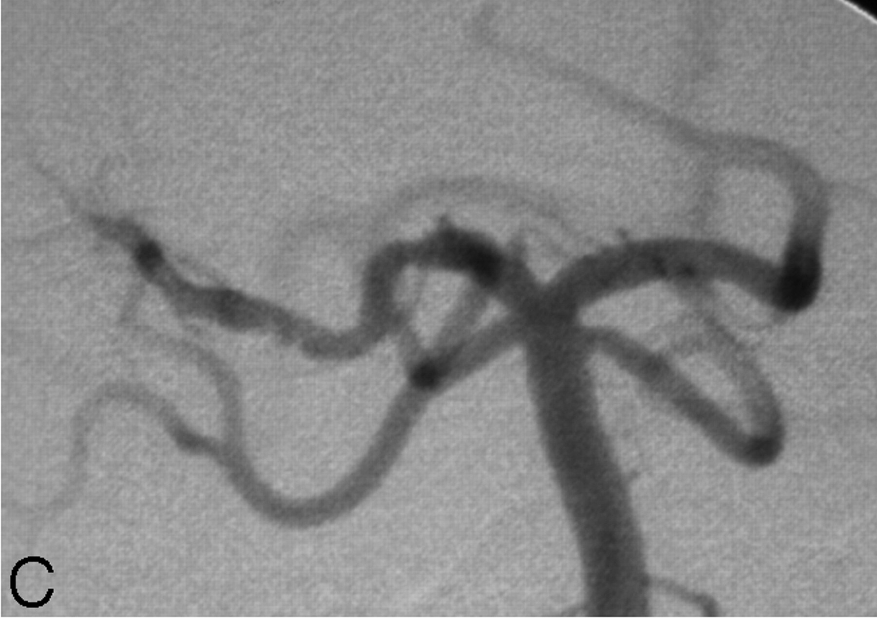
C, DSA, left vertebral artery injection, transfacial image demonstrating a focal narrowing and irregularity of the proximal right PCA.
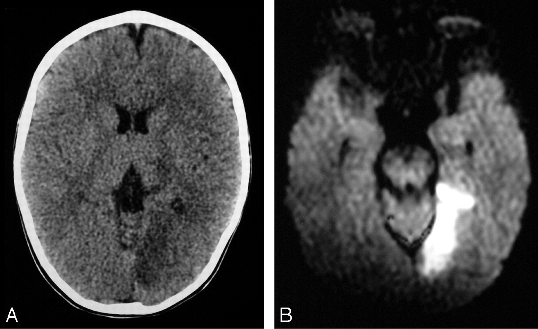
- Fig 3.
19-month old boy presenting with right hemiparesis.
A, Axial noncontrast head CT demonstrating hypoattenuation in the medial aspect of the left occipital lobe involving the cortex and subcortical white matter with loss of gray-white matter differentiation.
B, Axial DWI showing corresponding restricted diffusion compatible with infarct. There was respective low signal intensity on the apparent diffusion coefficient map (not shown).
C, DSA, right vertebral artery injection, transfacial image demonstrating an irregular and narrowed left proximal PCA.


