
Article Figures & Data
Figures
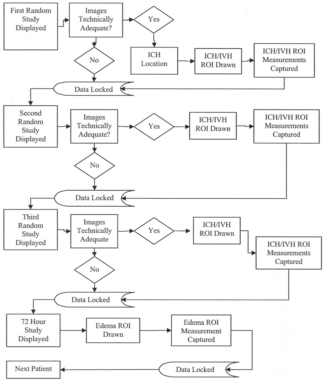
- Fig 1.
Masked read-flow diagram for each patient.
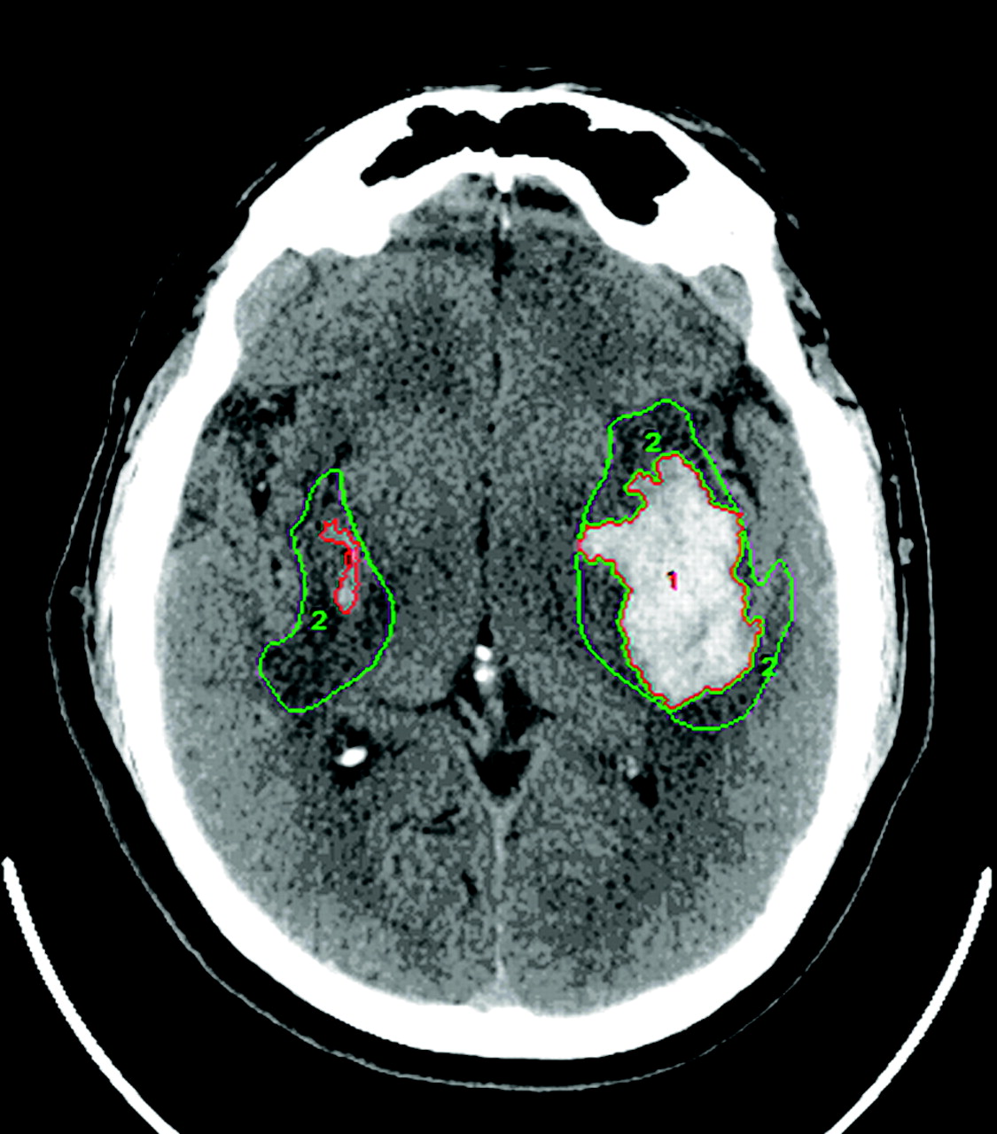
- Fig 2.
CT scan showing region of interest. Region of interest, area 1, defines zones of ICH. Region of interest, area 2, defines zones of edema. From these reader-defined regions, the Analyze software calculated statistics of each region of interest (area in square millimeters and volume in cubic millimeters).
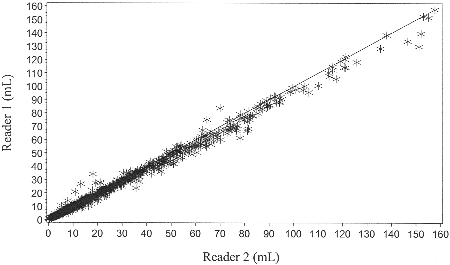
- Fig 3.
Graph shows agreement between reader 1 and reader 2 (ICH volume measurement).
- Fig 4.
Graph shows agreement between reader 1 and reader 2 (IVH volume measurement).
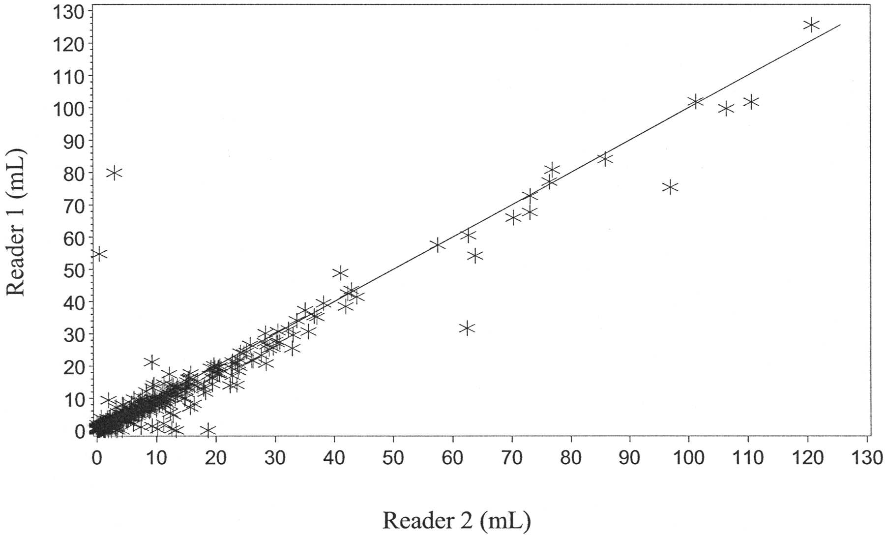
- Fig 5.
Graph shows agreement between reader 1 and reader 2 (edema volume measurement).


Tables
Intra-reader variation of ICH, IVH, and edema volumes by treatment
Placebo 40 mcg 80 mcg 160 mcg Total r2 ICH volume (mL) N 44 37 50 48 179 .9844 Mean (SD) −0.2 (1.6) 0.1 (0.5) −0.1 (2.0) −0.2 (2.5) −0.1 (1.9) IVH Volume (mL) N 44 37 50 48 179 .9918 Mean (SD) −0.1 (0.2) −0.1 (0.1) 0.4 (2.2) 0.0 (0.2) 0.1 (1.2) Edema volume (mL) N 14 14 10 12 50 .7266 Mean (SD) −12.6 (36.2) 0.0 (0.2) −0.2 (0.7) −0.6 (2.3) −3.7 (19.5) Note:—ICH indicates intracerebral hemorrhage; IVH, intraventricular volume.
In this issue


Jump to section
Related Articles
Cited By...
- 17p12 Influences Hematoma Volume and Outcome in Spontaneous Intracerebral Hemorrhage
- Accuracy of the ABC/2 Score for Intracerebral Hemorrhage: Systematic Review and Analysis of MISTIE, CLEAR-IVH, and CLEAR III
- Neuroimaging in Intracerebral Hemorrhage
- Intracranial-Derived Atherosclerosis Assessment: An In Vitro Comparison between Virtual Histology by Intravascular Ultrasonography, 7T MRI, and Histopathologic Findings
- Quantitative CT Densitometry for Predicting Intracerebral Hemorrhage Growth
- Cerebral Atrophy is an Independent Risk Factor for Unfavorable Outcome After Spontaneous Supratentorial Intracerebral Hemorrhage
- The Modified Graeb Score: An Enhanced Tool for Intraventricular Hemorrhage Measurement and Prediction of Functional Outcome
- Poor Prognosis in Warfarin-Associated Intracranial Hemorrhage Despite Anticoagulation Reversal
- Pharmacological Deep Vein Thrombosis Prophylaxis Does Not Lead to Hematoma Expansion in Intracerebral Hemorrhage With Intraventricular Extension
- Intraventricular Hemorrhage: Severity Factor and Treatment Target in Spontaneous Intracerebral Hemorrhage
- Density and Shape as CT Predictors of Intracerebral Hemorrhage Growth
- Intraventricular hemorrhage: Anatomic relationships and clinical implications
- Determinants of Intracerebral Hemorrhage Growth: An Exploratory Analysis