
Article Figures & Data
Figures
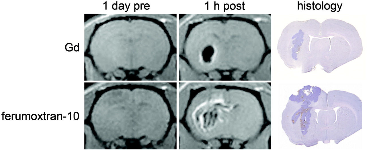
- Fig 1.
Imaging intratumor injection. Nude rats with 7-day intracerebral small cell lung carcinoma xenografts were scanned by MR imaging 1 day before and 1 hour after intratumor injection of either gadolinium (top) or ferumoxtran-10 (bottom) contrast agents. Axial T1-weighted MR images were obtained at 3T by using a custom rat head coil.9 One day after contrast injection, coronal 100-μm rat brain sections were evaluated for histology by hematoxylin staining. Delivery of the USPIO is highly heterogeneous throughout the tumor and brain around the tumor, in contrast to the more homogeneous distribution of gadolinium.
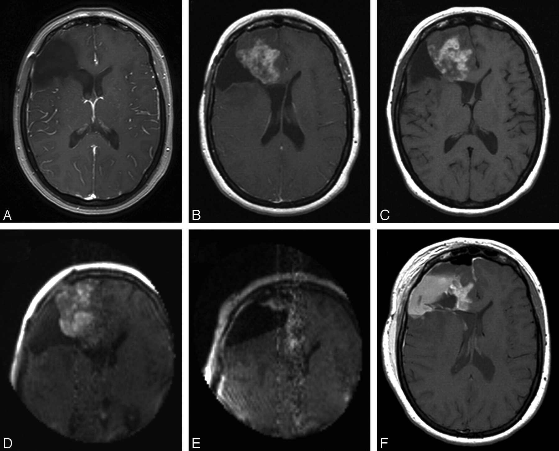
- Fig 2.
T1-weighted MR imaging in a patient with recurrence of a low-grade astrocytoma. The patient had stable disease after previous craniotomy (A) but showed recurrence on gadolinium images in March 2005 (B). The patient received ferumoxtran-10, and 24 hours later underwent preoperative MR imaging (C) as well as MR imaging with the 0.15T intraoperative scanner before (D) and during (E) craniotomy. Postoperative MR imaging (F) correlated with the residual areas of enhancing tumor seen on intraoperative MR imaging. High signal intensity in the tumor cavity, not present on intraoperative MR imaging, demonstrates the problem of differentiating residual enhancing tumor from blood products. Because the ferumoxtran-10 persists for 4–7 days, the postoperative MR images were exactly the same with or without the addition of gadolinium.
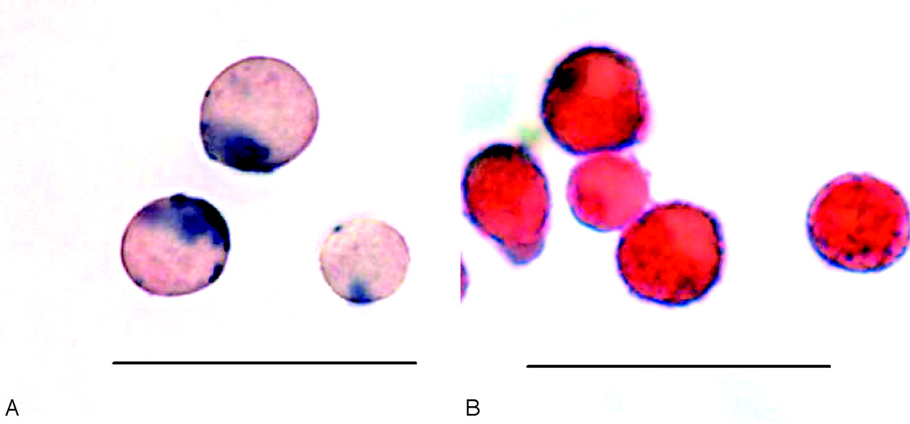
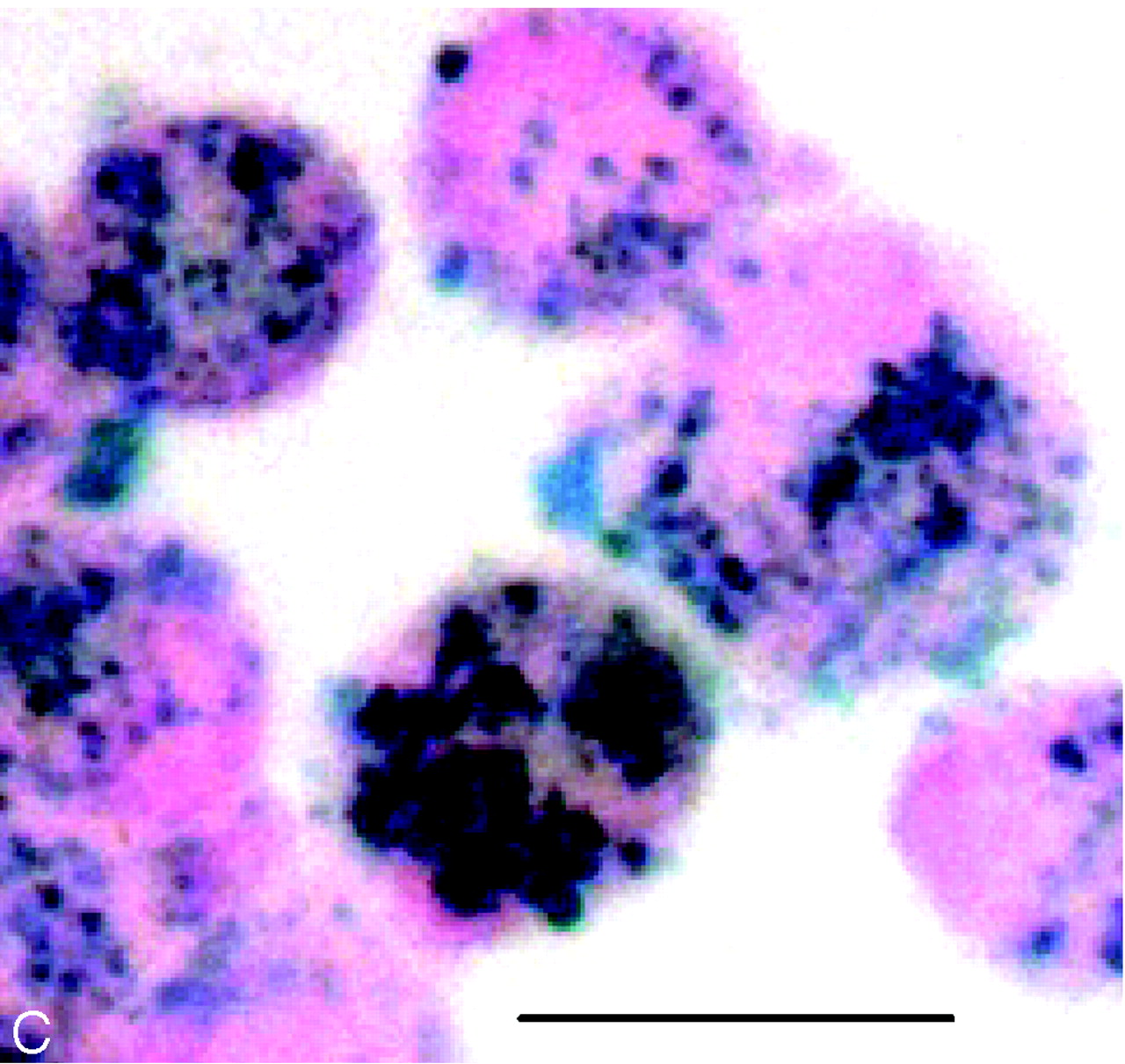
- Fig 3.
Examples of Prussian blue–stained histopathology of mouse Sca1+ hematopoietic stem cells labeled with ferumoxides/poly-l-lysine (A), human CD 34 AC 133 hematopoietic stem cells labeled with ferumoxides/protamine sulfate (B), and human mesenchymal stem cells labeled with ferumoxides/protamine sulfate (C). Both mouse and human hematopoietic stem cells have been used as endothelial precursor cells (Bar = 20 μm).
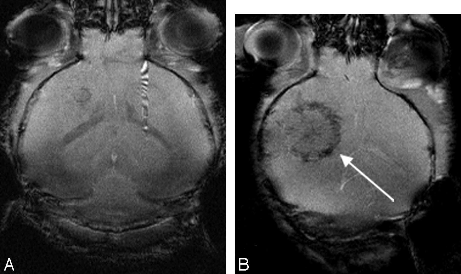
- Fig 4.
Axial T2*-weighted gradient-echo MR imaging (TR/TE, 450/4.2 ms, with resolution at 500 × 80 x 70 μm) at 7T performed 10 days after implantation of RT2 rat glioma into the brain of a mouse that received a tail vein infusion of 500,000 killed ferumoxides/poly-l-lysine–labeled Sca1+ endothelial precursor cells (A) or live 500,000 ferumoxides/poly-l-lysine–labeled Sca1+ endothelial precursor cells (B) 2 days before implantation of tumor. The hypointense ring (arrow) surrounding the tumor is clearly visible on the animal receiving magnetically labeled endothelial precursor cells and represents the incorporation of these cells into ongoing vasculogenesis and neovasculature of the growing tumor. Killed labeled cells served as a control and did not migrate to the tumor.17 MR imaging courtesy of Stasia A. Anderson, MD.
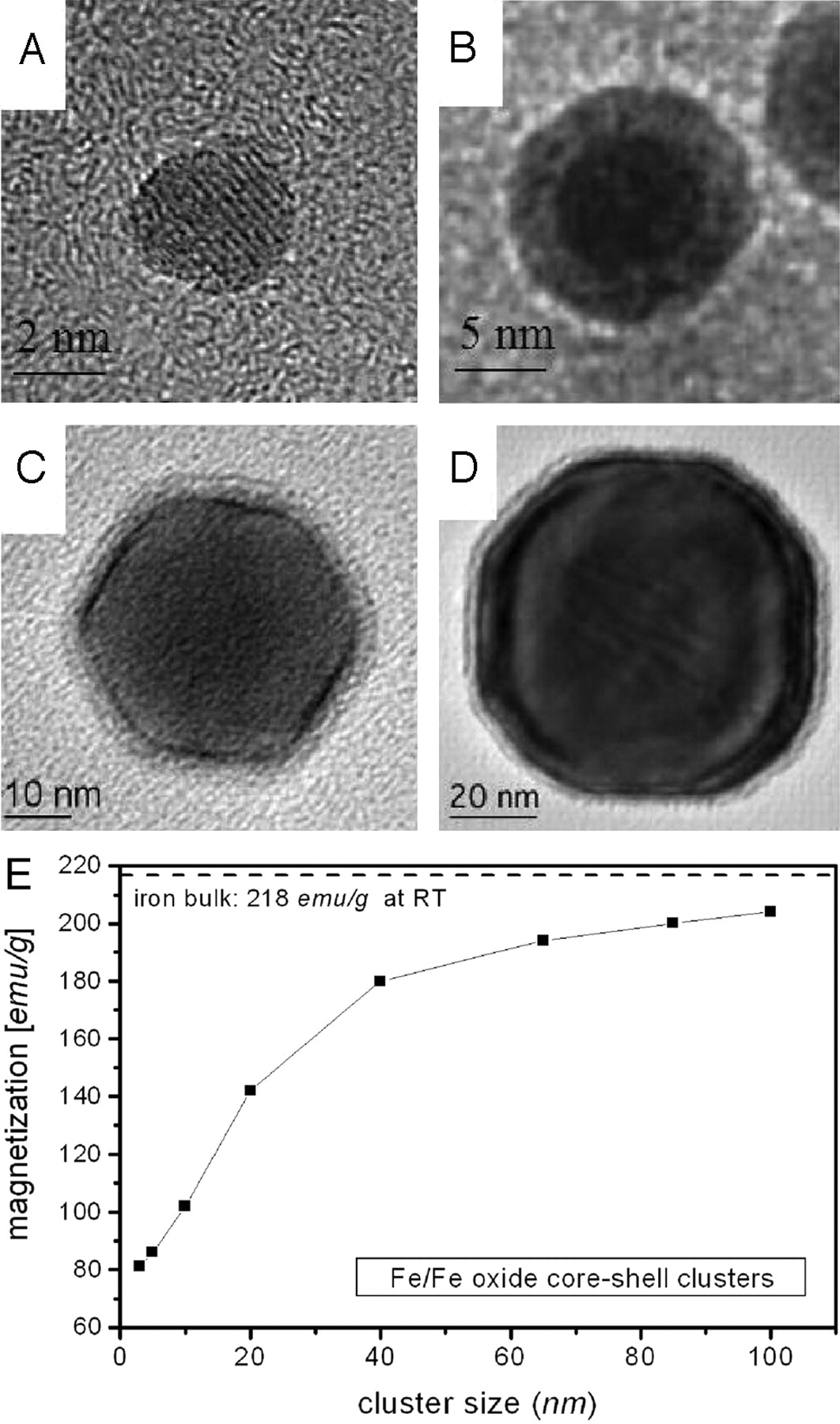
- Fig 5.
High-resolution transmission electron micrographs of the oxide-coated iron clusters with the diameters approximately 3–85 nm, prepared on carbon microgrids (A-D). The size-dependent-specific magnetic moments of iron oxide–coated iron nanoclusters are shown in panel E.


In this issue


Jump to section
- Article
- Abstract
- PET Imaging of Gliomas
- Imaging Delivery to the Brain
- Preclinical and Clinical Studies of CNS Imaging with Iron Oxide MR Imaging Agents
- Tracking Magnetically Labeled Stem Cells
- Nanotechnology, Nanobiology, and Nanomedicine
- Magnetic Nanoparticles for Biomedical Applications
- Functionalized Nanoparticles for Brain Tumor Imaging and Treatment
- Stealth Nanoparticle Delivery Across the BBB
- The Impact of the Nanobiotechnology Center at Cornell University: The Study of Synaptic Transmission
- A Future Direction: Nanotechnology for CNS Regeneration
- Conclusion
- Acknowledgments
- Footnotes
- References
- Figures & Data
- Info & Metrics
- Responses
- References
Related Articles
Cited By...
- No citing articles found.