
Article Figures & Data
Figures
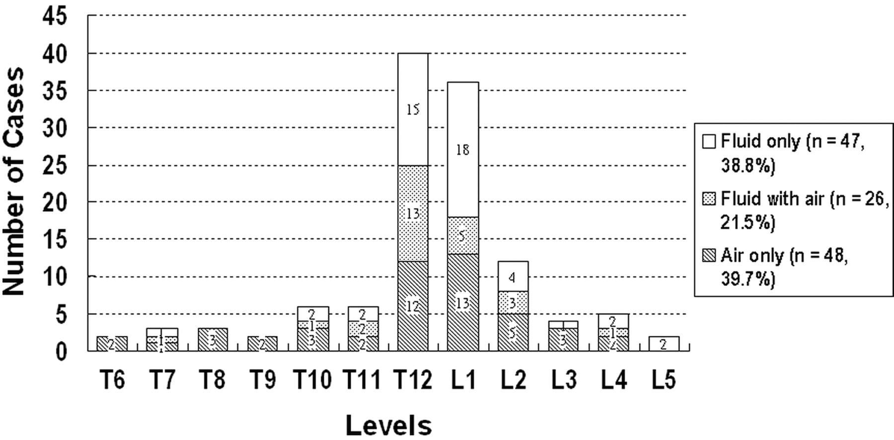
- Fig 1.
Chart showing the distribution of vertebral osteonecrosis at different levels. Changes of the content of affected vertebral bodies, intravertebral fluid only (blank bars), coexistence of both fluid and air (dot bars), and intravertebral air only (oblique line bars) are also shown. T12 (n = 40) and L1 (n = 36) were most often involved. Of 121 osteonecrotic vertebral bodies, 48 (39.7%) appeared with only intravertebral air, 47 (38.8%) appeared with only intravertebral fluid, and 26 (21.5%) appeared with both air and fluid.
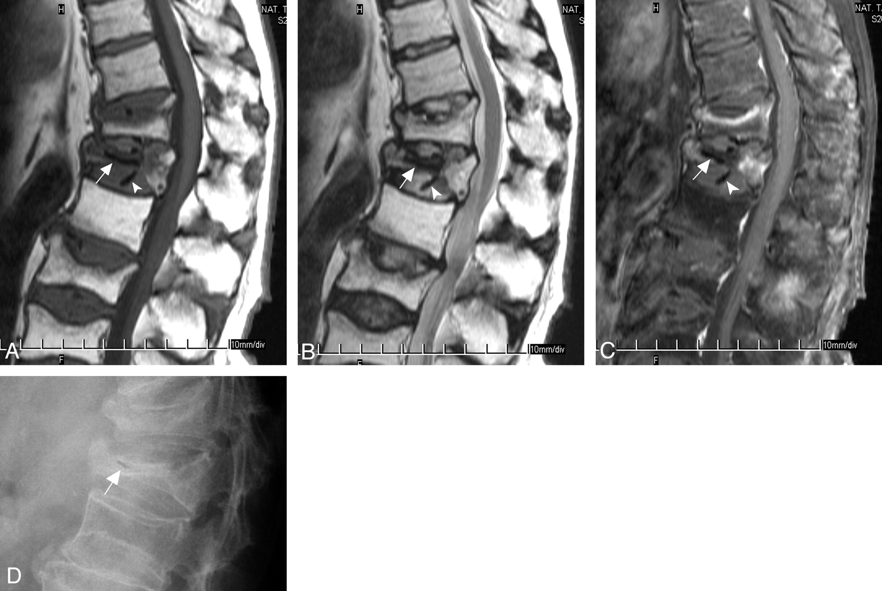
- Fig 2.
MR images and plain radiograph of a 73-year-old man who had compression fractures at T12, L1, L3, and L4 vertebral bodies and osteonecrosis at L1 vertebral body. There is intravertebral air in the severely collapsed T12 vertebral body and also intradiskal air in the adjacent T11–T12, T12–L1, and L1–L2 disks. In the center of the collapsed vertebral body is a horizontally oriented signal intensity void line (white arrows, panels A–C) on all pulse sequences. Also, several signal intensity void dots or rods in the adjacent disks (above and below) are shown on all pulse sequences (white arrowheads, panels A–C). A, Sagittal T1-weighted turbo spin-echo image (600/12, 4-mm section thickness) shows hypointensity in the most of the vertebral body; only the posterior fifth is relatively spared. B, Sagittal T2-weighted turbo spin-echo image (4000/120) shows some hypointensity at anterior four fifths of the collapsed vertebral body. C, Sagittal contrast-enhanced T1-weighted, fat-suppressed turbo spin-echo image (690/12, 4-mm section thickness) shows nonenhancement portion at middle three fifths of the collapsed vertebral body. D, Lateral view of plain radiograph shows severely collapsed vertebral body with a short and horizontally oriented air cleft at the anterior second fifth of the vertebral body (white arrow).
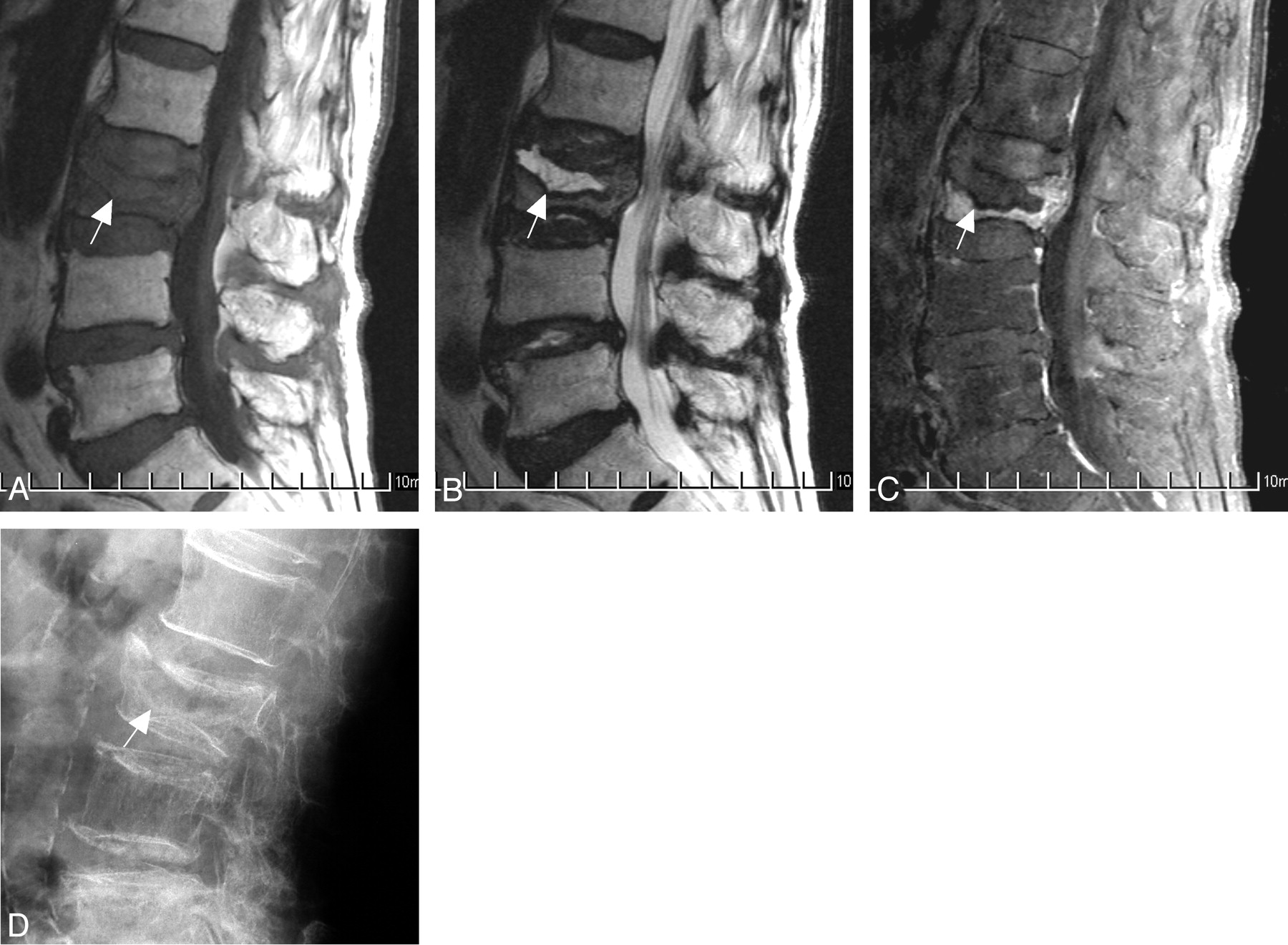
- Fig 3.
MR images and plain radiograph of an 82-year-old woman who had compression fractures and osteonecrosis at the L3 vertebral body. There is only intravertebral fluid in the mildly collapsed vertebral body. A, Sagittal T1-weighted turbo spin-echo image (600/12, 4-mm section thickness) shows complete bone marrow replacement by low signal intensity (arrow). B, Sagittal T2-weighted turbo spin-echo image (4000/128) shows homogeneous hyperintensity at the anterior superior portion of the vertebral body (arrow). The margin of the hyperintense area is well demarcated. C, Sagittal contrast-enhanced T1-weighted fat-suppressed turbo spin-echo image (700/12, 4-mm section thickness) shows nonenhancement of the anterior and superior portions of the vertebral body (arrow). The nonenhancing area corresponds to the hyperintense area of the T2WI in panel B. The remaining portion of this vertebral body had faint enhancement. D, Lateral view of plain radiograph shows faint radiolucent area at anterior superior portion of the vertebral body (arrow). The radiolucent area corresponds to the T2 hyperintnese area in the panel B.
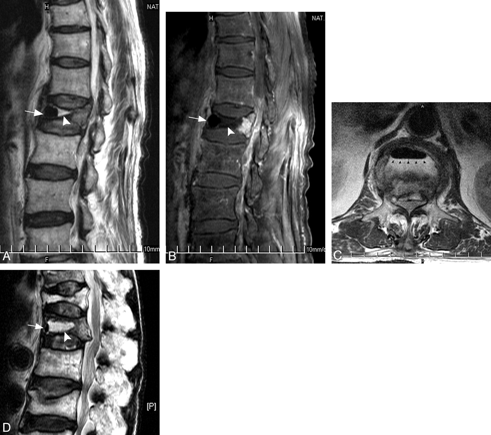
- Fig 4.
A–D, MR images of an 83-year-old man who was diagnosed with osteonecrosis at the L1 vertebral body. There coexists both intravertebral fluid and air in the affected vertebral body. A, Sagittal T2-weighted turbo spin-echo image (4,000/110) shows hyperintensity at middle third (arrowhead) and signal intensity void at anterior third (arrow) of the vertebral body. B, Sagittal contrast-enhanced T1-weighted, fat-suppressed turbo spin-echo image (550/12, 4-mm section thickness) shows nonenhancement of the anterior two thirds of the vertebral body (arrow and arrowhead). Only the posterior one third of vertebra had enhancement. C, Transverse T2-weighted turbo spin-echo image (4000/110) shows an ellipsoidal signal intensity void area (arrow) anterior to the hyperintensity within the vertebral body. The interface between these 2 components revealed an air–fluid level (small black arrowheads). D, Sagittal T2-weighted turbo spin-echo MR image of another 76-year-old man who was diagnosed with osteonecrosis at T12 vertebral body. There coexists both intravertebral fluid (most, arrowhead) and air (minority, arrow) in the affected vertebral body.


Tables
Age (y) Men Women Total 51–60 1 5 6 61–70 8 28 36 71–80 13 36 49 81–90 8 13 21 Total 30 82 112 Comparison in Categories Severe* (n = 67) Mild* (n = 54) P Value Air only (n = 48) 43 (90) 5 (10) Fluid only (n = 47) 13 (28) 34 (72) <.05 Presence of fluid† (n = 73) 24 (33) 49 (67) Absence of fluid (n = 48) 43 (90) 5 (10) <.05 Presence of air‡ (n = 74) 54 (73) 20 (27) Absence of air (n = 47) 13 (28) 34 (72) <.05 Numbers in parentheses are percentages.
* Vertebrae with >50% preserved height were categorized as mild collapse, and those with <50% preserved height were categorized as severe collapse.
† Presence of fluid represents the sum of case numbers of fluid only and fluid with air.
‡ Presence of air represents the sum of case numbers of air only and fluid with air.
- Table 3:
Changes of content of adjacent and next-to-adjacent disks according to different manifestation of vertebral osteonecrosis
Changes of Adjacent Disks Changes in Affected Vertebrae Air Only (n = 96) Fluid with Air (n = 52) Fluid Only (n = 94) P Value Air in adjacent disks 41 11 <.05 Upper 21 6 0 Lower 20 5 0 Air in disks next to adjacent disks 12 3 <.05 Next to upper 8 1 0 Next to lower 4 2 0


