
Article Figures & Data
Figures
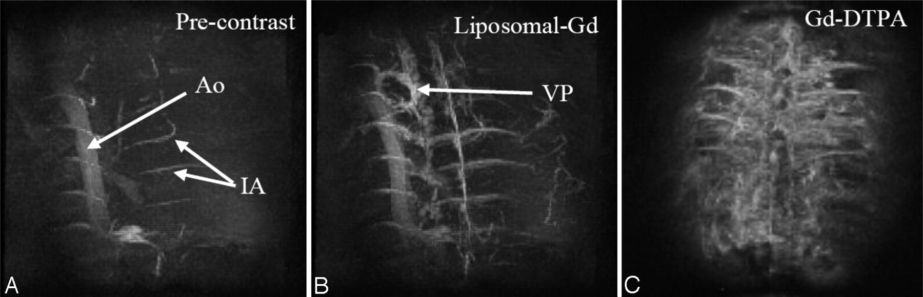
- Fig 1.
Coronal MIP images of the thoracolumbar spine region acquired precontrast (A), post–liposomal-Gd (B), and post–Gd-DTPA (C). The images were acquired using the following parameters: TR/TE, 18.3/2.8 ms; flip angle, 30°; image matrix, 128 × 128 × 64; FOV, 41 × 28 × 25 mm; no. of signal intensity average, 1. The total scan time was 2.5 minutes. The descending aorta (Ao), and intercostal arteries (IA) are demonstrated in the precontrast image (A). The epidural venous plexus (VP) and more details about the intercostal arteries are demonstrated in post–liposomal-Gd image (B). The tissue enhancement due to diffusion of Gd-DTPA into the extravasacular space is shown in post-Gd-DTPA image (C). Post–liposomal-Gd and post–Gd-DTPA images were acquired in different animals.
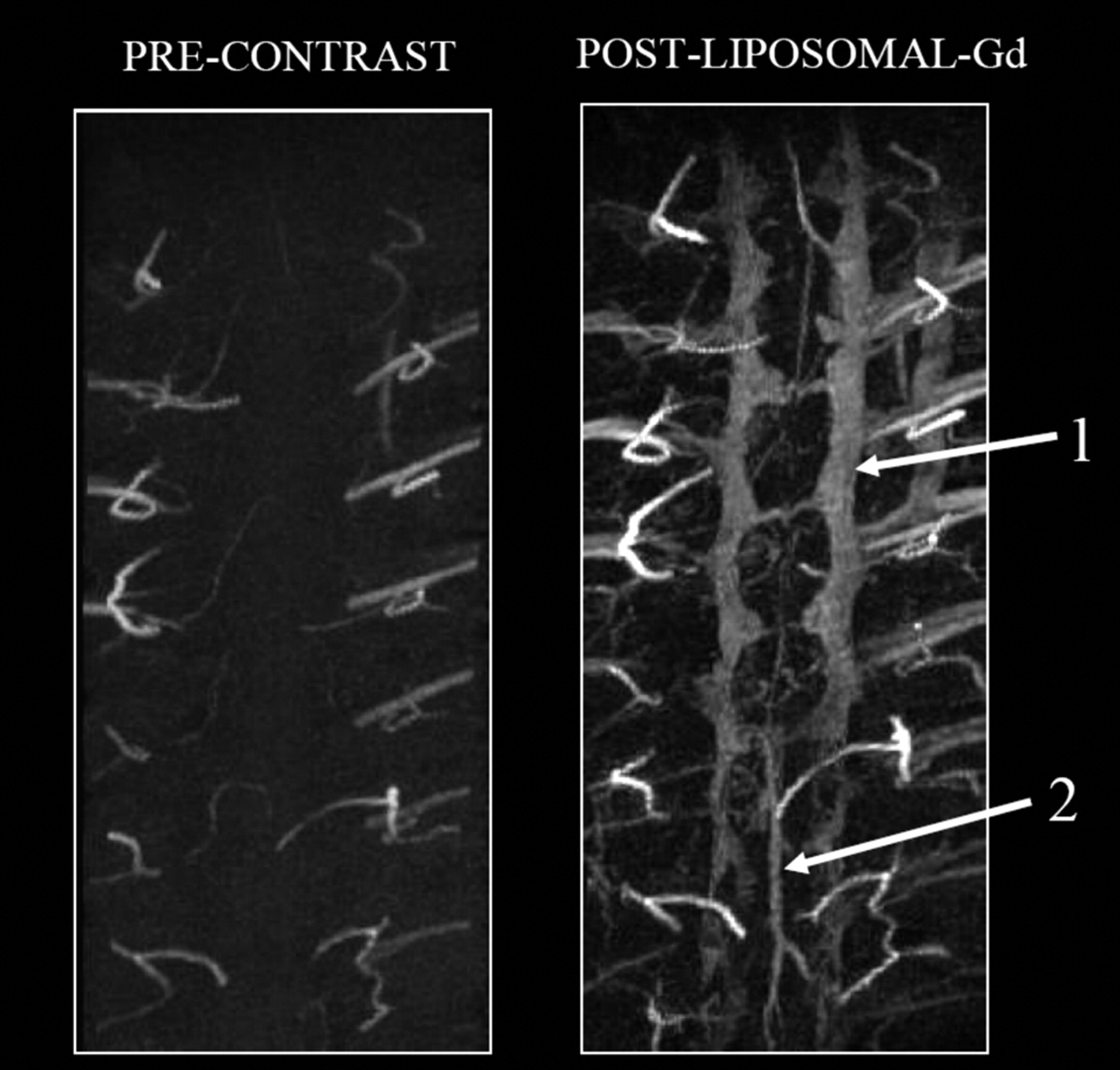
- Fig 2.
Coronal MIP images of the thoracolumbar spine region acquired before (left) and after (right) administration of liposomal-Gd. The images were acquired using the following parameters: TR/TE, 18.3/2.8 ms; flip angle, 30°; image matrix, 256 × 256 × 256; FOV, 41 × 28 × 25 mm, no. of signal intensity average, 3. The epidural venous plexus (1) and the posterior spinal vein (2) are clearly visible in the post–liposomal-Gd image.
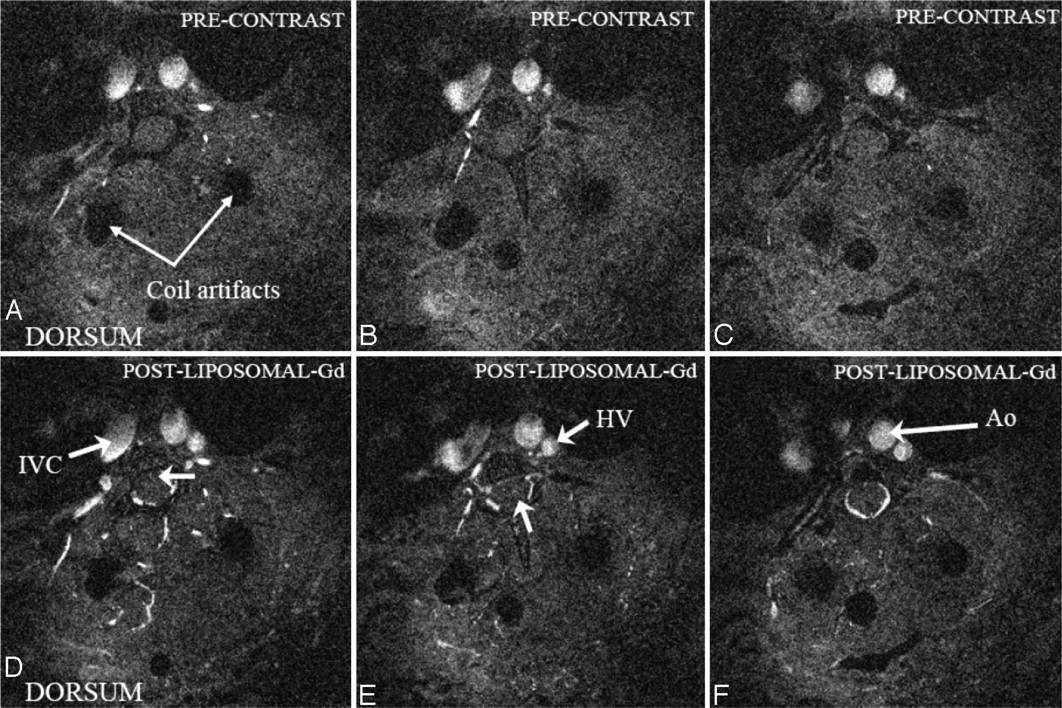
- Fig 3.
Precontrast (top row) and postcontrast (bottom row) axial sections at different locations through the thoracolumbar spine region showing several important vascular features (Ao, aorta, IVC, inferior vena cava; HV, hemiazygous vein). Blood vessels penetrating the spinal cord can be visualized in D and E (arrow). The epidural venous plexus is demonstrated in F. The artifacts due to the implantation of radio-frequency coil on the dorsal side are also visible. These images are acquired after administration of liposomal-Gd.
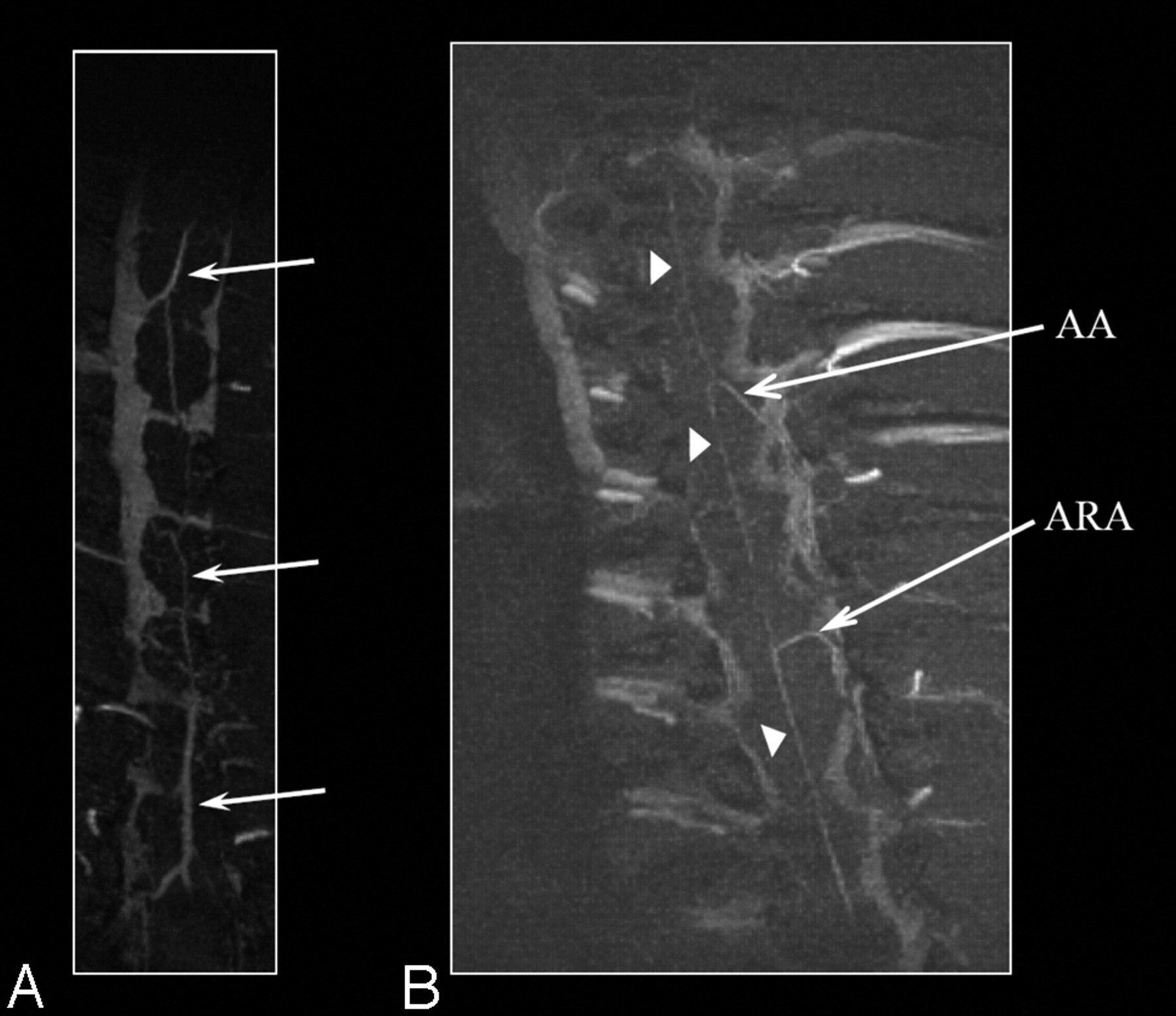
- Fig 4.
Coronal MIP images of the thoracolumbar spine region obtained with liposomal-Gd. The posterior spinal vein (PSV) is clearly seen in A (arrows). The anterior spinal artery (arrowheads), the artery of Adamkiewicz (AA) and an accessory anterior radicular artery (ARA) can be clearly seen in B.


Tables
- Table 1:
Contrast-to-noise ratio (CNR) for anatomic features in standard-resolution images obtained with Gd-DTPA to images obtained with liposomal-Gd
Anatomic Feature Gd-DTPA Liposomal-Gd Epidural venous plexus 8.2 21.0 Intercostal arteries 13.2 22.0 Hemiazygous vein 19.8 36.4 Aorta 8.3 18.2 Note:—Gd-DTPA indicates gadolinium-diethylene-triaminepentaacetic acid; Liposomal-Gd, gadolinium-encapsulated long-circulating liposomes.
- Table 2:
Contrast-to-noise ratio (CNR) for anatomic features in high-resolution images obtained precontrast and after administration of liposomal-Gd
Anatomic Feature Precontrast Liposomal-Gd Epidural venous plexus −0.1 6.5 Posterior spinal vein −0.5 2.3 Anterior spinal artery 1.4 4.1 Radicular artery (of Adamkiewicz) 3.2 6.8 Note:—Liposomal-Gd indicates gadolinium-encapsulated long-circulating liposomes.
In this issue


Jump to section
Related Articles
Cited By...
- Dual-Energy Computed Tomography Imaging of Atherosclerotic Plaques in a Mouse Model Using a Liposomal-Iodine Nanoparticle Contrast Agent
- Activatable Magnetic Resonance Imaging Agent Reports Myeloperoxidase Activity in Healing Infarcts and Noninvasively Detects the Antiinflammatory Effects of Atorvastatin on Ischemia-Reperfusion Injury